Abstract
Rectus sheath hematoma is a clinical entity characterized by the presence of blood within rectus abdominis muscle sheath. The aim of this study was to analyze clinical characteristics, diagnostic approach, treatment strategy, and outcomes of patients with rectus sheath hematoma. Patients diagnosed and treated for spontaneous rectus sheath hematoma between March 2010 and March 2014 were included in the study. A total of 10 patients were diagnosed as spontaneous rectus sheath hematoma. The mean age was 66.5 ± 16.9 years, and the mean hospital stay was 4.4 ± 1.8 days. There was no mortality. Six patients were using anticoagulant or antiplatelet agents. Eight patients recovered after conservative treatment. Two patients underwent surgery. Spontaneous rectus sheath hematoma is associated with anticoagulant therapy. Cases with abdominal pain and a non-pulsatile abdominal mass particularly in elderly women should be kept in mind. Treatment is mostly based on supportive care to preserve hemodynamic stability.
Keywords: Hematoma, Rectus sheath, Surgery, Conservative approach
Introduction
Rectus sheath hematoma (RSH) is a clinical entity characterized by the presence of blood within the rectus abdominis muscle sheath. It may be caused by damaged epigastric vessels or rectus muscle [1]. With the increasing use of antiplatelet and anticoagulant therapy, the frequency of spontaneous rectus sheath hematoma is also increasing [2]. Acute abdominal pain and a palpable abdominal mass with history of straining of the abdominal muscles, such as trauma, coughing, sneezing, and twisting, are typical features of RSH [3, 4]. In RSH, early diagnosis and treatment are essential to minimize further complications, including hemodynamic instability, abdominal compartment syndrome, multiple organ dysfunction syndrome, and death [5]. Both ultrasonography and computerized tomography (CT) are being used to differentiate a diagnosis between rectus sheath hematoma and other pathologies with a remarkable degree of success [6].
The aim of this study is to analyze clinical characteristics, our diagnostic approach, treatment strategy, and outcomes of patients with spontaneous rectus sheath hematoma (SRSH).
Patients and Methods
Patients who were admitted to our emergency surgery clinic between March 2010 and March 2014 were evaluated. Patients who were diagnosed and treated for spontaneous rectus sheath hematoma were included in the study. We retrospectively inspected patient files for all demographic characteristics, comorbidities, laboratory parameters, findings of physical examination, and radiological tests. We also tried to identify predisposing factors for spontaneous RSH. Treatment modalities, follow-up parameters, length of hospital stay, and outcome were also assessed.
Results
During the study period, a total of 10 patients were diagnosed with SRSH in our institution. the female/male ratio was 4:1 (eight women and two men). The mean age of these patients was 66.5 ± 16.9 years. The mean hospital stay was 4.4 ± 1.8 days. There was no any mortality. Patient characteristics are presented in Table 1.
Table 1.
Detailed information about our patients with rectus sheath hematoma
| Case | Age/gender | History | Medication | INR (normal range 0.9–1.17) | Location | Size (mm) | Diagnostic tool | Hospital staya (days) | Treatment |
|---|---|---|---|---|---|---|---|---|---|
| 1 | 25/M | Suspected BD | None | 0.9 | LRQ | 130 × 40 | USG | 2 | Cons. |
| 2 | 73/F | AF, CVD | Warfarin | 3.0 | LLQ | 13 × 26 | USG | 6 | Cons. + ET |
| 3 | 80/F | HT, cough | None | 1.0 | LLQ | 52 × 50 | USG | 3 | Cons. |
| 4 | 80/M | CAD | ASA | 0.95 | LRQ | 65 × 25 | USG | 3 | Cons |
| 5 | 78/F | None | None | 1.0 | LRQ | 60 × 55 | USG | 2 | Cons. |
| 6 | 74/F | DM, HT, COPD | None | 0.97 | LLQ | 20 × 24 | USG | 6 | Cons. |
| 7 | 61/F | MI | ASA, clopidogrel | 1.0 | LLQ | 130 × 100 | Surgery | 6 | Surgery |
| 8 | 62/F | MS | Enoxaparin | 0.98 | LLQ | 20 × 50 | CT | 6 | Surgery + ET |
| 9 | 76/F | CVD, CAD | ASA, clopidogrel | 0.99 | LRQ | 140 × 100 | CT | 4 | Cons. |
| 10 | 56/F | AF, CVD | Warfarin | 2.8 | LRQ | 140 × 110 | CT | 6 | Cons. + ET |
F female, M male, BD bleeding disorder, AF atrial fibrillation, CVD cerebrovascular disease, HT hypertension, CAD coronary artery disease, DM diabetes mellitus, COPD chronic obstructive pulmonary disease, MI myocardial infarction, MS mitral valve stenosis, ASA acetylsalicylic acid, LRQ lower right quadrant, LLQ lower left quadrant, USG ultrasonography, CT computerized tomography, Cons conservative treatment, ET erythrocyte transfusion, INR international normalized ratio
aLength of hospital stay
One of our patients was a 25-year-old male patient who had a previous medical history of prolonged bleeding after childhood circumcision. Six of our patients had a history of anticoagulant or antiplatelet drug use due to cardiac or cerebrovascular diseases. Two patients without anticoagulant medication use had a history of persistent cough and hypertension. One patient suffered from asthma, and the other patient had cough because of acute pharyngitis. Our last patient was a 78-year-old female without any obvious etiology or predisposing factor for SRSH.
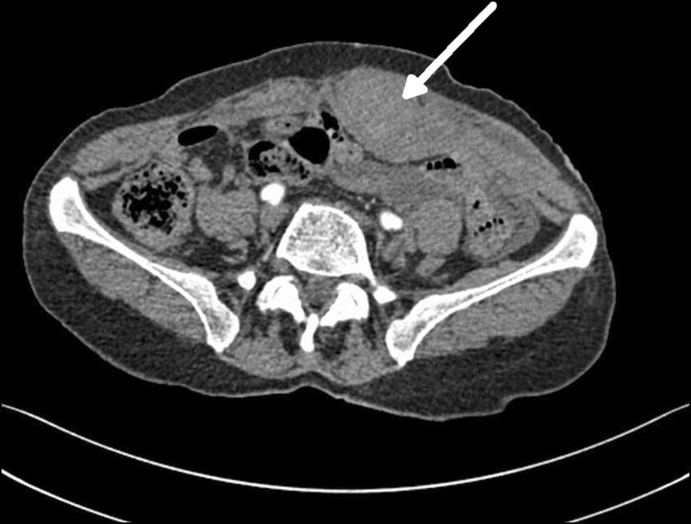
Sudden onset of abdominal pain was experienced by eight out of 10 patients. Two patients had no significant physical tension on rectus muscle. Clinical suspicion of rectus hematoma was due to palpable tender mass on the abdominal wall which was confirmed by ultrasound in all cases. Abdominal CT was used for further diagnostic purposes in three patients who had greater hematomas in a size of 14–20 cm (Fig. 1).
Fig. 1.
A typical image of a rectus hematoma that belongs to case 7. The arrow shows left-sided rectus hematoma
Eight of our patients responded to conservative treatment. The remaining two patients underwent surgery. Ligation of an epigastric vein was performed for one of these patients who had intact inferior epigastric artery, whereas only drainage was performed for the second patient since no bleeding vessel could be found. Packed red cell transfusion was required in three cases.
Discussion
RSH is one of the uncommon but clinically important causes of acute abdominal pain [7]. RSH is the presence of blood inside the sheath of the rectus abdominis muscle. Its physiopathology is based on torn epigastric vessels or ruptured muscle fibers [8]. RSH occur more frequently in women and especially in the fifth decade [9]. As a result of inadequate posterior support of abdominal fascias below umbilicus, RSH develops more commonly in lower quadrants of the abdomen [10]. Although hypovolemic shock may be the initial presentation of these patients, a non-pulsatile palpable mass with a positive Carnett’s test is the most common presentation. Carnett described in 1926 that maneuvers tensing abdominal muscles, like lifting the head and shoulders, would decrease pain in patients with intra-abdominal pathologies. In contrast, there would be an increase (or at least no decrease) in tenderness in patients with abdominal wall pathologies.
Our series demonstrate that female gender, older age, and lower abdominal quadrant location dominate in RSH patients.
Causes of RSH described in the literature are (a) anticoagulant usage, (b) abdominal trauma, (c) previous surgery, (d) asthma, (e) stretching,(f) hypertension, (g) pregnancy, (h) intra-abdominal injection, and (i) iatrogenic injury during laparoscopy [11]. Anticoagulant or antiplatelet use was the leading cause in our series. One patient had an undiagnosed bleeding disorder, and one patient had asthma, thus chronic bouts of coughing.
CT scan and ultrasonography are non-invasive and effective diagnostic procedures and usually avoid unnecessary surgical procedures. Ultrasonography is a useful initial test secondary to its wide availability. However, with its high sensitivity and specificity, CT scan is widely used as the primary diagnostic modality for RSH [12]. Additionally, CT can be used for follow-up of hematoma size and activity and can be a useful guide for surgical decision making [6].
Universally, a vast majority of RSH patients are treated conservatively. Conservative therapy defines bed rest, intravenous hydration, and analgesia. With conservative therapy, hematomas are resolved spontaneously [13, 14]. Coagulation disorders should also be treated, and the use of anticoagulation should be discontinued [15]. Conservative therapy and treatment of bleeding disorders were successfully applied to eight of our patients (80 %) [10]. All patients were hospitalized for treatment in our series.
Surgery is the preferred option for hemodynamically unstable patients. The aim of surgery is to quickly explore the rectus sheath for locating and ligating the bleeding vessel [16, 17]. Drainage would be the only intervention when no bleeding vessel could be found intra-operatively, as in one of our cases. Active bleeding can also be managed radiologically with catheter embolization [18].
Conclusion
RSH is often associated with anticoagulant therapy and trauma. RSH should be kept in mind in cases with abdominal pain and a non-pulsatile abdominal mass particularly in elderly women. Treatment is mostly based on supportive care to preserve hemodynamic stability. Transfusion of packed red cells may be lifesaving. Ultrasound can be used as an initial test, but CT is useful in follow-up comparison. Early diagnosis and sufficient supportive treatment are essential to avoid morbidity and surgical interventions. Surgery should be performed for cases with hemodynamic instability.
References
- 1.Siu WT, Tang CN, Law BK, Chau CH, Li MK. Spontaneous rectus sheath hematoma. Can J Surg. 2003;46:390. [PMC free article] [PubMed] [Google Scholar]
- 2.Alla VM, Karnam SM, Kaushik M, Porter J. Spontaneous rectus sheath hematoma. West J Emerg Med. 2010;11:76–9. [PMC free article] [PubMed] [Google Scholar]
- 3.Klingler PJ, Oberwalder MP, Riedmann B, DeVault KR. Rectus sheath hematoma clinically masquerading as sigmoid diverticulitis. Am J Gastroenterol. 2000;95:555–6. doi: 10.1111/j.1572-0241.2000.t01-1-01804.x. [DOI] [PubMed] [Google Scholar]
- 4.Bene J, Lassman D, Solomon SA. Rectus sheath haematoma in elderly patients: a diagnostic challenge. Age Ageing. 1998;27:512–4. doi: 10.1093/ageing/27.4.512. [DOI] [PubMed] [Google Scholar]
- 5.Osinbowale O, Bartholomew JR. Rectus sheath hematoma. Vasc Med. 2008;13:275–9. doi: 10.1177/1358863X08094767. [DOI] [PubMed] [Google Scholar]
- 6.Donaldson J, Knowles CH, Clark SK, Renfrew I, Lobo MD. Rectus sheath haematoma associated with low molecular weight heparin: a case series. Ann R Coll Surg Engl. 2007;89:309–12. doi: 10.1308/003588407X179152. [DOI] [PMC free article] [PubMed] [Google Scholar]
- 7.Kapan S, Turhan AN, Alis H, Kalayci MU, Hatipoglu S, Yigitbas H, et al. Rectus sheath hematoma: three case reports. J Med Case Rep. 2008;2:22. doi: 10.1186/1752-1947-2-22. [DOI] [PMC free article] [PubMed] [Google Scholar]
- 8.Dag A, Ozcan T, Turkmenoglu O, Colak T, Karaca K, Canbaz H, et al. Spontaneous rectus sheath hematoma in patients on anticoagulation therapy. Ulus Travma Acil Cerrahi Derg. 2011;17:210–4. doi: 10.5505/tjtes.2011.84669. [DOI] [PubMed] [Google Scholar]
- 9.Costello J, Wright J. Rectus sheath haematoma: a diagnostic dilemma? Emerg Med J. 2005;22:523–4. doi: 10.1136/emj.2004.015834. [DOI] [PMC free article] [PubMed] [Google Scholar]
- 10.Carnett J (1926) Intercostal neuralgia as a cause of abdominal pain and tenderness. Surg Gynecol Obstet 625–32
- 11.Cherry WB, Mueller PS. Rectus sheath hematoma: review of 126 cases at a single institution. Medicine. 2006;85:105–10. doi: 10.1097/01.md.0000216818.13067.5a. [DOI] [PubMed] [Google Scholar]
- 12.Moreno Gallego A, Aguayo JL, Flores B, Soria T, Hernandez Q, Ortiz S, et al. Ultrasonography and computed tomography reduce unnecessary surgery in abdominal rectus sheath hematoma. Br J Surg. 1997;84:1295–97. doi: 10.1046/j.1365-2168.1997.02803.x. [DOI] [PubMed] [Google Scholar]
- 13.Dubinsky IL. Hematoma of the rectus abdominis muscle: case report and review of the literature. J Emerg Med. 1997;15:165–7. doi: 10.1016/S0736-4679(96)00344-7. [DOI] [PubMed] [Google Scholar]
- 14.Adeonigbagbe O, Khademi A, Karowe M, Gualtieri N, Robilotti J. Spontaneous rectus sheath hematoma and an anterior pelvic hematoma as a complication of anticoagulation. Am J Gastroenterol. 2000;95:314–5. doi: 10.1111/j.1572-0241.2000.01718.x. [DOI] [PubMed] [Google Scholar]
- 15.Ozaras R, Yilmaz MH, Tahan V, Uraz S, Yigitbasi R, Senturk H. Spontaneous hematoma of the rectus abdominis muscle: a rare cause of acute abdominal pain in the elderly. Acta Chir Belg. 2003;103:332–3. doi: 10.1080/00015458.2003.11679435. [DOI] [PubMed] [Google Scholar]
- 16.Fitzgerald JE, Fitzgerald LA, Anderson FE, Acheson AG. The changing nature of rectus sheath haematoma: case series and literature review. Int J Surg. 2009;7:150–4. doi: 10.1016/j.ijsu.2009.01.007. [DOI] [PubMed] [Google Scholar]
- 17.Salemis NS, Gourgiotis S, Karalis G. Diagnostic evaluation and management of patients with rectus sheath hematoma. A retrospective study. Int J Surg. 2010;8:290–3. doi: 10.1016/j.ijsu.2010.02.011. [DOI] [PubMed] [Google Scholar]
- 18.Zissin R, Gayer G, Kots E, Ellis M, Bartal G, Griton I. Transcatheter arterial embolisation in anticoagulant-related haematoma—a current therapeutic option: a report of four patients and review of the literature. Int J Clin Pract. 2007;61:1321–7. doi: 10.1111/j.1742-1241.2006.01207.x. [DOI] [PubMed] [Google Scholar]


