Abstract
Sitafloxacin (STFX) is a newly developed quinolone that has robust antimicrobial activity against periodontopathic bacteria. We previously reported that oral administration of STFX during supportive periodontal therapy was as effective as conventional mechanical debridement under local anesthesia microbiologically and clinically for 3 months. The aim of the present study was to examine the short-term and long-term microbiological and clinical effects of systemic STFX and azithromycin (AZM) on active periodontal pockets during supportive periodontal therapy. Fifty-one patients receiving supportive periodontal therapy were randomly allocated to the STFX group (200 mg/day of STFX for 5 days) or the AZM group (500 mg/day of AZM for 3 days). The microbiological and clinical parameters were examined until 12 months after the systemic administration of each drug. The concentration of each drug in periodontal pockets and the antimicrobial susceptibility of clinical isolates were also analyzed. The proportions of red complex bacteria, i.e., Porphyromonas gingivalis, Treponema denticola, and Tannerella forsythia, which are the representative periodontopathic bacteria, were significantly reduced at 1 month and remained lower at 12 months than those at baseline in both the STFX and AZM groups. Clinical parameters were significantly improved over the 12-month period in both groups. An increase in the MIC of AZM against clinical isolates was observed in the AZM group. These results indicate that monotherapy with systemic STFX and AZM might be an alternative treatment during supportive periodontal therapy in patients for whom invasive mechanical treatment is inappropriate. (This study has been registered with the University Hospital Medical Information Network-Clinical Trials Registry [UMIN-CTR] under registration number UMIN000007834.)
INTRODUCTION
Periodontitis is caused by periodontopathic bacteria and characterized by gingival inflammation, periodontal pocket formation, and alveolar bone resorption (1). Periodontal infection and inflammation are known to affect systemic health and are associated with diseases such as cardiovascular disease and diabetes mellitus (2–4). Although periodontal infection is polymicrobial, Porphyromonas gingivalis, Treponema denticola, and Tannerella forsythia are known as representative periodontopathic bacteria and are called the red complex bacteria (RCB) (5). Successful periodontal treatment results in the reduction of periodontal pocket depth and the proportion of pathogenic bacteria such as RCB in the periodontal pockets (6, 7). Mechanical debridement is the principal strategy in periodontal treatment, although systemic antimicrobials are the most effective as a monotherapy for treatment of various bacterial infectious disorders. The use of antimicrobials is recognized as an adjunctive therapy to achieve greater and more longitudinal effects of mechanical debridement, especially in patients who have severe periodontitis or aggressive periodontitis (8, 9).
For various reasons, such as disease severity and complicated tooth anatomy, a fraction of patients exhibit residual periodontal pockets after active periodontal therapy consisting of scaling and root planing and periodontal surgery and require supportive periodontal therapy to keep such sites stable. The supportive periodontal therapy regimen includes periodic professional mechanical tooth cleaning, a procedure to remove free and biofilm dental plaque from the supra- and subgingival tooth surfaces using ultrasonic and micromotor devices without local anesthesia. Despite receiving supportive periodontal therapy, a fraction of patients experience exacerbation or recurrence of periodontal destruction because residual periodontal pockets provide suitable niches for periodontopathic bacteria. Bleeding on probing from periodontal pockets is considered to be a disease activation marker in advance of periodontal destruction (10). Mechanical debridement of the subgingival area under local anesthesia is a standard treatment for these active periodontal pockets in the maintenance phase also (11) and is often repeated periodically in the same disease site. As populations are aging in developed countries, such as Japan, there are concerns regarding the possible adverse effects of repeated local anesthesia and mechanical tissue injury in patients with particular systemic complications.
New quinolones possess improved bactericidal activity against a wide range of bacteria within a biofilm and demonstrate good tissue penetration. Sitafloxacin (STFX), a newly developed quinolone, has stronger antimicrobial activity against a variety of organisms found in the oral cavity than conventional quinolones (12). We previously reported that oral administration of STFX in patients receiving supportive periodontal therapy was microbiologically and clinically as effective as scaling and root planing under local anesthesia over a 3-month period (13). Recently, Tomita et al. demonstrated that STFX was the most potent against the clinical isolates from acute periodontal lesions among the antimicrobials tested (14). Therefore, it is suggested that this drug may be useful for longer maintenance of residual periodontal pockets without exacerbation.
Azithromycin (AZM), a macrolide antibiotic, is highly effective against a wide range of bacteria, including the common periodontopathic bacteria (15). A number of studies have supported the clinical and microbiological advantages of systemically administered AZM as an adjunct to scaling and root planing (16–20). The use of AZM in periodontal treatment has become popular in Japan (21) instead of metronidazole (22) and clindamycin (23), both of which are commonly used for periodontal therapy in the United States and Europe, but are not approved in Japan due to government regulations.
The systemic use of antibiotics may induce antimicrobial resistance. If a single regimen of antibiotic treatment has long-lasting effects, then this treatment modality may be suitable for elderly compromised patients. However, the long-term effect of a single regimen of antimicrobial treatment is not known.
The aim of the present study was to elucidate the short- and long-term microbiological and clinical effects of systemic STFX on active periodontal pockets during supportive periodontal therapy. As a reference, we also evaluated the effect of AZM, which is broadly used in periodontal therapy worldwide, especially in Japan. The antimicrobial susceptibilities of the clinical isolates and the drug concentrations in periodontal pockets were also examined. In addition, we discuss whether this type of therapy might serve as an effective alternative treatment for patients with systemic disorders and/or severe periodontitis in whom repeated scaling and root planing treatments under local anesthesia are contraindicated.
MATERIALS AND METHODS
Patients.
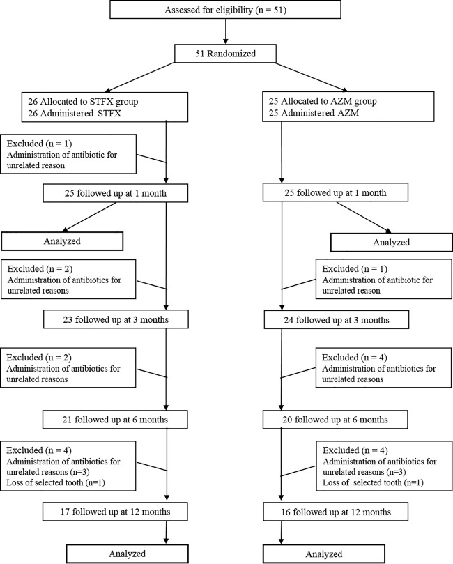
The subjects were recruited from the patients attending the Departments of Periodontics and General Dentistry, Niigata University Medical and Dental Hospital. Fifty-one patients, aged 36 to 78 years old on the day of baseline examination, were included in this study (Fig. 1). The patients had all been previously treated for chronic or aggressive periodontitis (24) and were placed on a supportive periodontal therapy regimen for at least 6 months. The patients met the following inclusion criteria: (i) good general health without any remarkable history except well-controlled hypertension and dyslipidemia, (ii) the presence of at least 15 remaining teeth, (iii) a history of chronic or aggressive periodontitis, (iv) the presence of at least 2 teeth with probing pocket depth (measurement of the depth from the gingival margin to the epithelial attachment in unhealthy gingival tissue by a periodontal probe) of ≥5 mm with concomitant bleeding on probing (bleeding that is induced by gentle manipulation of the tissue at the depth of the periodontal pocket by a periodontal probe), and (v) provision of informed consent. The exclusion criteria were the following: (i) the use of systemic or local antimicrobials in the past 3 months, (ii) scaling and root planing treatment under local anesthesia for the target teeth in the previous 3 months, (iii) subgingival mechanical debridement for the target teeth in the previous 1 month, and (iv) allergies to conventional quinolone, macrolide, and ketolide agents. The experimental protocol was approved by the Institutional Review Board (IRB) of Niigata University Medical and Dental Hospital (approval number NH23-010) and registered with the University Hospital Medical Information Network-Clinical Trials Registry (UMIN-CTR) under registration number UMIN000007834).
FIG 1.
Flow diagram showing the progress of the study.
Experimental design and treatment.
Subjects were randomly assigned to one of the experimental groups using a random-number table. The random-number table was kept by a research fellow who was not directly involved in the experiment. The clinicians in charge were blinded to the treatment allocation until the registration procedure and the examinations for baseline data were completed. The clinical and microbiological examinations were performed at two periodontal pocket sites with a probing pocket depth of ≥5 mm with concomitant bleeding on probing (here referred to as sampling sites), in each subject. Subsequently, oral hygiene instruction was given, and supragingival professional mechanical tooth cleaning was performed. The administration of the antimicrobials was initiated within 1 month after the baseline visit. Each patient allocated to the STFX group took STFX (100 mg, Gracevit; Daiichi Sankyo Co. Ltd., Tokyo, Japan) orally twice a day for 5 days, and each patient allocated to the AZM group took AZM (500 mg, Zithromac; Pfizer Japan Inc., Tokyo, Japan) orally once a day for 3 days. Clinical and microbiological examinations were performed at 1, 3, 6, and 12 months after administration of the drug (here called the examinations at 1, 3, 6, and 12 months), followed by oral hygiene instructions and professional mechanical tooth cleaning. During all of the examination visits, oral hygiene instructions were given and professional mechanical tooth cleaning was performed after the sampling and examination procedures. Professional mechanical tooth cleaning was performed only in the supragingival area before the examination at 3 months, and professional mechanical tooth cleaning in the subgingival area was restarted after the examination at 3 months.
Microbiological assessment of subgingival plaque samples.
Subgingival plaque samples were taken from the two sampling sites in each subject at baseline and at 1, 3, 6, and 12 months. After isolation of the tooth with cotton rolls, drying, and removal of supragingival plaque, subgingival plaque samples were taken with two 35 paper points (VDW GmbH, Munich, Germany) from each sampling site. The counts of total bacteria and the presence of Porphyromonas gingivalis, Aggregatibacter actinomycetemcomitans, Treponema denticola, Tannerella forsythia, and Prevotella intermedia were determined using real-time PCR (GC Corp., Tokyo, Japan). Subsequently, the proportion of each periodontopathic bacterium in the total number of bacteria was calculated. The percentage of the red complex bacteria (RCB), i.e., P. gingivalis, T. denticola, and T. forsythia, in the total bacterial counts was calculated.
Antimicrobial susceptibility testing of clinical isolates.
Subgingival plaque samples taken at baseline and 1 month were also transported to a microbiology laboratory (LSI Medience Corporation, Tokyo, Japan) for identification of plaque bacteria and subsequent determination of antimicrobial susceptibility to antimicrobials. The MIC was defined as the lowest concentration of the antimicrobial agent which completely inhibited visible bacterial growth. The MIC50 and MIC90 values indicate the concentrations inhibiting 50% and 90% of the isolates, respectively. Susceptible, intermediate, and resistant categories were described if an appropriate breakpoint was defined by the Clinical and Laboratory Standards Institute (CLSI).
Clinical measurements.
The following clinical outcome variables were assessed at baseline and at 1, 3, 6, and 12 months by two well-trained periodontists (T.N. and H.I.): the O'Leary plaque index (an index used for estimating the status of oral hygiene by measuring dental plaque that occurs in the areas adjacent to the gingival margin), the probing pocket depth, the clinical attachment level (an estimate of the periodontal support around the tooth as measured with a periodontal probe), and bleeding on probing. Probing was performed at 6 sites per tooth for all teeth present using a CP-12 color-coded probe (Hu-Friedy Mfg. Co., LLC, Chicago, IL, USA). The examiners were calibrated for a probing pressure of 25 g. Bleeding on probing was assessed 15 s after probing. Clinical parameters were the secondary outcome variables.
Analysis of the drug concentration in the periodontal pockets.
In order to examine the drug concentration in periodontal pockets, gingival crevicular fluid (GCF) samples were taken twice from the sampling sites in each subject: the 1st gingival crevicular fluid sampling was 2 h after the first administration of each antimicrobial in both groups; the 2nd sampling was 2 h after the medication on the last day of administration. After isolation of the tooth with cotton rolls, drying, and removal of supragingival plaque, a paper strip (PTM kit strips; Shofu Inc., Kyoto, Japan) was placed into each target site and left in place for 10 s. The gingival crevicular fluid volume in the paper strip was determined on the basis of measurements using a Periotron 8000 (Oraflow Inc., Plainview, NY, USA) and a calibration graph. The paper strip was harvested in a tube with 220 μl of phosphate-buffered saline. The procedure was repeated, and a total of 3 paper strips were harvested in a tube from each sampling site. After agitation for 10 min, the eluates were centrifuged for 5 min at 12,000 × g to remove plaque and cellular elements, and then supernatants were harvested and frozen at −80°C until concentration analysis. The concentration of the drug was measured by liquid chromatography-tandem mass spectometry (LC-MS/MS) methods by LSI Medience Corporation (Tokyo, Japan).
Statistical analysis.
The primary endpoints of this study were the changes in the proportions of RCB from baseline to 1 month in each experimental group. The practical decrease was set at >5% reduction in the RCB proportion in the present study. Our previous study examined subjects with characteristics similar to those of the present study and demonstrated that the proportion of RCB was decreased from a mean of approximately 7% at baseline to 1.0% and 2.0% by scaling and root planing and oral administration of STFX, respectively, with good clinical improvement (13). The secondary outcome variables included the following: changes in (i) the numbers of RCB, (ii) the proportions of P. gingivalis, T. forsythia, T. denticola, P. intermedia, and A. actinomycetemcomitans, (iii) the numbers of total bacterial counts, (iv) the probing pocket depth, (v) the clinical attachment level; and (iv) bleeding on probing in the target sites. Age and clinical parameters at baseline were compared between two treatment groups using the Welch t test. The significance of differences over time in each treatment group was analyzed using a paired t test and one-way repeated-measures analysis of variance (ANOVA) with the Tukey-Kramer test for probing pocket depth and clinical attachment level, the Pearson chi-square test for bleeding on probing, and the Wilcoxon signed-rank test and the Friedman test with Dunn's multiple-comparison test for microbiological parameters. Statistical significance was determined as a P value of <0.05. All statistical analyses were performed with GraphPad Prism 5 (GraphPad Software, Inc., San Diego, CA, USA).
RESULTS
Subject recruitment started in June 2012 and was completed by the end of May 2013. All of the 12-month follow-up visits were completed by the end of May 2014. Figure 1 shows the flow diagram of the study progress. All of the 51 patients who met the criteria were randomly assigned to the STFX or AZM group and then were given STFX or AZM. All of the subjects declared that they had taken the antimicrobials as prescribed. Before the 1-month examination, 1 patient was excluded because of systemic antimicrobial administration for reasons other than the experimental purpose. Therefore, 50 subjects had complete clinical and microbiological data from baseline through 1 month, with 25 patients each in the STFX and AZM groups. After the examination at 1 month, 17 patients were excluded for the following reasons: systemic antimicrobial administration for reasons other than the experimental purpose (n = 15) and loss of one of the selected teeth due to progression of periodontitis (n = 2). Therefore, 33 final subjects had complete data through 12 months, 17 patients in the STFX group and 16 patients in the AZM group. The baseline profiles of the study population whose analyses were completed at the 1-month and 12-month follow-ups are shown separately in Table 1, with no significant differences in age, the number of teeth, and the O'Leary plaque index between the groups. Nine patients in the STFX group and 7 patients in the AZM group had mild diarrhea but recovered immediately after the administration period. No serious adverse effects were observed or reported in the patients in either group.
TABLE 1.
Characteristics of the patients whose microbiological and clinical analyses were completed at the 1-month and 12-month follow-ups
| Characteristic | Follow-up period |
|||
|---|---|---|---|---|
| 1 mo |
12 mo |
|||
| STFXa | AZMb | STFX | AZM | |
| No. of subjects | 25 | 25 | 17 | 16 |
| Age (yr)c | 62.5 ± 8.6 | 64.6 ± 9.1 | 61.9 ± 8.5 | 64.9 ± 9.6 |
| Age range (yr) | 41–78 | 36–76 | 41–74 | 36–74 |
| Male/female (no.) | 12/13 | 9/16 | 7/10 | 8/8 |
| Smoker/nonsmoker (no.) | 1/24 | 1/24 | 0/17 | 1/15 |
| Hypertension (no.) | 7 | 6 | 6 | 4 |
| Dyslipidemia (no.) | 5 | 3 | 3 | 1 |
| No. of teethc | 24.0 ± 4.3 | 23.8 ± 3.3 | 24.8 ± 3.8 | 23.8 ± 2.8 |
| O'Leary plaque index (%)c | 15.2 ± 9.7 | 16.7 ± 10.1 | 16.9 ± 9.2 | 15.1 ± 9.8 |
STFX, sitafloxacin.
AZM, azithromycin.
Age, the number of teeth, and the O'Leary plaque index are expressed as means ± SD.
Microbiological findings up to 1 month.
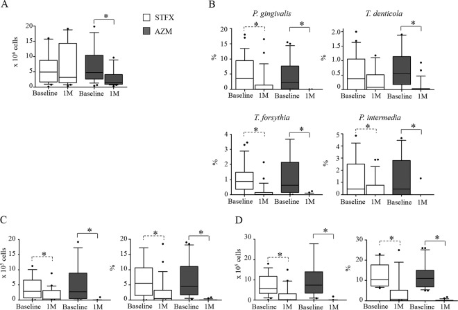
Both agents decreased the numbers of total bacteria (Fig. 2A) and the proportions of P. gingivalis, T. denticola, T. forsythia, and P. intermedia (Fig. 2B). A. actinomycetemcomitans was rarely detected in either group: 2 of 25 patients in the STFX group and 5 of 25 patients in the AZM group were A. actinomycetemcomitans positive at baseline. At 1 month, A. actinomycetemcomitans was undetectable in the STFX group, but the bacterium was detected in 1 patient in the AZM group. The number and percentage of RCB were significantly reduced at 1 month in both groups (Fig. 2C).
FIG 2.
Median changes in total bacterial counts (A) and proportions of each periodontopathic bacterium (B) and numbers and percentages of red complex bacteria (RCB) in all sampling sites (C) and those of sampling sites with >5% RCB proportions at baseline (D) through the 1-month (1M) experimental period. The bars indicate the upper and lower interquartile ranges. The error bars indicate the 90th and 10th percentile ranges. The dots indicate the maximum and minimum values. The significance of differences within groups was determined by the Wilcoxon signed-rank test: *, P < 0.05.
Changes in the proportions of RCB from baseline to 1 month are the primary endpoints. Both groups demonstrated significant reductions in the proportions of RCB (median reductions of 4.97% and 4.54% in the STFX and AZM groups, respectively) (Fig. 2C); however, the reductions were <5%, which was the reference value set as the practical decrease at the beginning of the research. Because only 24 of 50 sites in the STFX group and 23 of 50 sites in the AZM group showed >5% RCB existence at baseline, further analysis was performed for those sites. This analysis showed 9.59% and 10.99% reductions in RCB as medians in the STFX (10.35% at baseline to 0.76% at 1 month) and AZM (11.00% at baseline to 0.01% at 1 month) groups, respectively, and most sites had RCB at a less than detectable level (Fig. 2D).
Clinical findings up to 1 month.
Table 2 presents the mean values for the clinical parameters of the sampling sites at baseline and 1 month in the two experimental groups. There were no significant differences in the baseline clinical parameters between the groups. In both groups, the probing pocket depth was significantly decreased and the clinical attachment level was significantly increased at the 1-month examination compared with those at baseline. The number of instances of bleeding on probing in the STFX group and in the AZM group had reduced by 52% and 34% at 1 month, respectively. The concentrations of STFX and AZM in the gingival crevicular fluid samples are shown in Table 3.
TABLE 2.
Clinical parameters of the sampling sites at the baseline and 1-month examinations
| Parametera | Results at baseline | Results at 1 mo |
|---|---|---|
| PPD (mm) | ||
| STFX | 5.9 ± 0.8 | 5.1 ± 0.7b |
| AZM | 6.2 ± 1.1 | 4.9 ± 1.3b |
| CAL (mm) | ||
| STFX | 7.0 ± 1.3 | 6.3 ± 1.3b |
| AZM | 6.9 ± 1.5 | 5.8 ± 1.8b |
| BOP | ||
| STFX | 50/50 | 26/50 |
| AZM | 50/50 | 17/50 |
| Plaque | ||
| STFX | 19/50 | 16/50 |
| AZM | 14/50 | 10/50 |
PPD, probing pocket depth; CAL, clinical attachment level; BOP, bleeding on probing; plaque, dental plaque; STFX, sitafloxacin; AZM, azithromycin. PPD and CAL are expressed as means ± SD. BOP and plaque are expressed as the number of positive sites/all examined sites.
P < 0.05 versus baseline (paired t test).
TABLE 3.
Concentrations of drugs in gingival crevicular fluid obtained from the sampling sites of patients whose data are shown in Table 2
| Druga | Mean concentration ± SD (μg/ml) at: |
|
|---|---|---|
| 1st samplingb | 2nd samplingc | |
| STFX | 0.60 ± 0.45 | 1.17 ± 0.79 |
| AZM | 0.50 ± 0.69 | 5.49 ± 3.22 |
STFX, sitafloxacin; AZM, azithromycin.
Performed at 2 h after the first administration of each antimicrobial in both groups.
Performed at 2 h after medication on the last day of administration in both groups.
Microbiological findings up to 12 months.
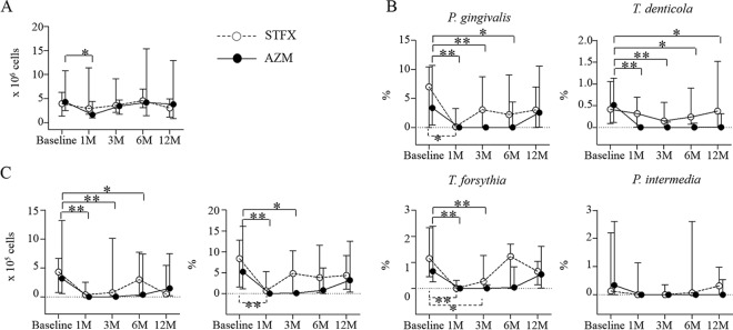
Figure 3 shows the longitudinal changes in the counts and the percentages of the total and the periodontopathic bacteria. The STFX group demonstrated significantly lower percentages of RCB at 1 month than at baseline. The AZM group demonstrated significantly lower percentages of RCB at 1 and 3 months than at baseline (Fig. 3C). At 12 months, both groups still showed decreased levels of RCB proportions compared to those at baseline, but without statistical significance.
FIG 3.
Median changes in total bacterial counts (A) and proportions of each periodontopathic bacterium (B) and numbers and percentages of red complex bacteria (RCB) (C) through the 12-month period. Error bars indicate the upper and lower interquartile ranges. The significance of differences compared to baseline was determined by the Friedman test with Dunn's multiple-comparison test: *, P < 0.05; **, P < 0.01.
Clinical findings up to 12 months.
Table 4 presents the mean values of the clinical parameters of the sampling sites at baseline and at 1, 3, 6, and 12 months in the two experimental groups. No significant differences in the baseline clinical parameters were evident between the STFX and AZM groups. In both groups, probing pocket depth, clinical attachment level, and bleeding on probing were significantly improved at all examination time points compared to those at baseline. However, it should be noted that within 6 to 12 months, one subject in each group lost a tooth due to exacerbation of periodontitis.
TABLE 4.
Clinical parameters of the sampling sites during the 12-month experimental period
| Parametera | Results at: |
||||
|---|---|---|---|---|---|
| Baseline | 1 mo | 3 mo | 6 mo | 12 mo | |
| PPD (mm) | |||||
| STFX | 5.8 ± 0.8 | 5.0 ± 0.7b | 4.8 ± 1.0b | 4.6 ± 1.0b | 4.6 ± 1.1b |
| AZM | 5.9 ± 0.7 | 4.7 ± 1.1b | 4.6 ± 0.9b | 4.6 ± 0.9b | 4.6 ± 1.0b |
| CAL (mm) | |||||
| STFX | 6.9 ± 1.4 | 6.1 ± 1.3b | 5.9 ± 1.2b | 5.7 ± 1.3b | 5.7 ± 1.2b |
| AZM | 6.4 ± 1.1 | 5.4 ± 1.4b | 5.3 ± 1.2b | 5.2 ± 1.2b | 5.4 ± 1.2b |
| BOP | |||||
| STFX | 34/34 | 19/34 | 19/34 | 12/34 | 19/34 |
| AZM | 32/32 | 6/32 | 11/32 | 15/32 | 12/32 |
| Plaque | |||||
| STFX | 16/34 | 12/34 | 15/34 | 16/34 | 19/34 |
| AZM | 8/32 | 6/32 | 8/32 | 8/32 | 15/32 |
PPD, probing pocket depth; CAL, clinical attachment level; BOP, bleeding on probing; plaque, dental plaque; STFX, sitafloxacin; AZM, azithromycin. PPD and CAL are expressed as means ± SD. BOP and plaque are expressed as the number of positive sites/all examined sites.
P < 0.001 versus baseline (one-way repeated-measures ANOVA with the Tukey-Kramer test).
Antimicrobial susceptibility of clinical isolates.
Clinical isolates at baseline and 1 month were categorized into two bacterial groups: viridans group streptococci and anaerobes. The MIC frequency distributions of the clinical isolates in the STFX group and AZM group are shown in Tables 5 and 6, respectively. In the STFX group, the antimicrobial susceptibilities for STFX and AZM were comparable before and after STFX administration (Table 5). In the AZM group, the antimicrobial susceptibility for STFX was similar before and after AZM administration, although that for AZM clearly changed. The percentage of AZM-resistant viridans group streptococci was markedly elevated (from 27.3% to 50.0%), and the MIC50 and MIC90 of AZM for anaerobes were increased 8-fold and >8-fold, respectively, in the AZM group (Table 6).
TABLE 5.
MIC frequency distribution of antimicrobials against viridans group streptococci and anaerobes isolated from patients in the sitafloxacin group
| Drug, bacteria, timea | No. (cumulative %) of isolates with indicated MIC (μg/ml) of: |
MIC50 (μg/ml) | MIC90 (μg/ml) | S/I/R (%)b | ||||||||||||||
|---|---|---|---|---|---|---|---|---|---|---|---|---|---|---|---|---|---|---|
| ≤0.004 | 0.008 | 0.015 | 0.03 | 0.06 | 0.12 | 0.25 | 0.5 | 1 | 2 | 4 | 8 | 16 | 32 | >32 | ||||
| STFX | ||||||||||||||||||
| Viridans group streptococci | ||||||||||||||||||
| Baseline | 1 (2.27) | 3 (9.09) | 15 (43.18) | 16 (79.54) | 6 (93.18) | 1 (95.45) | 1 (97.72) | 1 (100) | 0.06 | 0.12 | ||||||||
| 1 mo | 1 (2.24) | 2 (7.12) | 11 (33.95) | 18 (77.85) | 3 (85.17) | 2 (90.05) | 3 (97.37) | 1 (100) | 0.06 | 0.25 | ||||||||
| Anaerobes | ||||||||||||||||||
| Baseline | 1 (2.44) | 7 (19.51) | 18 (63.41) | 7 (80.48) | 6 (95.11) | 1 (97.55) | 1 (100) | 0.03 | 0.12 | |||||||||
| 1 mo | 2 (6.06) | 10 (36.36) | 6 (54.54) | 9 (81.81) | 3 (90.9) | 1 (93.93) | 1 (96.96) | 1 (100) | 0.03 | 0.12 | ||||||||
| AZM | ||||||||||||||||||
| Viridans group streptococci | ||||||||||||||||||
| Baseline | 6 (13.64) | 2 (18.19) | 5 (29.55) | 3 (36.37) | 1 (38.64) | 3 (45.46) | 1 (47.73) | 5 (59.09) | 7 (75.0) | 6 (88.64) | 1 (90.91) | 4 (100) | 1 | 16 | 47.7/11.4/40.9 | |||
| 1 mo | 2 (4.88) | 1 (7.32) | 5 (19.52) | 3 (26.84) | 5 (39.04) | 2 (43.92) | 3 (52.24) | 3 (58.56) | 4 (68.32) | 7 (85.39) | 4 (95.15) | 1 (97.59) | 1 (100) | 0.25 | 4 | 58.5/9.8/31.7 | ||
| Anaerobes | ||||||||||||||||||
| Baseline | 1 (2.44) | 2 (7.32) | 8 (26.83) | 6 (41.46) | 6 (56.09) | 11 (82.92) | 3 (90.24) | 1 (92.68) | 2 (97.56) | 1 (100) | 1 | 4 | ||||||
| 1 mo | 1 (3.03) | 1 (6.06) | 2 (12.12) | 2 (18.18) | 7 (39.39) | 6 (57.57) | 9 (84.84) | 1 (87.87) | 1 (90.9) | 2 (96.96) | 1 (100) | 1 | 8 | |||||
STFX, sitafloxacin; AZM, azithromycin.
Susceptible (S), intermediate (I), and resistant (R) categories are defined in CLSI document M100-S24 (28).
TABLE 6.
MIC frequency distribution of antimicrobials against Streptococcus viridans and anaerobes isolated from patients in the azithromycin group
| Drug, bacteria, timea | No. (cumulative %) of isolates with indicated MIC (μg/ml) of: |
MIC50 (μg/ml) | MIC90 (μg/ml) | S/I/R (%)b | ||||||||||||||
|---|---|---|---|---|---|---|---|---|---|---|---|---|---|---|---|---|---|---|
| ≤0.004 | 0.008 | 0.015 | 0.03 | 0.06 | 0.12 | 0.25 | 0.5 | 1 | 2 | 4 | 8 | 16 | 32 | >32 | ||||
| STFX | ||||||||||||||||||
| Viridans group streptococci | ||||||||||||||||||
| Baseline | 3 (9.09) | 15 (54.54) | 9 (81.81) | 2 (87.87) | 1 (90.9) | 2 (96.96) | 1 (100) | 0.03 | 0.25 | |||||||||
| 1 mo | 5 (13.89) | 18 (63.89) | 8 (86.11) | 3 (94.44) | 1 (97.22) | 1 (100) | 0.03 | 0.12 | ||||||||||
| Anaerobes | ||||||||||||||||||
| Baseline | 6 (14.29) | 21 (64.29) | 9 (85.72) | 4 (95.24) | 1 (97.62) | 1 (100) | 0.03 | 0.12 | ||||||||||
| 1 mo | 3 (13.64) | 8 (50.0) | 4 (68.18) | 5 (90.91) | 1 (95.46) | 1 (100) | 0.03 | 0.12 | ||||||||||
| AZM | ||||||||||||||||||
| Viridans group streptococci | ||||||||||||||||||
| Baseline | 1 (3.03) | 4 (15.15) | 2 (21.21) | 1 (24.24) | 2 (30.3) | 1 (33.33) | 1 (36.36) | 5 (51.51) | 7 (72.72) | 4 (84.84) | 2 (90.9) | 1 (93.93) | 1 (96.96) | 1 (100) | 0.5 | 4 | 51.5/21.2/27.3 | |
| 1 mo | 1 (2.78) | 1 (5.56) | 3 (13.89) | 1 (16.67) | 3 (25.0) | 9 (50.0) | 4 (61.11) | 8 (83.33) | 1 (86.11) | 2 (91.67) | 3 (100) | 1 | 32 | 25.0/25.0/50.0 | ||||
| Anaerobes | ||||||||||||||||||
| Baseline | 4 (9.52) | 7 (26.19) | 8 (45.24) | 6 (59.53) | 9 (80.96) | 5 (92.86) | 1 (95.24) | 1 (97.62) | 1 (100) | 1 | 4 | |||||||
| 1 mo | 1 (4.55) | 2 (13.64) | 1 (18.19) | 4 (36.37) | 3 (50.01) | 5 (72.74) | 1 (77.29) | 5 (100) | 8 | >32 | ||||||||
STFX, sitafloxacin; AZM, azithromycin.
Susceptible (S), intermediate (I), and resistant (R) categories are defined in CLSI document M100-S24 (28).
DISCUSSION
The present study demonstrated that both systemic STFX and AZM monotherapy significantly decreased RCB in active periodontal pockets during the supportive periodontal therapy period. The practical microbiological effectiveness was set at >5% reduction of RCB as a primary endpoint. Both groups showed significant decreases of RCB in the sites where RCB comprised >5% of the total microbiota composition at baseline, suggesting that both antimicrobials are microbiologically effective for active periodontal pockets. The STFX and AZM groups both demonstrated significant improvement in clinical parameters at 1 month. We previously reported that systemic STFX and scaling and root planing with local anesthesia, which is the conventional treatment for periodontal pockets, had comparable clinical and microbiological effects during the supportive periodontal therapy phase by the noninferior test within a short follow-up period (13). The results showed that the decrease in the proportions of RCB in the STFX group was not less than the decrease in the scaling and root planing group. The present study cannot determine the superiority or inferiority of this method compared either to conventional treatment or between groups, because no conventional treatment group was included as a direct control and because of the small sample size. This study aimed to evaluate the effectiveness of systemic STFX and AZM as alternative treatments for patients in whom conventional therapy could not be applied, by using the practical endpoint referring to the previous study. Therefore, not only systemic STFX but also AZM seems to have microbiological and clinical effects similar to those of scaling and root planing in the short term.
Regarding the prolonged effects, the STFX and AZM groups both maintained significantly improved clinical parameters at the 12-month examination compared to those at baseline. From the microbiological point of view, both groups demonstrated decreased proportions of RCB at 12 months, although the difference did not reach a statistically significant level. It is reported that bacterial recolonization of the periodontal pocket occurs with subgingival bacterial counts being restored almost to pretreatment values 3 to 7 days after scaling and root planing in untreated patients, although the changes induced in the composition of the microflora, such as the higher proportion of nonperiodontopathic bacteria and lower proportion of periodontopathic bacteria, last longer (25). Our previous report demonstrated that the scaling and root planing group showed rebound of the proportion of RCB at 3 months, as did the systemic STFX group, although the clinical effects were maintained at 3 months (13). The level of recolonization of periodontopathic bacteria during 12 months after systemic STFX or AZM is low enough to keep the clinical condition stable. The two agents seem to have prolonged microbiological and clinical effects comparable to those of conventional mechanical treatment.
One tooth from each group exfoliated without any symptoms except severe mobility. Those teeth demonstrated severe bone loss and mobility at baseline. Although the inclusion of these teeth in this study was not appropriate, this did not seem to have affected the results from other subjects.
Antimicrobial susceptibility testing of the clinical isolates revealed interesting findings. The MIC90 for STFX (0.12 to 0.25 μg/ml) was lower than the concentration of STFX in GCF (mean, 1.20 μg/ml) in both the STFX and AZM groups at baseline and 1 month, suggesting good antimicrobial activity of STFX in periodontal lesions. The excellent antimicrobial activity was confirmed by the study showing that the MIC90 for clinical isolates from acute periodontal lesions was very low for STFX (0.12 μg/ml) (14). On the other hand, the MIC90 for AZM was comparable to or higher than the concentration of AZM in GCF. The MIC90 for AZM was remarkably increased after AZM administration: 4 μg/ml at baseline to 32 μg/ml at 1 month for viridans group streptococci and 4 μg/ml at baseline to >32 μg/ml at 1 month for anaerobes. According to the CLSI breakpoint, the percentage of AZM-resistant viridans group streptococci increased from 27.3% to 50.0%. These results suggest that AZM might remove susceptible anaerobes together with nonpathogenic bacteria effectively. However, AZM induces strong antimicrobial selective pressure simultaneously and may result in the selection of some strains with decreased susceptibility.
Quinolones are the most potent antimicrobials in vitro for A. actinomycetemcomitans (26, 27), the pathogen associated with aggressive periodontitis. Although the detection rates of A. actinomycetemcomitans at baseline were too low to analyze the effect of the drugs, undetectable levels of A. actinomycetemcomitans after 1 and 3 months were only seen with STFX treatment. This finding is consistent with the results of our previous study (13), in which systemic STFX, but not mechanical scaling and root planing, eliminated A. actinomycetemcomitans from active periodontal pockets. Therefore, STFX may be more beneficial for clinical cases in which A. actinomycetemcomitans is involved.
The advantage of systemic administration of antibacterial agents is that all periodontal lesions, including the sites with complicated anatomical features such as root furcations, can be treated by only one visit, without invasive procedures. On the other hand, the disadvantages of antimicrobials, i.e., the risks of systemic adverse effects, the patient's noncompliance, microbial substitution, and bacterial resistance, should also be considered. In this study, no serious systemic adverse effect was observed, and all patients had good compliance. Considering that a fraction of clinical isolates have decreased susceptibility to AZM, probably due to past use of macrolide antibiotics, continuous efforts should be made to control overuse of antimicrobials.
During active periodontal therapy when moderate to heavy calculus deposits are present on the surface of teeth, the use of systemic antimicrobials is recommended as an adjunct to conventional periodontal treatment such as scaling and root planing and periodontal surgery. On the other hand, during supportive periodontal therapy, etiological agents are substantially decreased as a result of active periodontal therapy and continuous professional mechanical tooth cleaning; therefore, the dental plaque biofilm is the main target of treatment. Many recently developed antimicrobials have good penetration into biofilm, suggesting that a single use of antimicrobials may be effective in some cases. Furthermore, many patients receiving supportive periodontal therapy are aged and have systemic complications, making invasive treatment not suitable for them.
The present study clearly demonstrated that STFX and AZM monotherapy with appropriate personal and professional plaque control in the supportive periodontal therapy phase was effective microbiologically and clinically, and the clinical effect was maintained for 12 months. These results indicate that the use of systemic STFX and AZM would serve as an effective alternative therapy in combination with subgingival debridement when disease activity is increased during supportive periodontal therapy, especially in patients for whom invasive mechanical treatment is inappropriate.
Funding Statement
This work was supported in part by Daiichi Sankyo Co. Ltd. (Tokyo, Japan) and Japan Society for the Promotion of Science (JSPS) KAKENHI grants 23390476, 25670882, and 25463216. The funders had no role in study design, data collection and interpretation, or the decision to submit the work for publication.
REFERENCES
- 1.Okui T, Aoki-Nonaka Y, Nakajima T, Yamazaki K. 2014. The role of distinct T cell subsets in periodontitis—studies from humans and rodent models. Curr Oral Health Rep 1:114–123. doi: 10.1007/s40496-014-0013-z. [DOI] [Google Scholar]
- 2.Cullinan MP, Seymour GJ. 2013. Periodontal disease and systemic illness: will the evidence ever be enough? Periodontol 2000 62:271–286. doi: 10.1111/prd.12007. [DOI] [PubMed] [Google Scholar]
- 3.Lalla E, Papapanou PN. 2011. Diabetes mellitus and periodontitis: a tale of two common interrelated diseases. Nat Rev Endocrinol 7:738–748. doi: 10.1038/nrendo.2011.106. [DOI] [PubMed] [Google Scholar]
- 4.Nakajima T, Yamazaki K. 2009. Periodontal disease and risk of atherosclerotic coronary heart disease. Odontology 97:84–91. doi: 10.1007/s10266-009-0104-9. [DOI] [PubMed] [Google Scholar]
- 5.Socransky SS, Haffajee AD. 2002. Dental biofilm: difficult therapeutic targets. Periodontol 2000 28:12–55. doi: 10.1034/j.1600-0757.2002.280102.x. [DOI] [PubMed] [Google Scholar]
- 6.Haffajee AD, Teles RP, Socransky SS. 2006. The effect of periodontal therapy on the composition of the subgingival microbiota. Periodontol 2000 42:219–258. doi: 10.1111/j.1600-0757.2006.00191.x. [DOI] [PubMed] [Google Scholar]
- 7.Teles RP, Haffajee AD, Socransky SS. 2006. Microbiological goals of periodontal therapy. Periodontol 2000 42:180–218. doi: 10.1111/j.1600-0757.2006.00192.x. [DOI] [PubMed] [Google Scholar]
- 8.Herrera D, Alonso B, Leon R, Roldan S, Sanz M. 2008. Antimicrobial therapy in periodontitis: the use of systemic antimicrobials against the subgingival biofilm. J Clin Periodontol 35:45–66. doi: 10.1111/j.1600-051X.2008.01260.x. [DOI] [PubMed] [Google Scholar]
- 9.Walker CB, Karpinia K, Baehni P. 2004. Chemotherapeutics: antibiotics and other antimicrobials. Periodontol 2000 36:146–165. doi: 10.1111/j.1600-0757.2004.03677.x. [DOI] [PubMed] [Google Scholar]
- 10.Lang NP, Joss A, Orsanic T, Gusberti FA, Siegrist BE. 1986. Bleeding on probing. A predictor for the progression of periodontal disease? J Clin Periodontol 13:590–596. [DOI] [PubMed] [Google Scholar]
- 11.Ramfjord SP. 1987. Maintenance care for treated periodontitis patients. J Clin Periodontol 14:433–437. doi: 10.1111/j.1600-051X.1987.tb02247.x. [DOI] [PubMed] [Google Scholar]
- 12.Fujikawa K, Chiba M, Tanaka M, Sato K. 2005. In vitro antibacterial activity of DX-619, a novel des-fluoro(6) quinolone. Antimicrob Agents Chemother 49:3040–3045. doi: 10.1128/AAC.49.7.3040-3045.2005. [DOI] [PMC free article] [PubMed] [Google Scholar]
- 13.Nakajima T, Okui T, Miyauchi S, Honda T, Shimada Y, Ito H, Akazawa K, Yamazaki K. 2012. Effects of systemic sitafloxacin on periodontal infection control in elderly patients. Gerodontology 29:e1024–e1032. doi: 10.1111/j.1741-2358.2011.00605.x. [DOI] [PubMed] [Google Scholar]
- 14.Tomita S, Kasai S, Ihara Y, Imamura K, Kita D, Ota K, Kinumatsu T, Nakagawa T, Saito A. 2014. Effects of systemic administration of sitafloxacin on subgingival microflora and antimicrobial susceptibility profile in acute periodontal lesions. Microb Pathog 71–72:1–7. doi: 10.1016/j.micpath.2014.04.003. [DOI] [PubMed] [Google Scholar]
- 15.Hirsch R, Deng H, Laohachai MN. 2012. Azithromycin in periodontal treatment: more than an antibiotic. J Periodont Res 47:137–148. doi: 10.1111/j.1600-0765.2011.01418.x. [DOI] [PubMed] [Google Scholar]
- 16.Haas AN, de Castro GD, Moreno T, Susin C, Albandar JM, Oppermann RV, Rösing CK. 2008. Azithromycin as an adjunctive treatment of aggressive periodontitis: 12-months randomized clinical trial. J Clin Periodontol 35:696–704. doi: 10.1111/j.1600-051X.2008.01254.x. [DOI] [PubMed] [Google Scholar]
- 17.Haffajee AD, Torresyap G, Socransky SS. 2007. Clinical changes following four different periodontal therapies for the treatment of chronic periodontitis: 1-year results. J Clin Periodontol 34:243–253. doi: 10.1111/j.1600-051X.2006.01040.x. [DOI] [PubMed] [Google Scholar]
- 18.Mascarenhas P, Gapski R, Al-Shammari K, Hill R, Soehren S, Fenno JC, Giannobile WV, Wang HL. 2005. Clinical response of azithromycin as an adjunct to non-surgical periodontal therapy in smokers. J Periodontol 76:426–436. doi: 10.1902/jop.2005.76.3.426. [DOI] [PubMed] [Google Scholar]
- 19.Oteo A, Herrera D, Figuero E, O'Connor A, Gonzalez I, Sanz M. 2010. Azithromycin as an adjunct to scaling and root planing in the treatment of Porphyromonas gingivalis-associated periodontitis: a pilot study. J Clin Periodontol 37:1005–1015. doi: 10.1111/j.1600-051X.2010.01607.x. [DOI] [PubMed] [Google Scholar]
- 20.Smith SR, Foyle DM, Daniels J, Joyston-Bechal S, Smales FC, Sefton A, Williams J. 2002. A double-blind placebo-controlled trial of azithromycin as an adjunct to non-surgical treatment of periodontitis in adults: clinical results. J Clin Periodontol 29:54–61. doi: 10.1034/j.1600-051x.2002.290109.x. [DOI] [PubMed] [Google Scholar]
- 21.Yashima A, Gomi K, Maeda N, Arai T. 2009. One-Stage full-mouth versus partial-mouth scaling and root planing during the effective half-life of systemically administered azithromycin. J Periodontol 80:1406–1413. doi: 10.1902/jop.2009.090067. [DOI] [PubMed] [Google Scholar]
- 22.Soares GM, Mendes JA, Silva MP, Faveri M, Teles R, Socransky SS, Wang X, Figueiredo LC, Feres M. 2014. Metronidazole alone or with amoxicillin as adjuncts to non-surgical treatment of chronic periodontitis: a secondary analysis of microbiological results from a randomized clinical trial. J Clin Periodontol 41:366–376. doi: 10.1111/jcpe.12217. [DOI] [PubMed] [Google Scholar]
- 23.Gordon J, Walker C, Hovliaras C, Socransky S. 1990. Efficacy of clindamycin hydrochloride in refractory periodontitis: 24-month results. J Periodontol 61:686–691. doi: 10.1902/jop.1990.61.11.686. [DOI] [PubMed] [Google Scholar]
- 24.Armitage GC. 1999. Development of a classification system for periodontal diseases and conditions. Ann Periodontol 4:1–6. doi: 10.1902/annals.1999.4.1.1. [DOI] [PubMed] [Google Scholar]
- 25.Petersilka GJ, Ehmke B, Flemmig TF. 2002. Antimicrobial effects of mechanical debridement. Periodontol 2000 28:56–71. doi: 10.1034/j.1600-0757.2002.280103.x. [DOI] [PubMed] [Google Scholar]
- 26.Müller HP, Holderrieth S, Burkhardt U, Hoffler U. 2002. In vitro antimicrobial susceptibility of oral strains of Actinobacillus actinomycetemcomitans to seven antibiotics. J Clin Periodontol 29:736–742. doi: 10.1034/j.1600-051X.2002.290810.x. [DOI] [PubMed] [Google Scholar]
- 27.Núñez Otero V, Limeres Posse J, Carmona IT, Diz Dios P. 2009. Efficacy of fluoroquinolones against pathogenic oral bacteria. Mini Rev Med Chem 9:1147–1158. doi: 10.2174/138955709789055243. [DOI] [PubMed] [Google Scholar]
- 28.Clinical and Laboratory Standards Institute. 2014. Performance standards for antimicrobial susceptibility testing; 24th informational supplement. CLSI document M100-S24. Clinical and Laboratory Standards Institute, Wayne, PA. [Google Scholar]


