Abstract
The Wnt/β-catenin signaling is an evolutionarily conserved pathway that regulates a wide range of physiological functions, including embryogenesis, organ maintenance, cell proliferation and cell fate decision. Dysregulation of Wnt/β-catenin signaling has been implicated in various cancers, but its role in cell death has not yet been fully elucidated. Here we show that activation of Wg signaling induces cell death in Drosophila eyes and wings, which depends on dFoxO, a transcription factor known to be involved in cell death. In addition, dFoxO is required for ectopic and endogenous Wg signaling to regulate wing patterning. Moreover, dFoxO is necessary for activated Wg signaling-induced target genes expression. Furthermore, Arm is reciprocally required for dFoxO-induced cell death. Finally, dFoxO physically interacts with Arm both in vitro and in vivo. Thus, we have characterized a previously unknown role of dFoxO in promoting Wg signaling, and that a dFoxO-Arm complex is likely involved in their mutual functions, e.g. cell death.
The Wnt/β-catenin signaling represents one of the most intensively studied pathways, which is tightly associated with cancers, especially colorectal cancer1,2. Upon binding of Wnt ligands to the receptor Frizzled (Fz) and co-receptor LDL-receptor-related protein (LRP), the extracellular signals are transduced via Disheveled (Dsh) to the destruction complex composed of Axin (Axn), Adenomatous polyposis coli (APC) and glycogen synthase kinase 3 (GSK3), thereby preventing the proteasomal degradation of the transcriptional coactivator β-catenin. Stabilized β-catenin thus translocates into the nucleus and associates with the T cell factor (TCF)/Lymphoid-enhancing factor (LEF) family of transcription factors to activate the transcription of target genes3,4,5. Through this mechanism, Wnt/β-catenin signaling plays a wide range of functions, including embryogenesis, organ maintenance, cell proliferation and cell fate decisions3,6,7,8. Wnt signaling is also reported to regulate cell death during Drosophila retina and ommatidia development9,10,11, yet the underlying mechanism has not been fully elucidated, and the downstream regulators remain elusive.
The Forkhead box O (FoxO) transcription factors belong to the large forkhead family proteins, which are characterized by a winged helix DNA binding domain called ‘Forkhead box’12,13. The FoxO proteins have been conserved from C. elegans to mammals14. While there is only one FoxO gene in invertebrates, four members have been identified in mammals including FoxO1, FoxO3a, FoxO4 and FoxO613,15. FoxO1, 3a, and 4 are ubiquitously expressed, whereas FoxO6 expression is confined to the brain12. FoxO activity is negatively regulated by the Insulin/PI3K/Akt signaling pathway. Activation of Akt (also known as protein kinase B) phosphorylates FoxO and results in nuclear exclusion of FoxO, thereby inhibits its transcriptional activity14,15,16. However, stress conditions, such as high levels of reactive oxygen species (ROS) or deprivation of growth factors, promote the nuclear localization of FoxO and induce its target genes expression14. FoxO is critically involved in a variety of physiological processes including cell cycle, cell death and differentiation, DNA repair, oxidative stress response and longevity12,17,18,19. Dysregulation of FoxO has been associated with many diseases, including immune defects, malignancy, diabetes and Alzheimer’s disease (AD)13,20.
β-catenin has been reported to interact with FoxO and enhance its transcriptional activity in mammalian cells and C. elegans21. On the other hand, FoxO inhibits Wnt signaling by diverting the limited pool of β-catenin from Wnt/TCF to FoxO22,23, leading to embryogenesis and bone formation defects24,25.
Here using Drosophila melanogaster, which has reduced genome redundancy and many available genetic tools, we found that activation of Wingless (Wg, Drosophila Wnt homolog) signaling induces intensive cell death in Drosophila eyes and wings, which depends on dFoxO (Drosophila FoxO homolog). In addition, dFoxo is required for Wg signaling to activate target genes expression and execute its endogenous functions in wing patterning. Furthermore, loss of armadillo (arm, encoding Drosophila β-catenin homolog) or pangolin (pan, encoding Drosophila TCF homolog) could also suppress dFoxO-triggered cell death, suggesting a reciprocal effect. Finally, dFoxO physically interacts with Arm, providing a molecular mechanism for the role of dFoxO in promoting Wg signaling. Thus, contradict to the previous studies that FoxO proteins inhibit Wnt signaling, our data point to a positive role of dFoxO in modulating the canonical Wg signaling in Drosophila, suggesting FoxO differentially regulates Wnt signaling in a cell context-dependent manner.
Results
Activation of Wg signaling induces cell death in Drosophila
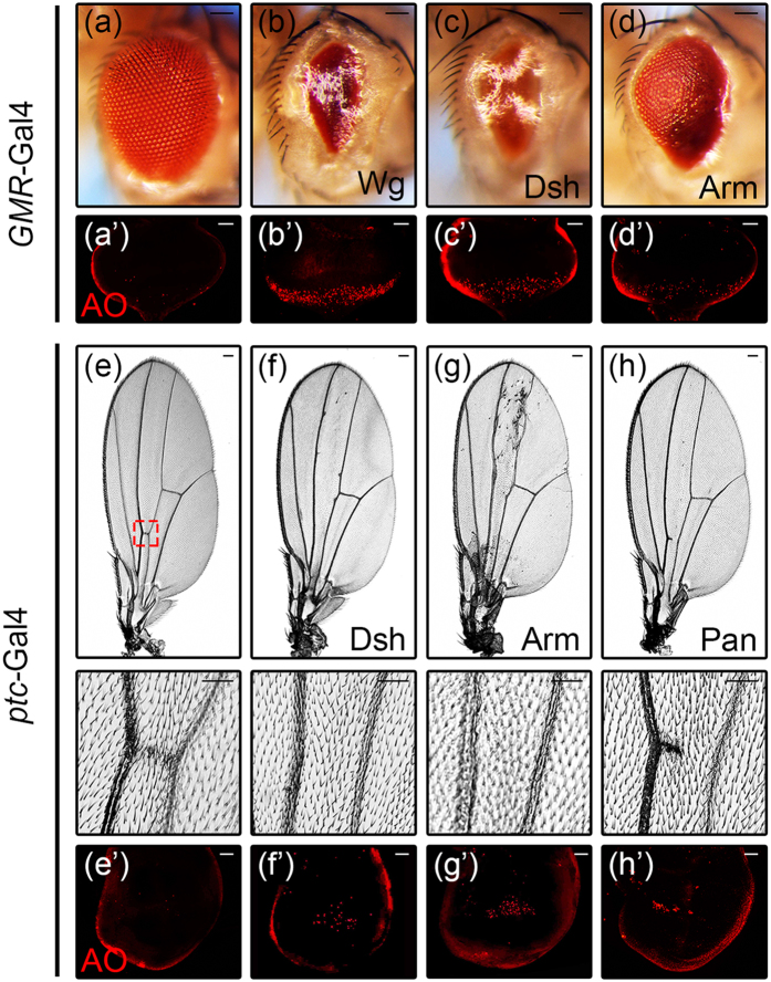
To investigate the role of Wg signaling in cell death, we expressed the representative components of this pathway in the Drosophila eye, and found that ectopic expression of Wg, Dsh or Arm resulted in massive cell death in 3rd instar eye discs as revealed by acridine orange (AO) staining (Fig. 1a’–d’), and produced eyes with reduced size (Fig. 1a–d). In addition, ectopic expression of Dsh, Arm and Pan under ptc-Gal4 produced a loss of anterior cross vein (ACV) phenotype (Fig. 1e–h), accompanied by increased cell death in wing discs (Fig. 1e’–h’). Furthermore, ectopic expression of Dsh or Arm in wing discs by additional drivers including en-Gal4, omb-Gal4 and sd-Gal4 resulted in extensive cell death in wing pouches, as indicated by AO or TUNEL staining (Fig. S1). Collectively, these results indicate that activation of Wg signaling is able to induce cell death in Drosophila.
Figure 1. Activation of Wg signaling induces cell death in Drosophila.
Light micrographs of Drosophila adult eyes and wings, and fluorescent micrographs of 3rd instar eye and wing discs are shown. Compared with the GMR-Gal4 control (a,a’), expression of Wg, Dsh or Arm induces cell death in eye discs indicated by AO staining (b’–d’) and produces adult eyes with reduced size (b–d). Compared with the ptc-Gal4 control (e,e’), expression of Dsh, Arm or Pan induces cell death in wing discs (f’–h’) and produces a loss-of-ACV phenotype in adult wings (f–h, the lower panels are high magnification of the boxed areas in upper panels). Scale bars: 100 μm in (a–d) and (e–h, upper panels); 50 μm in (a’–d’), (e–h, lower panels) and (e’–h’).
Wg signaling is necessary to provoke cell death in pupal retinas to sculpture the interommatidial lattice9,10. To investigate whether further activation of the Wg signaling could trigger more cell death in pupal retinas, we performed AO staining at 21 h after pupal formation (AFP). Consistent with previous reports9,10, extensive cell death was observed in GMR-Gal4 control retinas, mostly located in the anterior portion (Fig. S2a). Overexpression of Arm did not further increase cell death (Fig. S2b and S2c), suggesting the endogenous Wg signaling is optimally activated, and additional Wg signaling is not able to induce more cell death in this context.
Loss of dFoxO suppresses Arm-induced cell death
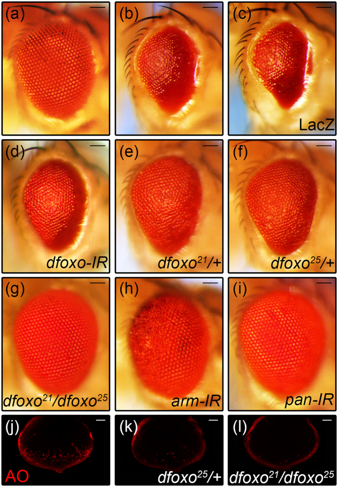
It has been reported that FoxO directly binds to β-catenin21, and competes with TCF for the limited pool of β-catenin, thereby inhibits Wnt signaling activity22,23. Thus we wonder whether dFoxO also inhibits Wg signaling-induced cell death in Drosophila. To our surprise, GMR > Arm-induced small eye phenotype (Fig. 2b) was suppressed partially by RNAi-mediated knocking-down of dfoxo (Fig. 2d), or in heterozygous dfoxo21 or dfoxo25 mutants (Fig. 2e,f), and fully in dfoxo21/dfoxo25 trans-heterozygous mutants (Fig. 2g), but remained unaffected by the expression of LacZ (Fig. 2c). As positive controls, loss of arm or pan fully suppressed GMR > Arm-induced small eye phenotype (Fig. 2h,i). Consistently, loss of dfoxo also suppressed GMR > Wg-induced small eye phenotype (Fig. S3). Moreover, GMR > Arm-induced AO staining (Fig. 2j) was suppressed partially in heterozygous dfoxo25 mutants (Fig. 2k), and fully in dfoxo21/dfoxo25 trans-heterozygous mutants (Fig. 2l). Thus, dFoxO is positively required for Wg signaling-induced cell death in Drosophila.
Figure 2. dFoxO is required for Arm-induced cell death.
Light micrographs of Drosophila adult eyes and fluorescent micrographs of 3rd instar eye discs are shown. Compared with the GMR-Gal4 control (a), expression of Arm triggers cell death and produces a small eye phenotype (b), which remains unaffected by expressing LacZ (c), but is partially suppressed by knocking-down of dfoxo (d), or in heterozygous dfoxo21 (e) or dfoxo25 (f) background, and fully suppressed in dfoxo21/dfoxo25 mutants (g). As positive controls, knocking-down of arm (h) or pan (i) suppresses the GMR > Arm small eye phenotype. GMR > Arm-induced AO staining (j) is suppressed partially in heterozygous dfoxo25 mutants (k), and fully in dfoxo21/dfoxo25 trans-heterozygous mutants (l). Sample numbers: a, 87; b, 110; c, 100; d, 75; e, 77; f, 56; g, 75; h, 97; i, 78; j, 22; k, 14; l, 17. Scale bars: 100 μm in (a–i) and 50 μm in (j–l).
Our previous study showed Wg mediates JNK signaling induced cell death26, and dFoxO is reported to be a downstream transcription factor of JNK signaling27,28,29. To test whether JNK is reciprocally required for Wg pathway induced cell death, we overexpressed Wg, Dsh or Arm by GMR-Gal4 in a compromised JNK background. We found activated Wg signaling induced small eye phenotype remained unaffected by blocking JNK signaling (Fig. S4a–S4r). As a positive control, GMR > Egr induced small eye phenotype was effectively suppressed by abrogating JNK activity (Fig. S4s–S4x). Thus, dFoxO acts independently of the JNK pathway to mediate Wg signaling induced cell death.
Loss of dFoxO suppresses Arm-induced apoptotic gene activation
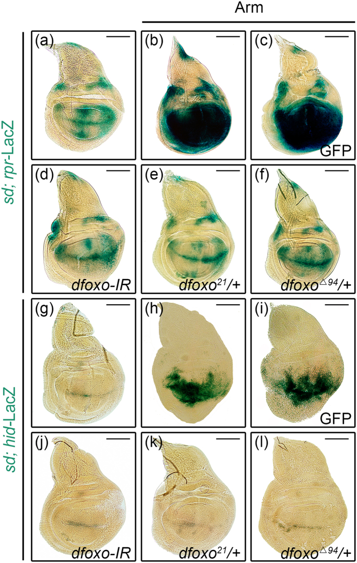
reaper (rpr) and head involution defective (hid) are important pro-apoptotic genes regulating cell death30. To examine whether activated Wg signaling induces cell death through up-regulation of rpr and hid, we ectopically expressed Arm in the wing pouch driven by sd-Gal4. Compared with the controls (Fig. 3a,g), rpr and hid expressions were significantly up-regulated by activated Wg signaling in the wing pouch (Fig. 3b,h), which were considerably suppressed by knocking-down of dfoxo or in heterozygous dfoxo mutants (Fig. 3d–f,j–l), but remained unaffected by the expression of GFP (Fig. 3c,i). Taken together, these results suggest that dFoxO is indispensable for Wg signaling induced cell death in Drosophila, which contradicts to the previous reports that FoxO impedes Wnt signaling in mammalian cells22,23. Thus, it is possible that FoxO may regulate Wnt signaling differently in a tissue or cell type specific manner.
Figure 3. dFoxO is required for Arm-induced hid and rpr expression.
Compared with the sd-Gal4 control (a,g), expression of Arm activates rpr–LacZ (b) and hid-LacZ (h) expression in the wing pouch, which remain unaffected by the expression of GFP (c,i), but are significantly suppressed by knocking-down of dfoxo (d,j), or heterozygous mutation of dfoxo21 (e,k) or dfoxoΔ94 (f,l). Scale bars: 100 μm.
dFoxO is required for the wing patterning functions of Wg signaling
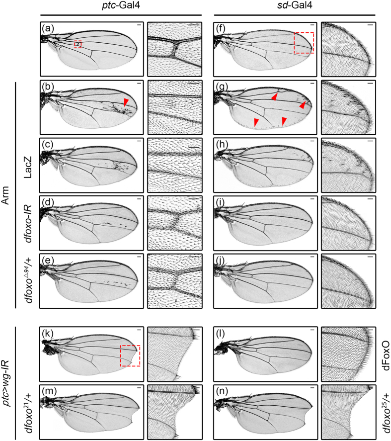
Wg signaling is one of the profound pathways that regulate wing pattern formation. Elevation of this pathway induces ectopic bristles, whereas inhibition of which results in wing margin notches31,32. Consistent with previous reports, we found that expression of Arm driven by ptc-Gal4 produced ectopic bristles between L3 and L4 veins (Fig. 4b, arrow head), and generated a loss-of-ACV phenotype (Fig. 4b). Intriguingly, a similar loss-of-ACV phenotype has been reported as a result of dFoxO overexpresion (Fig. S5b)20. We found that both the ectopic bristles and loss-of-ACV phenotypes were considerably suppressed by loss of dfoxo (Fig. 4d,e), but remained unchanged by the expression of LacZ (Fig. 4c). Similarly, sd > Arm induced ectopic bristles near the wing margin (Fig. 4g, arrow head) were fully suppressed by loss of dfoxo (Fig. 4i,j), but remained unaffected by the expression of LacZ (Fig. 4h). Hence, dFoxO is required for ectopic Wg signaling-induced extra brisltes and loss-of-ACV phenotypes in the wing.
Figure 4. dFoxO is required for the wing patterning functions of Wg signaling.
Light micrographs of Drosophila adult wings are shown. Compared with the ptc-Gal4 control (a, the right panel shows high magnification of the boxed area containing ACV in the left panel), expression of Arm induces ectopic bristles (indicated by the red arrow head) and loss-of-ACV phenotypes (b), which remain unaffected by the expression of LacZ (c), but are largely suppressed by RNAi-mediated knocking-down of dfoxo (d) or heterozygous mutation of dfoxoΔ94 (e). Compared with the sd-Gal4 control (f, the right panel shows high magnification of the boxed area containing anterior-distal wing margin in the left panel), expression of Arm induces ectopic bristles near the wing margin (g, indicated by red arrow heads), which remains unaffected by the expression of LacZ (h), but is suppressed by knocking-down of dfoxo (i) or heterozygous mutation of dfoxoΔ94 (j). knocking-down of wg by ptc-Gal4 generates a mild wing margin notch phenotype between the L3 and L4 veins (k, indicated by the red box and amplified in the right panel), which is rescued by the expression of dFoxO (l), but is enhanced by heterozygous mutation of dfoxo21 (m) or dfoxo25 (n). Sample numbers: a, 226; b, 201; c, 108; d, 197; e, 160; f,168; g, 166; h, 146; i, 130; j, 178; k,160; l, 62; m, 174; n, 226. Scale bars: 100 μm in (a–n) and 50 μm in high magnification figures.
Interestingly, despite cell death in wing discs, the adult wing sizes were not significantly altered upon Arm over expression (Fig. S6a and S6b). As Wg signaling also regulates cell proliferation, we speculate that cell death might be compensated by increased cell proliferation. To address this issue, we performed PH3 staining (Fig. S6c–S6f) and BrdU incorporation (Fig. S6g–S6j) in 3rd instar wing discs, but found no significant increase of cell proliferation in regions expressing Arm (Fig. S6k and S6l). Thus, other mechanism may exist to cope with ectopic Wg induced cell death and maintain tissue homeostasis in wing development. Alternatively, activation of cell death program may not necessary lead to cell loss, but instead, cell fate alteration. For instance, activation of apoptosis by ptc > Grim and ptc > pelle-IR produced the same loss-of-ACV phenotype19 as that of ptc > Dsh and ptc > Arm (Fig. 1f,g).
On the other hand, compromised Wg signaling impedes proliferation and results in loss of wing tissue, with wing margin notches as the most frequently observed phenotype31,33,34. Consistently, loss of wg between L3 and L4 veins by ptc > wg-IR generated a mild notch phenotype (Fig. 4k), which was suppressed by the expression of dFoxO (Fig. 4l), but enhanced in heterozygous dfoxo21 or dfoxo25 mutants (Fig. 4m,n). In addition, knock down dfoxo by two copies of dfoxo-IR also produced a weak notch phenotype in the wing margin (Fig. S5c). Hence, dFoxO is physiologically required for the wing patterning functions of endogenous Wg signaling.
dFoxO is required for the activation of Wg pathway target genes
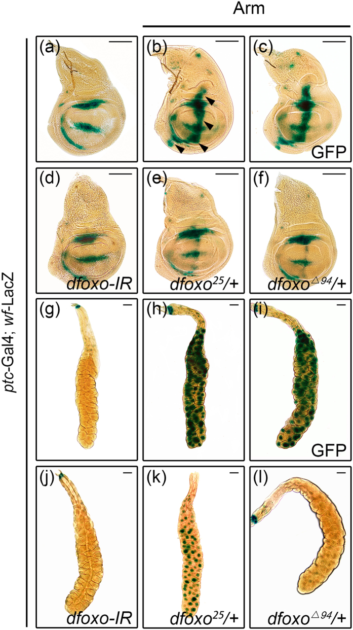
To further confirm that dFoxO is required for Wg signaling activity, we checked the expression of wingful (wf), a known Wg pathway target gene that mimic wg expression pattern, using a wf-LacZ reporter (Fig. 5a)35. Activation of Wg signaling by ptc > Arm along the A/P compartment boundary of wing discs strongly up-regulated wf transcription (Fig. 5b). The elevated wf expression was significantly suppressed by the expression of a dfoxo RNAi (Fig. 5d) or in heterozygous dfoxo mutants (Fig. 5e,f), but not that of GFP (Fig. 5c). Similar results were obtained in salivary glands where loss of dfoxo suppressed Arm-induced wf-LacZ expression (Fig. 5g–l). To verify the wf-LacZ reporter data, we carried out qRT-PCR experiments to analyze the transcription of endogenous wf and another Wg signaling target gene senseless (sens), which is required for bristle formation36. We confirmed that loss of dfoxo suppressed sd > Arm induced wf and sens expression in wing discs (Fig. S7). Thus, dFoxO is required for the transcriptional up-regulation of Wg pathway target genes.
Figure 5. dFoxO is required for Wg target gene activation.
Light micrographs of Drosophila 3rd instar wing discs (a–f) and salivary glands (g–l) with X-Gal staining are shown. Compared with the ptc-Gal4 control (a,g), ectopic Arm-induced wf-LacZ expression in the wing disc (b, indicated by black arrow heads) or salivary gland (h) remains unaffected by the expression of GFP (c and i), but is significantly suppressed by knocking-down of dfoxo (d,j), or heterozygous mutation of dfoxo25 (e,k) or dfoxoΔ94 (f,l). Scale bars: 100 μm in (a–f) and 200 μm in (g–l).
Arm binds to dFoxO and is reciprocally required for dFoxO-induced cell death
When Flag-tagged dFoxO and Myc-tagged Arm were co-expressed in Drosophila S2 cell, Flag-dFoxO could be immunoprecipitated with Myc-Arm, and vice versa (Fig. 6a). Furthermore, when Arm and GFP-dFoxO were co-expressed in the fly eye, Arm could be immunoprecipitated with GFP-dFoxO, and vice versa (Fig. 6b), suggesting dFoxO interacts with Arm both in vitro and in vivo. Furthermore, we co-expressed Flag-dFoxO, Myc-Arm and HA-Pan in S2 cells, and performed co-IP experiments with an anti-Flag antibody. We found that the antibody not only precipitated Flag-dFoxO, but also Myc-Arm and HA-Pan (Fig. S8), suggesting all three factors exist in the same complex.
Figure 6. Arm binds to dFoxO and is required for dFoxO-induced cell death.
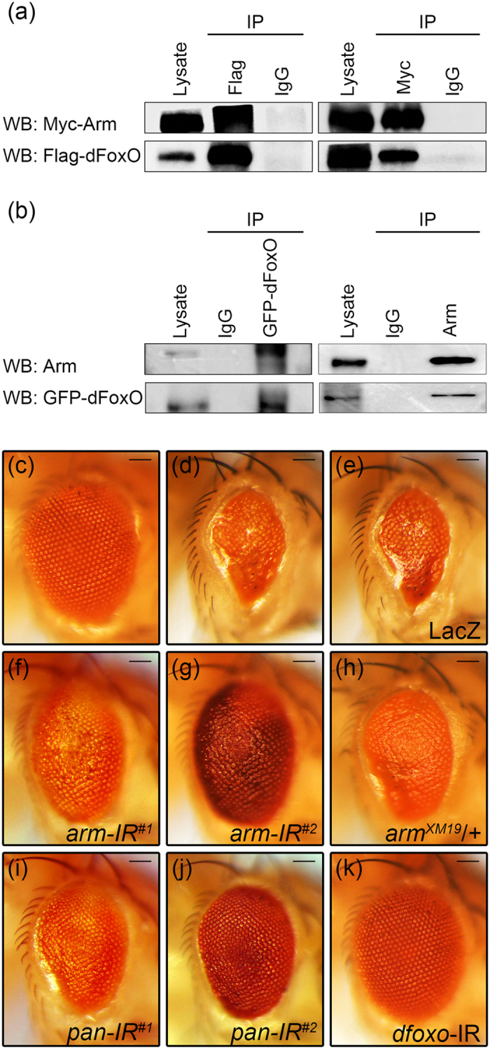
(a) Co-immunoprecipitation between transfected Myc-Arm and Flag-dFoxO in Drosophila S2 cells. (b) Co-immunoprecipitation between ectopically expressed Arm and GFP-dFoxO in Drosophila eye. (c–k) Light micrographs of Drosophila adult eyes are shown. Compared with the GMR-Gal4 control (c), expression of dFoxO triggers cell death and produces a small eye phenotype (d), which remains unaffected by the expression of LacZ (e), but is considerably suppressed by the expression of two independent arm RNAi (f,g), or heterozygous mutation of arm (h), or the expression of two independent pan RNAi (i,j). The GMR > dFoxO eye phenotype is also suppressed by knocking-down of dfoxo (k), which serves as a positive control. Sample numbers from c to k: c, 123; d, 104; e, 74; f, 67; g, 73; h, 64; i, 97; j, 106; k, 53. Scale bars: 100 μm.
Next we examined whether Arm is reciprocally required for the function of dFoxO. Expression of dFoxO in the eye driven by GMR-Gal4 also induced cell death and generated a small eye phenotype28 (Fig. 6d). The GMR > dFoxO-triggered small eye phenotype was suppressed by knocking-down of arm or arm mutation (Fig. 6f–h), but remained unaffected by the expression of LacZ (Fig. 6e). Consistently, loss of pan also suppressed dFoxO-induced small eye phenotype (Fig. 6i,j). As a positive control, this phenotype was fully suppressed by knocking-down of dfoxo (Fig. 6k). Therefore, dFoxO and Arm are reciprocally required for each other’s function, possibly through their physical interaction.
Discussion
In this work, we show that dFoxO promotes Wg signaling-induced cell death, wing pattern formation and target genes expression, while Arm is reciprocally required for dFoxO-induced cell death in vivo. Our data are consistent with previous study that β-catenin promotes FoxO activity21, but contradict to others that FoxO competes with TCF for the limited pool of β-catenin and thereby inhibits Wnt signaling activity22,23. A possible explanation for this discrepancy is that FoxO may regulate Wg/Wnt signaling in a tissue/cell type specific manner, depending on the presence of other transcriptional activating or repressing factor(s), or the different levels of FoxO and β-catenin/Arm in distinct cellular context. Importantly, while previous results were obtained mainly from the in vitro or gain-of-function studies, we have examined both loss-of-function and gain-of-function effects of dfoxo on ectopic and endogenous Wg signaling in vivo, and thus, the data are more likely to reflect the physiological situation. Moreover, the positive role of dFoxO in Wg signaling-induced cell death is in line with the well-known function of FoxO in promoting cell death.
The present study revealed for the first time that dFoxO acts as a vital regulator of Wg signaling-triggered cell death in vivo, which expands our knowledge of Wnt/Wg signaling function and the broad array of FoxOs’ regulatory activities. Both FoxO and Wnt/Wg signaling have been implicated in various cancers2,3,12,13,37, and a FOXO3a/β-catenin/GSK-3β signaling is essential for cell proliferation and apoptosis in prostate cancer cells38, further study is needed to understand their interaction in tumorigenesis.
Materials and Methods
Drosophila strains
The following lines have been described previously: UAS-Wg, UAS-Dsh, UAS-Arm, UAS-Pan, UAS-wg-IR, UAS-arm-IR#1, UAS-arm-IR#2, armXM19, UAS-pan-IR#1, GMR > Egr, UAS-BskDN, bsk1, en-Gal426; UAS-bsk-IR29; UAS-dFoxO-GFP-339; UAS-dfoxo-IR, dfoxoΔ94, ptc-Gal4, sd-Gal420; UAS-dFoxOP, UAS-dFoxOW2, dfoxo21, dfoxo25,40; UAS-LacZ41; UAS-GFP42; GMR-Gal443; omb-Gal419; hid-LacZ, rpr-LacZ44; wf-LacZ35. The UAS-Dcr2D2 (24650), UAS-ArmS2 (4783), UAS-pan-IR#2 (26743) and bsk2 (5283) lines were obtained from the Bloomington stock center.
X-Gal staining
X-Gal staining was done as described45, discs and salivary glands were dissected in PBST at the 3rd instar larva stage and fixed in 4% formaldehyde for 15 minutes, rinse the tissue once in PBST buffer containing 3.3 mM K3Fe(CN)6 and 3.3 mM K4Fe(CN)6, then incubate in the above PBST buffer with 0.2% X-gal at room temperature for 24 hours, photographs were taken under light microscope.
AO staining
AO staining was done as described46, discs were dissected at the 3rd instar larva stage, and stained in 1 × 10⁻⁵M AO solution for 5 minutes, photographs were taken under fluorescence microscope.
TUNEL staining
TUNEL staining was done as described26, discs were dissected at 3rd instar larva stage, and stained for TUNEL using the Fluorescein Cell Death Kit (Boster), photographs were taken under Leica confocal microscope.
Immunostaining
Discs were dissected at 3rd instar larva stage in PBS, fixed in 4% formaldehyde for 20 minutes and washed in 0.3% PBST for 3 times, then label with primary antibody overnight, and secondary antibody for 2 hours. Primary antibodies used were 1:100 mouse-anti-BrdU (Sigma) and 1:400 rabbit-anti-PH3 (Cell Signaling Technology, CST), secondary antibodies used were 1:1000 goat anti-mouse CY3 (CST) and 1:1000 goat anti- rabbit CY3 (CST). Photographs were taken under fluorescence microscope.
BrdU incorporation
BrdU incorporation was done as described47, discs were dissected at 3rd instar larva stage in Schneider’s medium (Sigma), incubated for 40 minutes in Schneider’s medium containing 0.2 mg/ml of BrdU, and fixed in 4% formaldehyde, then washed in PBST and hydrolyzed in 2 N HCl. After that the discs were blocked in 10% horse serum and labeled with mouse anti-BrdU and anti-mouse CY3 antibodies, photographs were taken under fluorescence microscope.
Co-immunoprecipitation
S2 cells were transfected with indicated plasmids for 48 h, immunoprecipitation and western analyses were performed as previously described43, pre-cleared cell lysates were incubated with the indicated antibodies followed by precipitation with protein G Sepharose beads (Sigma). Immune complexes were washed with lysis buffer, eluted in 2 × SDS sample buffer, and then subjected to western blot using corresponding antibodies. Fly heads were cut from indicated genotypes and homogenized in lysis buffer, immunoprecipitation and western analyses were performed as above. Antibodies used in this study were as follows: mouse anti-Arm antibody (DSHB), mouse anti-Flag (Sigma), rabbit anti-GFP (CST), mouse anti-Myc (CST), rabbit anti-HA (CST) and goat anti-mouse IgG-HRP (CST).
qRT-PCR
Wing discs were dissected at 3rd instar larva, for each genotype, more than 100 discs were collected and total RNA was isolated using TRIzol (Invitrogen). qRT-PCR was performed as previously described and rp49 was used as internal control48. Primer sequences were
wf fwd: AAGTCGAGCAATGGGCAATGAT,
rev: TGGAGGAGCGTGTCTTCTG;
sens fwd: CCGAAAAGGAGCATGAACTC,
rev: CGCTGTTGCTGTGGTGTACT36;
rp49 fwd: CATCCGCCCAGCATACAG,
rev: CCATTTGTGCGACAGCTTAG48.
Statistics
For pupal retina AO staining, wing size measurement and PH3 staining, unpaired t-text analysis was used. For qRT-PCR, one-way analysis of variance test followed by the post Dunnett test was used. A P value of less than 0.05 was considered significant.
Additional Information
How to cite this article: Zhang, S. et al. dFoxO promotes Wingless signaling in Drosophila. Sci. Rep. 6, 22348; doi: 10.1038/srep22348 (2016).
Supplementary Material
Acknowledgments
We thank Haiyun Song, Ernst Hafen, the Bloomington Drosophila Stock Center and the Vienna Drosophila RNAi Center for fly stocks, members of Xue lab for discussion and critical comments. This work is supported by the National Basic Research Program of China (973 Program) (2011CB943903), National Natural Science Foundation of China (31171413, 31371490, 31571516), the Specialized Research Fund for the Doctoral Program of Higher Education of China (20120072110023 and 20120072120030), the China Postdoctoral Science Foundation (2015M581659), and Shanghai Committee of Science and Technology (09DZ2260100, 14JC1406000).
Footnotes
Author Contributions S.Z., W.L. and L.X. conceived and designed the experiments. S.Z., X.G., C.C., Y.C., J.L., C.W., Y.Y. and W.L. performed the experiments. S.Z., Y.S., C.J., W.L. and L.X. analyzed the data, S.Z. and L.X. wrote the manuscript.
References
- Clevers H. & Nusse R. Wnt/beta-catenin signaling and disease. Cell 149, 1192–1205, doi: 10.1016/j.cell.2012.05.012 (2012). [DOI] [PubMed] [Google Scholar]
- Anastas J. N. & Moon R. T. WNT signalling pathways as therapeutic targets in cancer. Nature reviews. Cancer 13, 11–26, doi: 10.1038/nrc3419 (2013). [DOI] [PubMed] [Google Scholar]
- MacDonald B. T., Tamai K. & He X. Wnt/beta-catenin signaling: components, mechanisms, and diseases. Developmental cell 17, 9–26, doi: 10.1016/j.devcel.2009.06.016 (2009). [DOI] [PMC free article] [PubMed] [Google Scholar]
- Nusse R. Wnt signaling in disease and in development. Cell Res 15, 28–32, doi: 10.1038/sj.cr.7290260 (2005). [DOI] [PubMed] [Google Scholar]
- Miller J. R. The Wnts. Genome biology 3, REVIEWS3001 (2002). [DOI] [PMC free article] [PubMed] [Google Scholar]
- Swarup S. & Verheyen E. M. Wnt/Wingless signaling in Drosophila. Cold Spring Harbor perspectives in biology 4, doi: 10.1101/cshperspect.a007930 (2012). [DOI] [PMC free article] [PubMed] [Google Scholar]
- van Amerongen R. & Nusse R. Towards an integrated view of Wnt signaling in development. Development 136, 3205–3214, doi: 10.1242/dev.033910 (2009). [DOI] [PubMed] [Google Scholar]
- Cadigan K. M. & Nusse R. Wnt signaling: a common theme in animal development. Genes & development 11, 3286–3305 (1997). [DOI] [PubMed] [Google Scholar]
- Cordero J., Jassim O., Bao S. & Cagan R. A role for wingless in an early pupal cell death event that contributes to patterning the Drosophila eye. Mechanisms of development 121, 1523–1530, doi: 10.1016/j.mod.2004.07.004 (2004). [DOI] [PubMed] [Google Scholar]
- Lin H. V., Rogulja A. & Cadigan K. M. Wingless eliminates ommatidia from the edge of the developing eye through activation of apoptosis. Development 131, 2409–2418, doi: 10.1242/dev.01104 (2004). [DOI] [PubMed] [Google Scholar]
- Cordero J. B. & Cagan R. L. Canonical wingless signaling regulates cone cell specification in the Drosophila retina. Developmental dynamics: an official publication of the American Association of Anatomists 239, 875–884, doi: 10.1002/dvdy.22235 (2010). [DOI] [PMC free article] [PubMed] [Google Scholar]
- Greer E. L. & Brunet A. FOXO transcription factors at the interface between longevity and tumor suppression. Oncogene 24, 7410–7425, doi: 10.1038/sj.onc.1209086 (2005). [DOI] [PubMed] [Google Scholar]
- Maiese K., Chong Z. Z., Shang Y. C. & Hou J. A “FOXO” in sight: targeting Foxo proteins from conception to cancer. Medicinal research reviews 29, 395–418, doi: 10.1002/med.20139 (2009). [DOI] [PMC free article] [PubMed] [Google Scholar]
- Calnan D. R. & Brunet A. The FoxO code. Oncogene 27, 2276–2288, doi: 10.1038/onc.2008.21 (2008). [DOI] [PubMed] [Google Scholar]
- Zhao Y., Wang Y. & Zhu W. G. Applications of post-translational modifications of FoxO family proteins in biological functions. Journal of molecular cell biology 3, 276–282, doi: 10.1093/jmcb/mjr013 (2011). [DOI] [PubMed] [Google Scholar]
- Puig O., Marr M. T., Ruhf M. L. & Tjian R. Control of cell number by Drosophila FOXO: downstream and feedback regulation of the insulin receptor pathway. Genes & development 17, 2006–2020, doi: 10.1101/gad.1098703 (2003). [DOI] [PMC free article] [PubMed] [Google Scholar]
- Lam E. W., Francis R. E. & Petkovic M. FOXO transcription factors: key regulators of cell fate. Biochemical Society transactions 34, 722–726, doi: 10.1042/BST0340722 (2006). [DOI] [PubMed] [Google Scholar]
- Huang H. & Tindall D. J. Dynamic FoxO transcription factors. Journal of cell science 120, 2479–2487, doi: 10.1242/jcs.001222 (2007). [DOI] [PubMed] [Google Scholar]
- Wu C. et al. Pelle Modulates dFoxO-Mediated Cell Death in Drosophila. PLoS genetics 11, e1005589, doi: 10.1371/journal.pgen.1005589 (2015). [DOI] [PMC free article] [PubMed] [Google Scholar]
- Wang X. et al. FoxO mediates APP-induced AICD-dependent cell death. Cell Death and Disease 5, e1233, doi: 10.1038/cddis.2014.196 (2014). [DOI] [PMC free article] [PubMed] [Google Scholar]
- Essers M. A. et al. Functional interaction between beta-catenin and FOXO in oxidative stress signaling. Science 308, 1181–1184, doi: 10.1126/science.1109083 (2005). [DOI] [PubMed] [Google Scholar]
- Almeida M., Han L., Martin-Millan M., O’Brien C. A. & Manolagas S. C. Oxidative stress antagonizes Wnt signaling in osteoblast precursors by diverting beta-catenin from T cell factor- to forkhead box O-mediated transcription. The Journal of biological chemistry 282, 27298–27305, doi: 10.1074/jbc.M702811200 (2007). [DOI] [PubMed] [Google Scholar]
- Hoogeboom D. et al. Interaction of FOXO with beta-catenin inhibits beta-catenin/T cell factor activity. The Journal of biological chemistry 283, 9224–9230, doi: 10.1074/jbc.M706638200 (2008). [DOI] [PubMed] [Google Scholar]
- Xie X. W., Liu J. X., Hu B. & Xiao W. Zebrafish foxo3b negatively regulates canonical Wnt signaling to affect early embryogenesis. PloS one 6, e24469, doi: 10.1371/journal.pone.0024469 (2011). [DOI] [PMC free article] [PubMed] [Google Scholar]
- Iyer S. et al. FOXOs attenuate bone formation by suppressing Wnt signaling. The Journal of clinical investigation 123, 3409–3419, doi: 10.1172/JCI68049 (2013). [DOI] [PMC free article] [PubMed] [Google Scholar]
- Zhang S. et al. The canonical Wg signaling modulates Bsk-mediated cell death in Drosophila. Cell death & disease 6, e1713, doi: 10.1038/cddis.2015.85 (2015). [DOI] [PMC free article] [PubMed] [Google Scholar]
- Essers M. A. et al. FOXO transcription factor activation by oxidative stress mediated by the small GTPase Ral and JNK. The EMBO journal 23, 4802–4812, doi: 10.1038/sj.emboj.7600476 (2004). [DOI] [PMC free article] [PubMed] [Google Scholar]
- Luo X., Puig O., Hyun J., Bohmann D. & Jasper H. Foxo and Fos regulate the decision between cell death and survival in response to UV irradiation. The EMBO journal 26, 380–390, doi: 10.1038/sj.emboj.7601484 (2007). [DOI] [PMC free article] [PubMed] [Google Scholar]
- Wu C. et al. Toll pathway modulates TNF-induced JNK-dependent cell death in Drosophila. Open biology 5, 140171, doi: 10.1098/rsob.140171 (2015). [DOI] [PMC free article] [PubMed] [Google Scholar]
- Cashio P., Lee T. V. & Bergmann A. Genetic control of programmed cell death in Drosophila melanogaster. Seminars in cell & developmental biology 16, 225–235, doi: 10.1016/j.semcdb.2005.01.002 (2005). [DOI] [PubMed] [Google Scholar]
- Couso J. P., Bishop S. A. & Martinez Arias A. The wingless signalling pathway and the patterning of the wing margin in Drosophila. Development 120, 621–636 (1994). [DOI] [PubMed] [Google Scholar]
- Zhang J. & Carthew R. W. Interactions between Wingless and DFz2 during Drosophila wing development. Development 125, 3075–3085 (1998). [DOI] [PubMed] [Google Scholar]
- Phillips R. G. & Whittle J. R. wingless expression mediates determination of peripheral nervous system elements in late stages of Drosophila wing disc development. Development 118, 427–438 (1993). [DOI] [PubMed] [Google Scholar]
- Zeng Y. A., Rahnama M., Wang S., Lee W. & Verheyen E. M. Inhibition of Drosophila Wg signaling involves competition between Mad and Armadillo/beta-catenin for dTcf binding. PloS one 3, e3893, doi: 10.1371/journal.pone.0003893 (2008). [DOI] [PMC free article] [PubMed] [Google Scholar]
- Gerlitz O. & Basler K. Wingful, an extracellular feedback inhibitor of Wingless. Genes & development 16, 1055–1059, doi: 10.1101/gad.991802 (2002). [DOI] [PMC free article] [PubMed] [Google Scholar]
- Eivers E. et al. Mad is required for wingless signaling in wing development and segment patterning in Drosophila. PloS one 4, e6543, doi: 10.1371/journal.pone.0006543 (2009). [DOI] [PMC free article] [PubMed] [Google Scholar]
- Reya T. & Clevers H. Wnt signalling in stem cells and cancer. Nature 434, 843–850, doi: 10.1038/nature03319 (2005). [DOI] [PubMed] [Google Scholar]
- Li Y. et al. Regulation of FOXO3a/beta-catenin/GSK-3beta signaling by 3,3’-diindolylmethane contributes to inhibition of cell proliferation and induction of apoptosis in prostate cancer cells. The Journal of biological chemistry 282, 21542–21550, doi: 10.1074/jbc.M701978200 (2007). [DOI] [PubMed] [Google Scholar]
- Wagner C., Isermann K. & Roeder T. Infection induces a survival program and local remodeling in the airway epithelium of the fly. FASEB journal: official publication of the Federation of American Societies for Experimental Biology 23, 2045–2054, doi: 10.1096/fj.08-114223 (2009). [DOI] [PubMed] [Google Scholar]
- Junger M. A. et al. The Drosophila forkhead transcription factor FOXO mediates the reduction in cell number associated with reduced insulin signaling. Journal of biology 2, 20, doi: 10.1186/1475-4924-2-20 (2003). [DOI] [PMC free article] [PubMed] [Google Scholar]
- Hu Y., Han Y., Wang X. & Xue L. Aging-related neurodegeneration eliminates male courtship choice in Drosophila. Neurobiology of aging, doi: 10.1016/j.neurobiolaging.2014.02.026 (2014). [DOI] [PubMed] [Google Scholar]
- Ma X. et al. Src42A modulates tumor invasion and cell death via Ben/dUev1a-mediated JNK activation in Drosophila. Cell death & disease 4, e864, doi: 10.1038/cddis.2013.392 (2013). [DOI] [PMC free article] [PubMed] [Google Scholar]
- Ma X. et al. Bendless modulates JNK-mediated cell death and migration in Drosophila. Cell death and differentiation 21, 407–415, doi: 10.1038/cdd.2013.154 (2014). [DOI] [PMC free article] [PubMed] [Google Scholar]
- Ma X. et al. NOPO modulates Egr-induced JNK-independent cell death in Drosophila. Cell Res 22, 425–431, doi: 10.1038/cr.2011.135 (2012). [DOI] [PMC free article] [PubMed] [Google Scholar]
- Xue L. & Noll M. Dual role of the Pax gene paired in accessory gland development of Drosophila. Development 129, 339–346 (2002). [DOI] [PubMed] [Google Scholar]
- Ma X. et al. dUev1a modulates TNF-JNK mediated tumor progression and cell death in Drosophila. Developmental biology 380, 211–221, doi: 10.1016/j.ydbio.2013.05.013 (2013). [DOI] [PubMed] [Google Scholar]
- Peng F. et al. Loss of Polo ameliorates APP-induced Alzheimer’s disease-like symptoms in Drosophila. Scientific reports 5, 16816, doi: 10.1038/srep16816 (2015). [DOI] [PMC free article] [PubMed] [Google Scholar]
- Beck E. S. et al. Regulation of Fasciclin II and synaptic terminal development by the splicing factor beag. The Journal of neuroscience: the official journal of the Society for Neuroscience 32, 7058–7073, doi: 10.1523/JNEUROSCI.3717-11.2012 (2012). [DOI] [PMC free article] [PubMed] [Google Scholar]
Associated Data
This section collects any data citations, data availability statements, or supplementary materials included in this article.


