Abstract
Debate on the adaptive origins of primates has long focused on the functional ecology of the primate visual system. For example, it is hypothesized that variable expression of short- (SWS1) and middle-to-long-wavelength sensitive (M/LWS) opsins, which confer color vision, can be used to infer ancestral activity patterns and therefore selective ecological pressures. A problem with this approach is that opsin gene variation is incompletely known in the grandorder Euarchonta, that is, the orders Scandentia (treeshrews), Dermoptera (colugos), and Primates. The ancestral state of primate color vision is therefore uncertain. Here, we report on the genes (OPN1SW and OPN1LW) that encode SWS1 and M/LWS opsins in seven species of treeshrew, including the sole nocturnal scandentian Ptilocercus lowii. In addition, we examined the opsin genes of the Central American woolly opossum (Caluromys derbianus), an enduring ecological analogue in the debate on primate origins. Our results indicate: 1) retention of ultraviolet (UV) visual sensitivity in C. derbianus and a shift from UV to blue spectral sensitivities at the base of Euarchonta; 2) ancient pseudogenization of OPN1SW in the ancestors of P. lowii, but a signature of purifying selection in those of C. derbianus; and, 3) the absence of OPN1LW polymorphism among diurnal treeshrews. These findings suggest functional variation in the color vision of nocturnal mammals and a distinctive visual ecology of early primates, perhaps one that demanded greater spatial resolution under light levels that could support cone-mediated color discrimination.
Keywords: color vision, sensory ecology, Caluromys, Dendrogale, Euarchonta, Ptilocercus, Tupaia
Introduction
The color vision of mammals is based on the expression of two opsin genes (OPN1SW and OPN1LW) that encode short- (SWS1) and middle-to-long-wavelength sensitive (M/LWS) photopigments. Some variant of this dichromatic phenotype (Peichl 2005) is the probable ancestral state of therian mammals (Wakefield et al. 2008) and every successive lineage, such as primates, that subsequently lost or gained opsin genes (Jacobs 2013; Meredith et al. 2013; Veilleux et al. 2013). Among primates, OPN1LW has differentiated into multiple alleles (lemurs, most New World monkeys) or paralogs (howler monkeys, Old World primates), resulting in spectrally shifted photopigments that confer allelic or routine trichromatic vision, respectively (Jacobs 2009; Kawamura et al. 2012). The M/LWS opsin variation that causes allelic trichromacy is widespread among primates (Tan et al. 2005; Melin et al. 2013) but unknown outside the order. Limited sampling, however, has precluded a formal comparative analysis of opsin genes in the grandorder Euarchonta, that is, the orders Scandentia (treeshrews), Dermoptera (colugos), and Primates. It is therefore challenging to infer the ancestral state of the primate visual system.
Opsin Sensitivity and Spatial Resolution
The peak spectral absorbance (λmax) of opsins is sensitive to natural selection and varies in response to environmental conditions and natural histories (Parry et al. 2004; Davies et al. 2012; Hunt and Peichl 2014). Crucially, functional variation of OPN1SW is widespread among mammals (Emerling et al. 2015). Mutations can disable function completely (Jacobs 2013) or cause large shifts in λmax, conferring ultraviolet (UV) visual sensitivity (λmax = 360 nm) to a mouse or blue sensitivity (λmax = 444 nm) to a treeshrew, Tupaia belangeri (Jacobs and Neitz 1986; Jacobs et al. 2004).
The functional ecological significance of these spectral differences is uncertain, but a relationship with visual acuity has been reported. Douglas and Jeffery (2014) examined the ocular media of 38 mammalian species and found that UV transmission and sensitivity prevail in low-acuity visual systems. This result suggests that natural selection for greater visual acuity (spatial resolution) also favored UV-filtering ocular media and blue-sensitive SWS1 opsins. In theory, spectral convergence of the SWS1 and M/LWS opsins should enhance visual acuity by minimizing chromatic aberrations (Walls 1942; Thibos et al. 1990). This premise has particular relevance to primates, a lineage with exceptional levels of visual acuity (references in Moore et al. 2012; Moritz et al. 2014).
SWS1—a Lens on Primate Origins?
Degenerate OPN1SW opsin genes—resulting in monochromatic vision—are a common trait of nocturnal mammals, or those active under dark (scotopic) light conditions, such as fossorial, cave, or deep marine habitats (Jacobs et al. 1993; David-Grey et al. 2002; Zhao et al. 2009; Davies et al. 2011; Jacobs 2013; Meredith et al. 2013). At the same time, functional OPN1SW genes have been retained by purifying selection in some fruit bats (Wang et al. 2004) and nocturnal euarchontans (colugos: Moritz et al. 2013; primates: Kawamura and Kubotera 2004; Perry et al. 2007; Veilleux et al. 2013). Such differences are difficult to explain. Some authors view the dichromatic vision of nocturnal primates as evidence of evolutionary disequilibrium (Tan et al. 2005), whereas others propose an ecological function on the grounds that dim twilight or full moonlight is sufficient for cone-mediated color vision (Melin et al. 2012, 2013; Veilleux and Cummings 2012; Moritz 2015). This distinction between visual anachronism and adaptation is now a central topic in the debate on primate origins (Tan et al. 2005).
Thus, the functional preservation and λmax of SWS1 opsins are tandem traits that can speak to the evolution and ecology of high-acuity color vision, a key derivation of primates (Ravosa and Savakova 2004; Cartmill 2012; Sussman et al. 2013). Here, we examine opsin gene variation across Euarchonta in order to explore how, when, and why enhanced visual acuity evolved. We believe that this comparative, integrated approach has the potential to inform hypotheses on the origin and evolution of primates.
Present Study
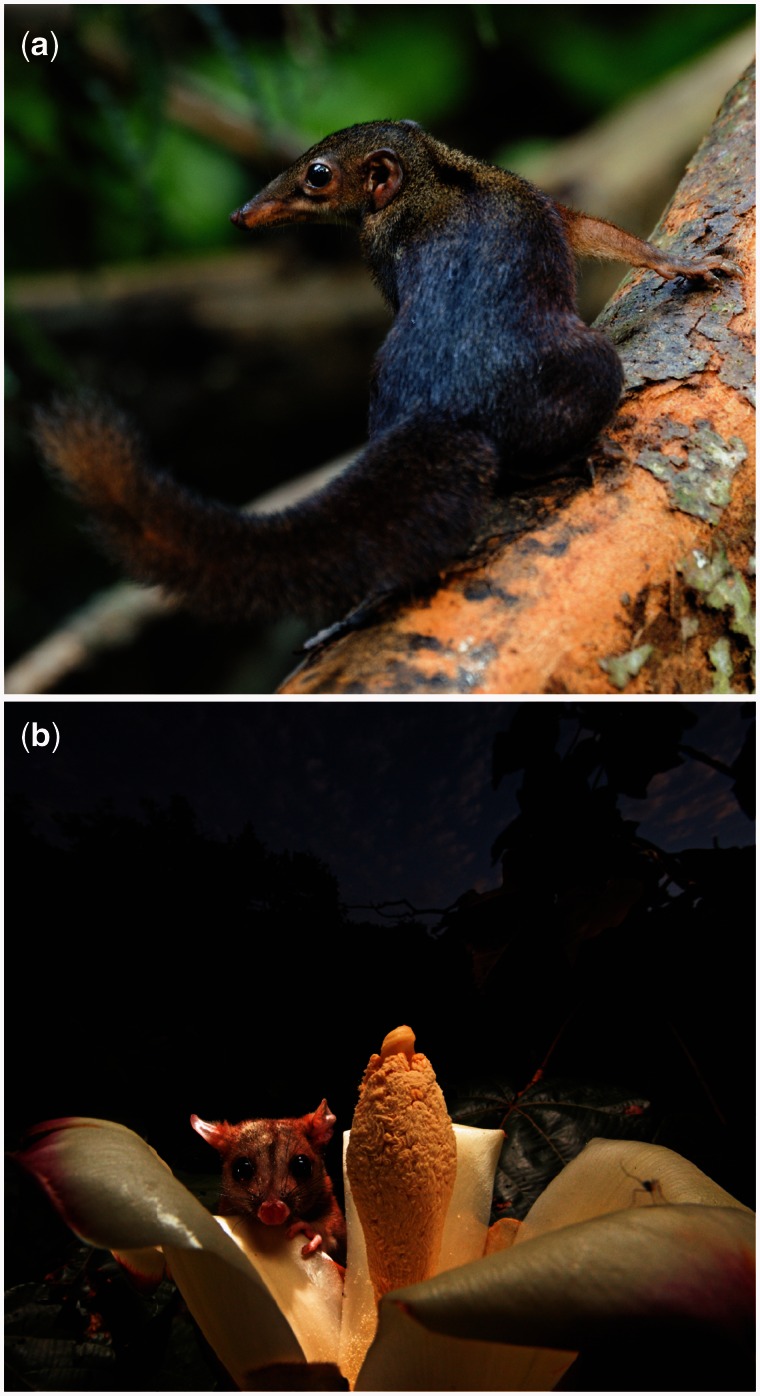
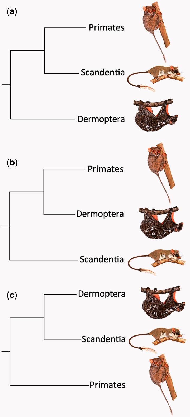
We report on the opsin genes of treeshrews (n = 7 species) and a woolly opossum (Caluromys derbianus) and analyze the gene sequences together with published data from other mammals. Treeshrews (fig. 1a) are sometimes described as “living models” of ancestral primates due to shared phyletic, morphological, and ecological affinities, albeit with a focus on locomotion (Tattersall 1984; Jenkins 1987; Martin 1990; Sargis 2004; Silcox et al. 2015; Li and Ni 2016). Similarly, C. derbianus (fig. 1b) is convergent toward primates in having a relatively large brain and eyes, small litters, and a slow life history. The arboreal agility and diet of this marsupial are therefore enduring topics in the debate on primate origins (Rasmussen 1990, 2002; Schmitt and Lemelin 2002; Gebo 2004; Sargis et al. 2007). The monophyly of treeshrews, colugos, and primates in Euarchonta is firmly established; however, the internal structure of Euarchonata is debated. Three phylogenetic hypotheses exist: 1) a sister-group relationship between treeshrews and primates (fig. 2a), 2) a sister-group relationship between colugos and primates, that is, Primatomorpha (fig. 2b); and, 3) both treeshrews and colugos as sister to primates, that is, Sundatheria (fig. 2c).
Fig. 1.
Treeshrews [(a) Tupaia tana] and woolly opossums [(b) Caluromys derbianus] are phyletic and/or ecological analogues of ancestral primates, factors that invite study of their opsin genes. Panel (b) also depicts the pollination of balsa (Ochroma pyramidale) under twilight conditions [Kays et al. 2012]. Photographs by Wong Tsu Shi and Christian Ziegler, respectively, and reproduced with permission.
Fig. 2.
The internal structure of Euarchonta is debated and revolves around three hypotheses: (a) a sister-group relationship between treeshrews and primates (Wible and Covert 1987; Kay et al. 1992), (b) a sister-group relationship between colugos and primates (Primatomorpha; Janečka et al. 2007; Meredith et al. 2011), or (c) both treeshrews and colugos as sister to primates (Sundatheria; Murphy et al. 2001; Sargis 2002; Bloch et al. 2007; O'Leary et al. 2013). Illustrations © The Sabah Society, reproduced with permission.
To estimate the λmax of extant and ancestral SWS1 opsins, we identified the amino acids at ten spectral tuning sites of the OPN1SW genes, of which two—Tyr86 and Val93, respectively—primarily determine sensitivity in the violet-blue (400–450 nm) region of the spectrum. We tested for purifying selection as a function of activity pattern; and, in the case of pseudogenes, estimated the antiquity of functional loss by comparing rates of substitution in coding versus noncoding regions of the gene. We estimated the λmax of extant and ancestral M/LWS opsins on the basis of three spectral tuning sites in OPN1LW; and finally, we explored the potential for allelic trichromatic vision in treeshrews.
Results
We succeeded in sequencing each exon of OPN1SW in Caluromys and all tupaiids, excepting exon 2 of Dendrogale melanura and Tupaia montana. Our initial OPN1SW polymerase chain reactions (PCRs) failed for all exons of P. lowii, thus necessitating the shotgun genome sequencing approach to reconstruct this gene sequence. Partial sequencing of OPN1LW was successful for all species—including P. lowii—although two of eight individuals of T. montana failed repeatedly, which we attributed to low DNA quality. Whole-genome sequence reads of P. lowii are deposited in the NCBI Sequence Read Archive (study accession no. SRP064536) and representative sequences of each species in GenBank (supplementary table S1, Supplementary Material online).
The most likely phylogenetic trees for Euarchonta based on the synonymous and intron sites of OPN1SW are consistent with the concept of Primatomorpha, a sister group relationship of primates and colugos (figs. 2b and 3). Further, the most likely phyletic relationship of opsin genes within sampled treeshrews agrees well with previously reported phylogenies (Luckett 1980; Roberts et al. 2011). Some study species are omitted from the intron tree due to the absence of overlapping sequence data.
Fig. 3.
Phylogenies of Euarchonta based on intron (a) and synonymous (b) sites of the OPN1SW. The evolutionary history was inferred by using the Neighbor-Joining method (Saitou and Nei 1987). The percentage of replicate trees in which the associated taxa clustered together in the bootstrap test (1,000 replicates) is shown next to the branches (Felsenstein 1985). The evolutionary distances are in units of the number of differences per site (Nei-Gojobori 1986). The analysis involved 9 (a) and 11 (b) nucleotide sequences. All ambiguous positions were removed for each sequence pair. There were a total of 774 (a) and 354 (b) positions in the final data set. Evolutionary analyses were conducted in MEGA6 (Tamura et al. 2013).
Signatures of Selection
The coding sequences of OPN1SW in the woolly opossum (C. derbianus) and diurnal treeshrews (D. melanura, Tupaia spp.), and the coding sequences of OPN1LW in all species, were free of indels (insertions/deletions), nonsense mutations, and premature stop codons, indicating strict conservation and functional preservation (fig. 4; supplementary fig. S1, Supplementary Material online). Greater amino acid divergence was present in OPN1SW (mean amino acid difference per sequence/total amino acids analyzed, SE over 1,000 bootstrap replicates: 36.53/349 = 10.47% divergence, 2.86) than in OPN1LW (15.46/228 = 6.78% divergence, 2.13; note: for trichromatic species, only the LWS allele/paralogue was assessed). However, this difference did not reach statistical significance (Fisher’s Exact Test, P = 0.14).
Fig. 4.
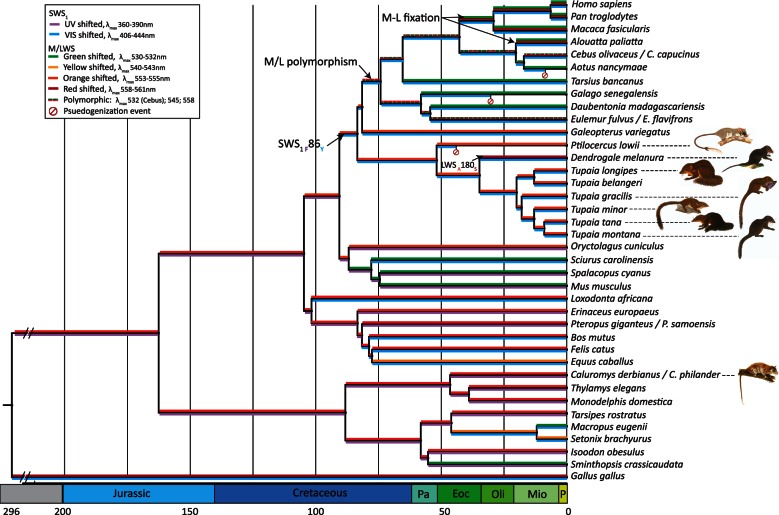
Phylogenetic relationships and divergence dates for Euarchonta and outgroups were based on TimeTree (Hedges et al. 2006; accessed June 2015) and published estimates (Prideaux and Warbuton 2010; Roberts et al. 2011; Fabrae et al. 2012; Song et al. 2012). Branch colors correspond with the presence and spectral tuning of opsin photopigments. Pseudogenization events are marked with a diagonally bisected circle. The inferred shift from UV to blue sensitivity in the SWS1 opsin of the ancestral euarchontan is marked with an arrow, along with the amino acids proposed to be responsible. Dashed branches indicate opsin polymorphism. The geological time scale is abbreviated as Pa, Paleocene; Eoc, Eocene; Oli, Oligocene; Mio, Miocene; P, Plio/Pleistocene. Treeshrew art © The Sabah Society, reproduced with permission.
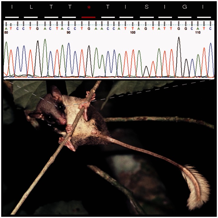
We detected a series of frameshift deletions in the OPN1SW sequence of P. lowii. We artificially corrected the frameshifts by filling the contig gaps with the consensus bases from other taxa, and repeated the translation. While correcting the frameshifts improved the amino acid alignment, when P. lowii is compared with the five Tupaia species with exon 2 sequences, a total of ten nonsynonymous mutations out of 117 nonsynonymous sites in exon 2 (gaps sites were omitted) were altered by to amino acids unique to Ptilocercus in the alignment, and two stop codons occurred within the Ptilocercus translation (fig. 5; supplementary fig. S1a, Supplementary Material online). There were also nine synonymous mutations out of 34 possible synonymous sites. Among Tupaia, which uniformly possessed intact OPN1SW, there were zero nonsynonymous differences and three synonymous differences. The divergence of OPN1SW of P. lowii explains why repeated attempts to amplify this gene via PCR were unsuccessful.
Fig. 5.
Ptilocercus lowii and a corresponding partial amino acid sequence to demonstrate one of several stop codons in the coding region of the OPN1SW pseudogene. Photograph by Annette Zitzmann, reproduced with permission.
For OPN1SW, purifying selection (dN < dS; P < 0.05) was favored over the null hypothesis of neutrality (dN = dS) for the majority of pairwise comparisons among the species in our complete data set. The notable exceptions to this pattern were the pairwise comparisons including P. lowii. Here, substitution rates were consistent with neutrality for the majority (25/35) of possible pairwise comparisons and nearly all (25/26) comparisons with eutherian mammals (supplementary table S2, Supplementary Material online). Taken together with the alignment results, these data indicate ancient pseudogenization of the OPN1SW in Ptilocercidae.
For OPN1LW, purifying selection (dN < dS; P < 0.05) was favored over the null hypothesis of neutrality (dN = dS) for the majority of paired comparisons (supplementary table S3, Supplementary Material online). Among tupaiids, neutrality was not uniformly rejected; however, the recent diversification of Tupaia (Roberts et al. 2011) and the short regions examined here (exons 3 and 5 only) suggest an underpowered analysis. Consistent with this interpretation is our finding that purifying selection is evident for the OPN1LW of T. belangeri, for which more sequencing data were available.
Pseudogenization of the SWS1 Opsin Gene of Ptilocercus
The ratio of the rate of nonsyonymous substitutions (dN) to presumably neutral substitutions at synonymous and intron sites (dS+I) among tupaiids, following divergence from other euarchontans, reveals moderate functional constraint (mean dN/dS+I = 0.0394/0.1060 = 0.37). Dendrogale melanura and T. montana are excluded from this analysis because we lacked sequence data for exon 2, the only region of reconstruction for P. lowii. The mean neutral mutation rate, k, of the tupaiid lineage, following the split with other euarchontans, was low (0.1060/83.43 Ma = 1.27 × 10⁻⁹ per site per year). A total of 277 nonsynonymous and 649 synonymous and intron sites were used in this analysis. The sequence data from P. lowii are excluded from our calculations of the neutral mutation rate because the shorter sequence recovered for this species would have unnecessarily constrained the data set used in the analysis.
We added P. lowii to the data set to calculate the dN of Ptilocercidae. The divergence of this lineage from Tupaiidae was previously estimated to be 60.19 Ma (Roberts et al. 2011). The dN value for P. lowii (0.0977) is relatively high, and the fN (fraction of neutral substitutions; 0.0977/0.0207 = 4.72) far exceeds 1, revealing an unusually high substitution rate at sites than would have been nonsynonymous in a functional gene. This result, together with the low rate of neutral evolution observed for tupaiid SWS1 opsins, prohibits an accurate estimation of the timing of pseudogenization in the lineage that gave rise to P. lowii. For example, using the methods of Chou et al. (2002), we calculate a date that precedes the estimated divergence of this species from other treeshrews [t1 = ((0.0977/1.27 × 10−9)−(0.37*60.19 Ma))/(1 − 0.37) = 86.75 Ma]. Analyses using alternative calculations of the timing of pseudogenization based on nucleotide position in the codon (Yokoyama et al. 2014) fare no better. Thus, the pseudogenization of OPN1SW cannot be dated with precision, but it is clear that the antiquity of monochromatic vision is great within Ptilocercidae.
Spectral Sensitivities of Opsins
OPN1SW Opsin Gene
Spectral tuning sites are invariant in Tupaiidae (supplementary fig. S1a, Supplementary Material online). The λmax of the opsin is therefore expected to resemble that of T. belangeri, which is calculated at 444 nm (based on electroretinogram (ERG) flicker photometry, Jacobs and Neitz 1986) or 428 ± 15 nm (based on microspectrophotometry, Petry and Harosi 1990). In contrast, the spectral tuning sites of C. derbianus (Phe86, Thr93) predict UV sensitivity (λmax = 360 nm), a result that agrees well with earlier findings from South American marsupials (Hunt et al. 2009; Palacios et al. 2010).
OPN1LW Opsin Gene
The three spectral tuning sites—180:A, 277:Y, 285:T—are invariant in Ptilocercus and Tupaia and we detected no intraspecific polymorphisms (supplementary figs. S1b and S2, Supplementary Material online). The inferred λmax is therefore 555 nm (Yokoyama et al. 2008), a result that agrees well with ERG flicker photometry- and microspectrophotometry-based findings (T. glis: Tigges et al. 1967; T. belangeri: Jacobs and Neitz 1986; Petry and Harosi 1990). The three-site composition of D. melanura differed (A180S) from other treeshrews, predicting a λmax shifted by 5–7 nm to 560–562 nm. Lastly, the three-site composition of C. derbianus (AYT; λmax = 555 nm) agrees with earlier findings from South American marsupials (Hunt et al. 2009; Palacios et al. 2010).
Ancestral States
Our phylogenies based on OPN1SW intron and synonymous sites are consistent with the concept of Primatomorpha, and we found that the ancestral state of all living and extinct (crown) euarchontans was unambiguous at eight of ten tuning sites: Phe46, Phe49, Thr52, Tyr86, Ser90, Ala114, Leu116, and Ser118. Maximum parsimony (MP) was ambiguous at two sites: Ile/Pro/Thr/Val93 and Ala/Asn97. Accordingly, we used maximum likelihood (ML) to estimate Thr as the ancestral amino acid at site 93, with shifts to Val in Scandentia, Ile in Dermoptera, and Pro in Primates. We reconstructed Ala as the ancestral amino acid at site 97, with retention in Primates and independent shifts to Asn in Dermoptera and Scandentia. Ancestral states based on a sister-group relationship between treeshrews and primates (fig. 2a) or Sundatheria (fig. 2c) are identical or nearly so, respectively. In the case of Sundatheria, the MP analysis is unambiguous concerning Ala97, but site 86 was ambiguous as either Phe or Tyr. In the ensuing ML analysis, Phe is reconstructed as the ancestral state. In all ML analyses, the posterior probabilities are greater than 0.9. Lastly, the ancestral state of the M/LWS opsin gene in crown Euarchonta is unambiguous for each spectral tuning site (Ala180, Tyr277, Thr285) in all configurations.
Discussion
Our primary conclusions are 4-fold: 1) Frameshift deletions in OPN1SW have an ancient origin in the lineage that gave rise to Ptilocercus lowii. The resulting phenotype (cone monochromacy) unites P. lowii with numerous mammals active under dark (scotopic) conditions (Jacobs 2013). 2) At the same time, we detected a signature of purifying selection in OPN1SW of C. derbianus, a nocturnal opossum. The preservation of a UV-sensitive SWS1 opsin in C. derbianus challenges the proposed incompatibility of nocturnality and dichromatic vision (Tan et al. 2005), and is consistent with other findings (Kawamura and Kubotera, 2004; Perry et al. 2007; Zhao et al. 2009). 3) The UV-sensitivity of SWS1 opsins was likely abolished at the base of Euarchonta, a result that partly diminishes the value of arboreal marsupials for modeling the visual ecology of early primates (Rasmussen 1990; Rasmussen and Sussman 2007). 4) A selective aversion to UV sensitivity in crown Euarchonta and Primates suggests a distinctive visual ecology or photic niche, that is, one that demanded greater spatial resolution under light levels that could support cone-mediated color discrimination (Emerling et al. 2015).
Such findings advance our understanding of primate origins by resolving and extending the antiquity of blue-sensitive shifts in the visual systems of Euarchonta. Previously, Carvalho et al. (2012) suggested that a substitution from phenylalanine to tyrosine at site 86 of OPN1SW evolved at the base of Primates. Our findings support this hypothesis and show that Phe86Tyr is the most parsimonious ancestral state of crown Euarchonta. Site 93 is also critical to λmax and our findings implicate Thr93 as the ancestral state. Combinations of Tyr86 and Thr93 exist in two Australian marsupials, the Tamar wallaby (Macropus eugenii) and quokka (Setonix brachyurus), and confer blue sensitivity (λmax = 424 nm; Deeb et al. 2003; Arrese et al. 2005). Independent shifts at site 93 occurred in Scandentia (Thr93Val), Dermoptera (Thr93Ile), and Primates (Thr93Pro). Further, tandem shifts of Phe86Tyr and Thr93Val evolved in at least one diurnal rodent, Sciurus carolinensis (Carvalho et al. 2006). The arboreal proclivities of these eutherians distinguish them from the Tamar wallaby and quokka, raising the possibility that the spectral tuning afforded by Tyr86 and Thr93 are unfavorable in an arboreal milieu. Alternatively, amino acid divergence at other sites, or incompatible interactions, may have favored multiple, independent replacements of Thr93 in the latter group following the Phe86Tyr transition. Such incompatibilities have been observed, for example, in mutant pigments created by in vitro by site-directed mutagenesis (Carvalho et al. 2012).
Future genome sequence of additional euarchontans and other mammals may resolve debate between Primatomorpha and Sundatheria. Sundatheria would implicate either Tyr86 (as above) or Phe86 as the ancestral state of crown Euarchonta. In this latter scenario, our ancestral state reconstruction indicates a transition of Phe86Tyr in the common ancestor of Scandentia and Dermoptera and the retention of Phe86 in Primates. It follows, then, that the aye-aye (Daubentonia madagascariensis) is either the sole extant primate to have retained the ancestral OPN1SW sequence at site 86 (λmax = 406 nm; Carvalho et al. 2012) or that a subsequent reversion of this amino acid occurred in the aye-aye lineage.
Implications for Primate Origins
We conclude that the color vision of ancestral primates is based on functional SWS1 and M/LWS opsins with estimated λmax values of approximately 424 and 555 nm, respectively. Although precise λmax values can be difficult to predict from sequence data alone (Hauser et al. 2014), blue sensitivity is strongly implicated by the inferred amino acid composition (Tyr86, Ser90, Thr93) of OPN1SW. Significantly, this phenotype is incompatible with the UV sensitivity of C. derbianus and impoverished color vision of P. lowii, and it follows that color vision in these species is unsuitable for evaluating the visual ecology of ancestral primates (pace Rasmussen 1990; Sargis 2004).
At the same time, differences in the nocturnality of C. derbianus and P. lowii are instructive, revealing the practical limits of the concept. The visual ecology of C. derbianus includes twilight (fig. 1b) and daylight activities (Reid 1997), whereas P. lowii is strictly nocturnal (Lyon 1913; Le Gros Clark 1926; Lim 1967; Gould 1978) and averse to moonlight (Emmons 2000). It is therefore likely that the dichromatic vision of ancestral primates is indicative of occasional or regular activity under dim (mesopic) to daylight (photopic) conditions.
The blue sensitivity of the SWS1 opsin is also telling, suggesting activities that demanded enhanced visual acuity (Douglas and Jeffrey 2014). The nature of these activities is uncertain, but the elimination of UV sensitivity could suggest that nectar was a supplemental, rather than primary, resource (cf. Sussman and Raven 1978; Gómez and Verdú 2012; Sussman et al. 2013). A diet premised on floral nectar unites most nocturnal mammals with UV-sensitive SWS1 opsins, perhaps to discriminate UV-reflecting flowers under dim light (Fleming et al. 2009). Our supposition does not refute the importance of nectar during primate evolution, but a shift to blue sensitivity suggests a diet that commanded greater spatial resolution than necessary for discriminating flowers (Veilleux and Kirk 2014).
This premise is supported by the convergent pseudogenization of OPN1SW in many primate taxa (lorisids; Allocebus, Cheirogaleus, Phaner, and Aotus) and other mammals (Jacobs 2013; Veilleux et al. 2013). As with P. lowii, a blue-sensitive SWS1 opsin is the inferred ancestral state among euarchontans, and it is tempting to speculate that natural selection favored pseudogenization of violet–blue sensitive (400–450 nm) SWS1 opsins more readily than a reversion to UV sensitivity (360–400 nm). It follows that most OPN1SW pseudogenes were formerly blue-sensitive and that pseudogenization is the result of shifts to scotopic, lunarphobic conditions rather than nocturnality per se, a pattern exemplified by flying squirrels (Carvalho et al. 2006). The advantage of cone monochromacy for detecting arboreal foods with high luminance contrasts (flowers, exudates; Moritz 2015) is an argument against relaxation, suggesting that blue-sensitive SWS1 opsins are deleterious under scotopic conditions. On balance, UV- or luminance-sensitive vision appears indispensable to a nocturnal diet dependent on floral resources or exudates.
Taken together, our results suggest that the visual ecology of ancestral primates required enhanced acuity under light levels that can support color vision. This interpretation is agnostic on the debate over nocturnal or diurnal origins (Heesy and Ross 2004; Tan et al. 2005), but it does stress the role of vision under dim light. Dim (mesopic) light is perhaps a better focus of debate than crude categorical concepts such as nocturnality and diurnality (Prugh and Golden 2014). Our results also speak to the importance of enhanced visual acuity at the base of Euarchonta and Primates. Many extant euarchontans have cone-dense retinae and high visual acuity, controlling for eye size, relative to other mammals (Müller and Peichl 1989; Müller et al. 1989; Veilleux and Kirk 2014). The evolution and retention of blue-sensitive SWS1 opsins is a fresh line of evidence to support the hypothesized importance of visual acuity to early primates for resolving chromatically cryptic foods or tracking fast moving prey (Allman 1977; Cartmill 1992, 2012; Crompton 1995).
Future Directions
Retention of intact opsin genes in nocturnal mouse lemurs (Microcebus; Cheirogaleidae) invites study of their visual ecology. The visually-mediated foraging of Microcebus in an arboreal milieu, particularly under mesopic conditions, is perhaps the best living model for testing hypotheses on primate origins. Further, the absence of the M/LWS polymorphism in Microcebus and other strongly nocturnal primates (Tan et al. 2005) unites them with all tupaiid (diurnal) treeshrews (present study) and fruit bats (Wang et al. 2004), including day-active species (Melin et al. 2014). Together, these results reaffirm that allelic trichromatic vision is exclusive to primates with significant daytime activity. The visual acuity and corresponding visual ecology of these species are therefore germane to determining the critical combination of diet and light that favored the evolution of trichromatic vision.
To explore this premise briefly, we note the measured or estimated visual acuities of Tupaia (1.2–4.7 cycles/deg; Petry et al. 1984; Veilleux and Kirk 2014) and Microcebus murinus (4.9 c/deg; Dkhissi-Benyahya et al. 2001) and compare them with Eulemur flavifrons (3.8–5.1 c/deg; Veilleux and Kirk 2009), a cathemeral lemur with an M/LWS polymorphism (Veilleux and Bolnick 2009). The comparable visual acuities of Microcebus and Eulemur are instructive, suggesting that a combination of 1) blue-sensitive SWS1 opsins; 2) a visual acuity of approximately 5 or more c/deg; and, 3) increasing levels of photopic activity, are contributing factors to the diversification of M/LWS opsins. Although speculative, this conjecture invites testing. Comparative study of the visual ecologies of Microcebus and Eulemur could shed insight on the critical light thresholds that favored the evolution and diversification of primate opsins.
Materials and Methods
Study Species and Sample Collection
Treeshrews comprise a single order, Scandentia, in which two families are recognized: Ptilocercidae, containing one nocturnal species, Ptilocercus lowii, and Tupaiidae, containing 19 diurnal species in four genera (Dendrogale, Anathana, Urogale, and Tupaia). The present study examines the opsin gene sequences of P. lowii, D. melanura, and five species of Tupaia (table 1). Treeshrews were live-captured in locally-made wire-mesh cage traps (external dimensions: 25 × 25 × 40 cm) at six lowland and two montane forest sites in Borneo (years: 2002–2010, as reported in Wells et al. 2007, 2011; with supplemental captures in years 2011–2012; table 1).
Table 1.
Taxonomy, Natural History, and Sampling Localities of Bornean Treeshrew Species.
| Sampling Localitiesa |
||||||||||||||
|---|---|---|---|---|---|---|---|---|---|---|---|---|---|---|
| Taxonomy | Natural Historyb |
Lowland |
Montane |
|||||||||||
| Species | Common Name | Activity Pattern | Foraging Height | Danum Valley | Luasong | Monggis | Poring | Tawau | Tumba-lang | Mesilou | Mount Alap | |||
| Ptilocercidae | Ptilocercus | 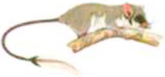 |
Ptilocercus lowii | Pen-tailed treeshrew | Nocturnal | Arboreal | x | |||||||
| Dendrogale | 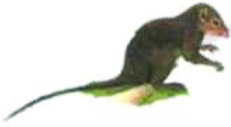 |
Dendrogale melanura | Smooth-tailed treeshrew | Diurnal | Terrestrial | x | ||||||||
| Tupaia | 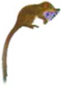 |
Tupaia gracilis | Slender treeshrew | Diurnal | Terrestrial | x | x | x | x | x | x | x | ||
| Tupaiidae | 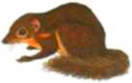 |
Tupaia longipes | Long-footed treeshrew | Diurnal | Terrestrial | x | x | x | x | x | x | |||
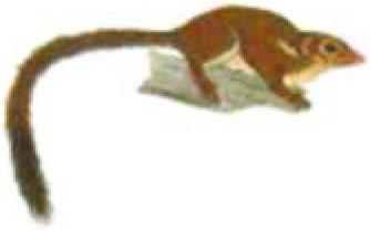 |
Tupaia minor | Lesser treeshrew | Diurnal | Arboreal | x | x | x | x | x | |||||
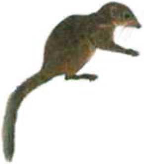 |
Tupaia montana | Montane treeshrew | Diurnal | Terrestrial | x | x | ||||||||
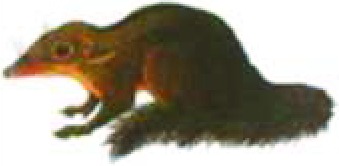 |
Tupaia tana | Large treeshrew | Diurnal | Terrestrial | x | x | x | x | x | x | ||||
Note.—Illustrations © The Sabah Society, reproduced with permission.
aLowland sites: Poring, Kinabalu National Park (06°02’N, 116°42′E); Danum Valley Conservation Area (4°57′N, 117°48′E); Tawau Hills National Park (04°23′N, 117°53′E); Luasong Field Centre (4°36′N, 117°23′E); Monggis (06°13′N, 116°45′E); Tumbalang (06°08′N, 116°53′E). Montane sites: Mesilou, Mount Kinabalu (6°00′N, 116°35′E); Mount Alap, Crocker Range National Park (5°49′N, 116°20′E).
bEmmons 2000, Wells et al. 2004, and Wiens et al. 2008 detail the natural history and foraging ecology of these species.
We collected tissue samples (2-mm ear biopsies) under two conditions: animals were 1) lightly anesthetized (diethyl ether or isoflurane; years: 2002–2010) or 2) restrained by hand in a cloth bag with a small opening for accessing the ear (years: 2011–2012). This latter procedure has the advantage of speed (<5 min of human handling). The tissue samples were stored in 99% ethanol (years: 2002–2010) or RNAlater stabilization reagent (Ambion; years: 2011–2012). All animals were released at capture site after individual marking with subcutaneous PIT tags. Recaptured animals showed no adverse effects from our protocol, which was approved by the Institutional Animal Care and Use Committee of Dartmouth College (protocol no. 11-06-07AT).
The Central American (Derby’s) woolly opossum (C. derbianus) is a small, arboreal (200–400 g) didelphid marsupial (Bucher and Hoffmann 1980). We biopsied muscle from a single specimen accessioned (KU 164643) in the University of Kansas Natural History Museum. This individual, a male, was found dead by RMT on December 26, 2006 at the La Selva Biological Station, Costa Rica (10°26′N, 84°0′W). Muscle tissue was preserved initially in 95% ethanol and later frozen. This protocol was approved by the Institutional Animal Care and Use Committee of the University of Kansas (protocol no. 132-06) and follows the guidelines of the American Society of Mammalogists (Sikes et al. 2011).
DNA Extraction, Amplification, and Sequencing
We extracted genomic DNA (DNeasy Blood and Tissue Kit, Qiagen) from eight females in each of the five species of Tupaia (n = 16 X chromosomes per species; n = 80 X chromosomes in total across Tupaia). We chose animals from across trapping sites in attempt to maximize the genetic diversity in our sample. Due to low capture rates, our study was limited to two individuals of P. lowii (sexes unknown) and one individual of D. melanura (a male).
We conducted a BLAST search of the T. belangeri (chinensis) whole-genome sequence (European Nucleotide Archive; WGS Sequence Set: ALAR00000000.1) using human opsin gene sequences to identify sequences of OPN1SW and OPN1LW, which were used to design PCR primers within introns (Primer3; http://primer3.sourceforge.net, last accessed January 16, 2016) (table 2). To estimate the functionality and peak spectral absorbance (λmax) of the SWS1 opsin, we amplified the spectral tuning sites in tandem with the entire coding region of the OPN1SW. Ten amino acid sites on exon 1—especially sites 86 and 93—are critical to λmax (Shi et al. 2001; Yokoyama et al. 2006; Hunt et al. 2007; Carvalho et al. 2012). We examined the OPN1SW of one individual per species as this autosomal gene has low levels of functional polymorphism (Shimmin et al. 1998).
Table 2.
Primers and Annealing Temperatures Used in Polymerase Chain Reactions to Amplify Partial OPN1SW and OPN1LW Opsin Genes in Treeshrews (Genera: Dendrogale, Ptilocercus, Tupaia) and Woolly Opossum (Genus: Caluromys).
| Gene | Genus | Region | Forward (5′ to 3′) | Reverse (5′ to 3′) | Tanneal°C |
|---|---|---|---|---|---|
| OPN1SW | Ptilocercus | Exon 2 and 3 | GCC TAA AGG CTT CAA GCA GGG GG | TGC CAC AGG TCT GGT GAT AGG CT | 64 |
| Dendrogale | Exon 1 | GTA CCA CCT TGC CCC TGT CT | CCT TTC CCC TGC AGT ACC T | 58 | |
| Exons 2 and 3 | GGT GAT AGG CTG GTC ATT GG | CCC AGC AGC TGA GAG TAG GA | 60 | ||
| Exon 4 | GCT CAG CAG CAG GAG TCA G | TTC ATG AAG CAG TAG ATG ATG G | 58 | ||
| Exon 5 | ATG AGG CGT CTT TTC CAC AC | TGG CTT TGT TAG CAG GAA GG | 60 | ||
| Tupaia | Exon 1 | AAG AAC ACA ATC GGC TTT GG | GTG GCG TAG TGT CCT TTG CT | 58 | |
| Exons 2 and 3 | CAG CCC AGC CTA GAA GTT TG | CCT GAC CCT CTC AAG ACC AC | 62 | ||
| Exon 4 | TAA TGA ATA AGG CGG GGT GA | CTG ACA AGT CAC TGG CGA GA | 58 | ||
| Exon 5 | ATG AGG CGT CTT TTC CAC AC | TGG CTT TGT TAG CAG GAA GG | 60 | ||
| Caluromys | Exons 1–3 | TGT CAG GGG ATG AGG AGT TC | GGC CAC ACG AGC ACT GTA | 62 | |
| OPN1LW | Dendrogale, Ptilocercus | Exon 3 | CAT CAC GGG GCT CTG GTC | CTG CTC CAA CCA AAG ATG G | 60 |
| Exon 5 | AGG CTG AGA AGG AGG TGA CA | GTG GCA CTT TTG GCG AAG TA | 60 | ||
| Tupaia | Exon 3 | TAC CTG TCT GCT CTT CCC TGT AG | GGT CCT AAA TGA GCC ACC CTT AC | 64 | |
| Exon 5 | TGC ACT GTC CCT GTC TCA CCC AG | GGC CTG CCG ATG GCC TTA CTT AC | 68 |
λmax of the M/LWS opsin is governed by five amino acid sites spanning exons 3–5 (Yokoyama et al. 2008), of which three are variable among primates (exon 3: 180; exon 5: 277, 285) and responsible for intra- and interspecific variation in color vision (Hiramatsu et al. 2005; Kawamura et al. 2012). We therefore amplified exons 3 and 5 of all scandentians in our sample in order to estimate λmax and to explore the potential for intraspecific variation.
Initial PCR amplification was poor for the two most basal treeshrews (P. lowii and D. melanura). We therefore used sequences from Tupaia to design primers based on highly conserved regions of each gene. This approach was successful with the exception of the OPN1SW of P. lowii (see below). The primers for C. derbianus (table 2) were designed from the conserved regions of the OPN1SW in a wide range marsupials: Isoodon obesulus, Monodelphis domestica, Setonix brachyurus, Sminthopsis crassicaudata, Tarsipes rostratus, and Thylamys elegans (supplementary table S1, Supplementary Material online). The OPN1LW sequence of C. philander was supplied by Professor David M. Hunt (University of Western Australia).
PCRs were carried out in 25 μl containing 12.5 μl of iProof HF 2x Master Mix (0.04 U/μl DNA polymerase, 2X HF buffer, 400 μM dNTPs [each]; Bio-Rad), 1.0 μM each of the forward and reverse primers, and 70–200 ng template DNA. Pure water was used as a negative control in each experiment. We carried out PCRs at 98 °C for 3 min followed by 35 cycles of 98 °C for 10 s, Tanneal (annealing temperature) for 30 s, 72 °C for 30 s, and concluding with 72 °C for 5 min. Tanneal was optimized for each gene/primer pair combination (table 2).
We purified amplicons with the QIAquick PCR Purification Kit or, when nontarget sequences were also amplified, the QIAquick Gel Extraction Kit (Qiagen). Purified DNA was sequenced directly using an Applied Biosystems Model 3100 automatic sequencer with the ABI PRISM BigDye Terminator Cycle Sequencing Ready Reaction Kits v3.0 with AmpliTaq DNA polymerase. Primers designed for PCR were used during sequencing reactions. DNA sequences were assembled and edited manually using Sequencher v 5.1 (Gene Codes). Heterozygous sites were scored manually using a single-letter nucleic acid code (IUPAC nomenclature) when chromatograms displayed peaks of nearly equal height.
Whole-Genome Sequencing
Amplification of the OPN1SW of P. lowii failed repeatedly. We therefore pursued massive parallel sequencing using whole-genome shotgun sequencing and a reference-assisted assembly strategy. We prepared an Illumina sequencing library (Meyer and Kircher 2010), and sequenced it to low (∼3–5×) coverage on a single lane of an Illumina HiSeq 2000 at the Penn State Genomics Core Facility. After quality control filtering, we formatted the short read data set as a BLAST database and queried the OPN1SW sequences of human and Tupaia, revealing reads that likely originated in the OPN1SW of P. lowii. We pairwise-aligned these candidate reads to the coding regions of the human OPN1SW using Geneious version 5.6.7, and manually extended high-confidence local contigs of P. lowii by querying the ends for overlapping regions in the short read data set until they reached coverage gaps on either side. Using this strategy, we recovered a 569-bp region of the OPN1SW sequence including the complete sequence of exon 2. Our contig aligned to the corresponding human OPN1SW region with 77.1% pairwise identity, whereas it failed to align meaningfully to other human opsin genes, strongly implicating an origin in the OPN1SW of P. lowii rather than similar regions.
To verify the contig sequence, we PCR-amplified a region containing the complete exon 2 using the KAPA HiFi Hotstart PCR Kit according to the manufacturer’s protocol. The resulting PCR product was purified with homemade Solid Phase Reversible Immobilization (SPRI) beads (Rohland and Reich 2012) and Sanger-sequenced from both ends using PCR primers on an Applied Biosystems 3730XL capillary system. We aligned the product (336 bp) with the short read contig containing exon 2; the two sequences were identical, confirming the fidelity of the SWS1 opsin gene sequence. The presence of premature stop codons and indels indicates pseudogenization and release from functional constraint. Degradation of this gene explains the failure of the initial experiments using conserved primers.
Opsin Sequence Analyses
For the OPN1SW (1,062 nt positions) and partial OPN1LW (exons 3–5; 692 nt positions) opsin genes, we constructed a multiple alignment with each species in our study, together with sequences from selected mammals (supplementary table S1, Supplementary Material online). We used congeners when species-level data for both opsins were unavailable. In the case of Spalacopus cyanus, sequence data for the OPN1LW were unavailable but the λmax of the opsin is known (Peichl et al. 2005). Multiple individuals per species were included in the analysis of treeshrew OPN1LW opsin genes to assess the potential for polymorphism. We compiled and aligned nucleotide sequences using the Clustal W procedure in MEGA6 (Tamura et al. 2013) and refined the alignment manually. The coding regions were then translated into amino acid sequences.
To study exon-wide patterns of genetic drift and natural selection, we calculated the rates of nonsynonymous substitutions per nonsynonymous site (dN) and synonymous (dS) substitutions per synonymous site (Nei and Gojobori 1986) using MEGA6 (Tamura et al. 2013). All ambiguous positions were removed for each sequence pair. We used the Z-test of purifying selection to assess whether differences between dN and dS differed significantly from 0.
Spectral Tuning Sites and Ancestral Character States
Ancestral tuning sites of OPN1SW and OPN1LW were inferred using the MP algorithm in MEGA6 with a 70% site coverage cutoff (Tamura et al. 2013). When results were ambiguous, we used ML analysis, using the Jones–Taylor–Thornton matrix-based model and assumed uniform rates among sites to select the most likely candidate amino acid. We then used TimeTree (Hedges et al. 2006; accessed June 2015) and published divergence dates (Prideaux and Warbuton 2010; Roberts et al. 2011; Fabrae et al. 2012; Song et al. 2012) to determine the most likely phylogenetic structure of Euarchonta and calculate divergence times. We tested all possible phylogenetic configurations of Euarchonta (i.e., phylogenetic structure following the Primatomorpha, Sundatheria, and treeshrew–primate sister group relationship hypotheses, respectively), as outlined in figure 2.
SWS1 Opsin Pseudogenization
The timing of pseudogenization of genes has been estimated in other studies by examining substitutions of nucleotides that would have been nonsynonymous in a functional gene, relative to the neutral mutation rate and calibrated against species divergence estimates (Chou et al. 2002; Stedman et al. 2004). We follow similar methods to estimate the loss of function of OPN1SW in P. lowii. Briefly, the nonsynonymous substitution rate in a functional gene is defined as the neutral mutation rate, k, multiplied by the fraction of neutral substitutions fN. fN is inversely related to the degree of functional constraint on a gene, where 1 = neutral evolution (no constraint) and small values indicate strong purifying selection. Following pseudogenization, fN = 1 and the substitution rate increases to k, a property we used to estimate the timing of pseudogenization. We defined t1 as the time of inactivation and t as the time of divergence between the taxon with the pseudogene and a related lineage with a functional orthologous gene. We estimated t1 using the formula: t1 = (dN (pseudogene)/k − fNt)/(1−fN) . In this study, k = mean per site substitutions at synonymous and intron sites (dS+I) among taxa/estimated divergence time. We combined intron data with synonymous data to increase the sample size. Although selective constraints on introns and synonymous sites might vary slightly, both are predominantly subject to neutral evolution. Analyses were conducted in PAML with codeml; dN was calculated for coding sequences only, whereas dS+I was calculated by concatenating coding sequences with the intron data set in which the nucleotides T and C were inserted before each intronic site using a custom Perl script. In this way, PAML treated the intron sites (x) as synonymous positions of Serine (TCx).
Supplementary Material
Supplementary figures S1 and S2 and tables S1–S3 are available at Molecular Biology and Evolution online (http://www.mbe.oxfordjournals.org/).
Acknowledgments
The authors dedicate this article to D.T. Rasmussen (1958–2014), whose work on C. derbianus motivated crucial aspects of this research. The authors acknowledge with gratitude the practical assistance of A. Fatah Bin Amir, A. Biun, R.M. Brown, M.S. Callahan, C.F. Danosi, L.H. Emmons, D.M. Hunt, R. Kays, M. Montague, C. Sendall, and F. Tuh Yit Yu. Collection and export of treeshrew tissues was approved by Sabah Parks, the Sabah Biodiversity Council, and the Economic Planning Unit of Malaysia (permit nos. JKM/MBS.1000-2/2(26) and JKM/MBS.1000-2/3(30)). This work was supported by the Natural Sciences and Engineering Research Council of Canada (PDF-420810-2012 to A.D.M), the German Academic Exchange Service (DAAD; scholarship to K.W.), the Cramer Fund of the Department of Biological Sciences, Dartmouth College (to G.L.M.), and the David and Lucile Packard Foundation (Fellowship in Science and Engineering no. 2007-31754 to N.J.D.).
References
- Allman J. 1977. Evolution of the visual system in early primates. Prog Psychobiol Physiol Psychol. 7:1–53. [Google Scholar]
- Arrese CA, Oddy AY, Runham PB, Hart NS, Shand J, Hunt DM, Beazley LD. 2005. Cone topography and spectral sensitivity in two potentially trichromatic marsupials, the quokka (Setonix brachyurus) and quenda (Isoodon obesulus). Proc R Soc Lond B Biol Sci. 272:791–796. [DOI] [PMC free article] [PubMed] [Google Scholar]
- Bloch JI, Silcox MT, Boyer DM, Sargis EJ. 2007. New Paleocene skeletons and the relationship of plesiadapiforms to crown-clade primates. Proc Natl Acad Sci U S A. 104:1159–1164. [DOI] [PMC free article] [PubMed] [Google Scholar]
- Bucher JE, Hoffmann RS. 1980. Caluromys derbianus. Mamm Species. 140:1–4. [Google Scholar]
- Cartmill M. 1992. New views on primate origins. Evol Anthropol. 1:105–111. [DOI] [PubMed] [Google Scholar]
- Cartmill M. 2012. Primate origins, human origins, and the end of higher taxa. Evol Anthropol. 21:208–220. [DOI] [PubMed] [Google Scholar]
- Carvalho LS, Cowing JA, Wilkie SE, Bowmaker JK, Hunt DM. 2006. Shortwave visual sensitivity in tree and flying squirrels reflects changes in lifestyle. Curr Biol. 16:R81–R83. [DOI] [PubMed] [Google Scholar]
- Carvalho LS, Davies WL, Robinson PR, Hunt DM. 2012. Spectral tuning and evolution of primate short-wavelength-sensitive visual pigments. Proc R Soc Lond B Biol Sci. 279:387–393. [DOI] [PMC free article] [PubMed] [Google Scholar]
- Chou HH, Hayakawa T, Diaz S, Krings M, Indriati E, Leakey M, Paabo S, Satta Y, Takahata N, Varki A. 2002. Inactivation of CMP-N-acetylneuraminic acid hydroxylase occurred prior to brain expansion during human evolution. Proc Natl Acad Sci U S A. 99:11736–11741. [DOI] [PMC free article] [PubMed] [Google Scholar]
- Crompton RH. 1995. “Visual predation,” habitat structure, and the ancestral primate niche In: Alterman L, Doyle GA, Izard MK, editors. Creatures of the dark: the nocturnal prosimians. New York: Plenum Press; p. 11–30. [Google Scholar]
- David-Gray ZK, Bellingham J, Munoz M, Avivi A, Nevo E, Foster RG. 2002. Adaptive loss of ultraviolet-sensitive/violet-sensitive (UVS/VS) cone opsin in the blind mole rat (Spalax ehrenbergi). Eur J Neurosci. 16:1186–1194. [DOI] [PubMed] [Google Scholar]
- Davies WL. 2011. Adaptive gene loss in vertebrates: photosensitivity as a model case In: Encyclopedia of life sciences (eLS). Chichester: John Wiley & Sons. [Google Scholar]
- Davies WL, Collin SP, Hunt DM. 2012. Molecular ecology and adaptation of visual photopigments in craniates. Mol Ecol. 21:3121–3158. [DOI] [PubMed] [Google Scholar]
- Deeb SS, Wakefield MJ, Tada T, Marotte L, Yokoyama S, Marshall Graves JA. 2003. The cone visual pigments of an Australian marsupial, the Tammar wallaby (Macropus eugenii): sequence, spectral tuning, and evolution. Mol Biol Evol. 20:1642–1649. [DOI] [PubMed] [Google Scholar]
- Dkhissi-Benyahya O, Szel A, Degrip WJ, Cooper HM. 2001. Short and mid-wavelength cone distribution in a nocturnal strepsirrhine primate (Microcebus murinus). J Comp Neurol. 438:490–504. [DOI] [PubMed] [Google Scholar]
- Douglas RH, Jeffery G. 2014. The spectral transmission of ocular media suggests ultraviolet sensitivity is widespread among mammals. Proc R Soc Lond B Biol Sci. 281:20132995. [DOI] [PMC free article] [PubMed] [Google Scholar]
- Emerling CA, Huynh HT, Nguyen MA, Meredith RW, Springer MS. 2015. Spectral shifts of mammalian ultraviolet-sensitive pigments (short wavelength-sensitive opsin 1) are associated with eye length and photic niche evolution. Proc R Soc Lond B Biol Sci. 282:20151817. [DOI] [PMC free article] [PubMed] [Google Scholar]
- Emmons LH. 2000. Tupai: a field study of Bornean treeshrews. Berkeley: University of California Press. [Google Scholar]
- Fabre P-H, Lionel H, Dimitrov D, Douzery EJP. 2012. A glimpse on the pattern of rodent diversification: a phylogenetic approach. BMC Evol Biol. 12:1–19. [DOI] [PMC free article] [PubMed] [Google Scholar]
- Felsenstein J. 1985. Confidence limits on phylogenies: an approach using the bootstrap. Evolution 39:783–791. [DOI] [PubMed] [Google Scholar]
- Fleming TH, Geiselman C, Kress WJ. 2009. The evolution of bat pollination: a phylogenetic perspective. Ann Bot. 104:1017–1043. [DOI] [PMC free article] [PubMed] [Google Scholar]
- Gebo DL. 2004. A shrew-sized origin for primates. Am J Phys Anthropol. 125:40–62. [DOI] [PubMed] [Google Scholar]
- Gómez JM, Verdú M. 2012. Mutualism with plants drives primate diversification. Syst Biol. 61:567–577. [DOI] [PubMed] [Google Scholar]
- Gould E. 1978. The behavior of the moonrat, Echinosorex gymnurus (Erinaceidae) and the pentail shrew, Ptilocercus lowi (Tupaiidae) with comments on the behavior of other Insectivora. Z Tierpsychol. 48:1–27. [Google Scholar]
- Hauser FE, van Hazel I, Chang BSW. 2014. Spectral tuning in vertebrate short wavelength sensitive 1 (SWS1) visual pigments: can wavelength sensitivity be inferred from sequence data? J Exp Zool B Mol Dev Evol. 322:529–539. [DOI] [PubMed] [Google Scholar]
- Hedges SB, Dudley J, Kumar S. 2006. TimeTree: a public knowledge-base of divergence times among organisms. Bioinformatics 22:2971–2972. [DOI] [PubMed] [Google Scholar]
- Heesy C, Ross CF. 2004. Mosaic evolution of activity pattern, diet and color vision in haplorhine primates In: Ross C, Kay RF, editors. Anthropoid origins: new visions. New York: Kluwer Academic/Plenum Press; p. 665–698. [Google Scholar]
- Hiramatsu C, Tsutsui T, Matsumoto Y, Aureli F, Fedigan LM, Kawamura S. 2005. Color-vision polymorphism in wild capuchins (Cebus capucinus) and spider monkeys (Ateles geoffroyi) in Costa Rica. Am J Primatol. 67:447–461. [DOI] [PubMed] [Google Scholar]
- Hunt DM, Carvalho LS, Cowing JA, Parry JW, Wilkie SE, Davies WL, Bowmaker JK. 2007. Spectral tuning of shortwave-sensitive visual pigments in vertebrates. Photochem Photobiol. 83:303–310. [DOI] [PubMed] [Google Scholar]
- Hunt DM, Chan J, Carvalho LS, Hokoc JN, Ferguson MC, Arrese CA, Beazley LD. 2009. Cone visual pigments in two species of South American marsupials. Gene 433:50–55. [DOI] [PubMed] [Google Scholar]
- Hunt DM, Peichl L. 2014. S cones: evolution, retinal distribution, development, and spectral sensitivity. Vis Neurosci. 31:115–138. [DOI] [PubMed] [Google Scholar]
- Jacobs GH. 2009. Evolution of colour vision in mammals. Philos Trans R Soc Lond B Biol Sci. 364:2957–2967. [DOI] [PMC free article] [PubMed] [Google Scholar]
- Jacobs GH. 2013. Losses of functional opsin genes, short-wavelength cone photopigments, and color vision–a significant trend in the evolution of mammalian vision. Visual Neurosci. 30:39–53. [DOI] [PubMed] [Google Scholar]
- Jacobs GH, Deegan JFII, Neitz J, Crognale MA, Neitz M. 1993. Photopigments and color vision in the nocturnal monkey, Aotus. Vision Res. 33:773–783. [DOI] [PubMed] [Google Scholar]
- Jacobs GH, Neitz J. 1986. Spectral mechanisms and color vision in the tree shrew (Tupaia belangeri). Vision Res. 26:291–298. [DOI] [PubMed] [Google Scholar]
- Jacobs GH, Williams GA, Fenwick JA. 2004. Influence of cone pigment coexpression on spectral sensitivity and color vision in the mouse. Vision Res. 44:1615–1622. [DOI] [PubMed] [Google Scholar]
- Janečka JE, Miller W, Pringle TH, Wiens F, Zitzmann A, Helgen KM, Springer MS, Murphy WJ. 2007. Molecular and genomic data identify the closest living relative of primates. Science 318:792–794. [DOI] [PubMed] [Google Scholar]
- Jenkins FA., Jr 1987. Tree shrew locomotion and the origins of primate arborealism In: Ciochon RL, Fleagle JG, editors. Primate evolution and human origins. New York: Aldine de Gruyter; p. 2–13. [Google Scholar]
- Kawamura S, Hiramatsu C, Melin AD, Schaffner CM, Aureli F, Fedigan LM. 2012. Polymorphic color vision in primates: evolutionary considerations In: Hirai H, Imai H, Go Y, editors. Post-genome biology of primates. Tokyo: Springer; p. 93–120. [Google Scholar]
- Kawamura S, Kubotera N. 2004. Ancestral loss of short wave-sensitive cone visual pigment in lorisiform prosimians, contrasting with its strict conservation in other prosimians. J Mol Evol. 58:314–321. [DOI] [PubMed] [Google Scholar]
- Kay RF, Thewissen JGM, Yoder AD. 1992. Cranial anatomy of Ignacius graybullianus and the affinities of the Plesiadapiformes. Am J Phys Anthropol. 89:477–498. [Google Scholar]
- Kays R, Rodríguez ME, Valencia LM, Horan R, Smith AR, Ziegler C. 2012. Animal visitation and pollination of flowering balsa trees (Ochroma pyramidale) in Panama. Mesoamericana 16:56–70. [Google Scholar]
- Le Gros Clark WE. 1926. On the anatomy of the pen-tailed tree-shrew (Ptilocercus lowii). Proc Zool Soc Lond. 96:1179–1309. [Google Scholar]
- Li Q, Ni X. 2016. An early Oligocene fossil demonstrates treeshrews are slowly evolving “living fossils”. Sci Rep. 6:18627. [DOI] [PMC free article] [PubMed] [Google Scholar]
- Lim BL. 1967. Note on the food habits of Ptilocercus lowii Gray (pentail tree-shrew) and Echinosorex gymnurus (Raffles) (moonrat) in Malaya with remarks on “ecological labelling” by parasite patterns. J Zool. 152:375–379. [Google Scholar]
- Luckett WP. (editor) 1980. Comparative biology and evolutionary relationships of tree shrews. New York: Plenum Press. [Google Scholar]
- Lyon MW. 1913. Treeshrews: an account of the mammalian family Tupaiidae. Washington (DC: ): Government Printing Office. [Google Scholar]
- Martin RD. 1990. Primate origins and evolution: a phylogenetic reconstruction. Princeton: Princeton University Press. [Google Scholar]
- Melin AD, Danosi C, McCracken G, Dominy NJ. 2014. Dichromatic vision in a fruit bat with diurnal proclivities, the Samoan flying fox (Pteropus samoensis). J Comp Physiol A. 200:1015–1022. [DOI] [PubMed] [Google Scholar]
- Melin AD, Matsushita Y, Moritz GL, Dominy NJ, Kawamura S. 2013. Inferred L/M cone opsin polymorphism of ancestral tarsiers sheds dim light on the origin of anthropoid primates. Proc R Soc Lond B Biol Sci. 280:20130189. [DOI] [PMC free article] [PubMed] [Google Scholar]
- Melin AD, Moritz GL, Fosbury RAE, Kawamura S, Dominy NJ. 2012. Why aye-ayes see blue. Am J Primatol. 74:185–192. [DOI] [PubMed] [Google Scholar]
- Meredith RW, Gatesy J, Emerling CA, York VM, Springer MS. 2013. Rod monochromacy and the coevolution of cetacean retinal opsins. PLoS Genet. 9:e1003432. [DOI] [PMC free article] [PubMed] [Google Scholar]
- Meredith RW, Janečka JE, Gatesy J, Ryder OA, Fisher CA, Teeling EC, Goodbla A, Eizirik E, Simao TL, Stadler T, et al. 2011. Impacts of the Cretaceous terrestrial revolution and KPg extinction on mammal diversification. Science 334:521–524. [DOI] [PubMed] [Google Scholar]
- Meyer Kircher 2010. Illumina sequencing library preparation for highly multiplexed target capture and sequencing. Cold Spring Harb Protoc. 2010: pdb.prot5448. doi: 10.1101/pdb.prot5448. [DOI] [PubMed] [Google Scholar]
- Moore BA, Kamilar JM, Collin SP, Bininda-Emonds ORP, Dominy NJ, Hall MI, Heesy CP, Johnsen S, Lisney TJ, Loew ER, et al. 2012. A novel method for comparative analysis of retinal specialization traits from topographic maps. J Vision. 12:13 1–24. [DOI] [PubMed] [Google Scholar]
- Moritz GL.2015. Primate origins through the lens of functional and degenerate cone opsins. Ph.D. dissertation. Hanover (NH): Dartmouth College.
- Moritz GL, Lim NT-L, Neitz M, Peichl L, Dominy NJ. 2013. Expression and evolution of short wavelength sensitive opsins in colugos: a nocturnal lineage that informs debate on primate origins. Evol Biol. 40:542–553. [DOI] [PMC free article] [PubMed] [Google Scholar]
- Moritz GL, Melin AD, Tuh Yit Yu F, Bernard H, Ong PS, Dominy NJ. 2014. Niche convergence suggests functionality of the nocturnal fovea. Front Integr Neurosci. 8:61 doi:10.3389/fnint.2014.00061. [DOI] [PMC free article] [PubMed] [Google Scholar]
- Müller B, Peichl L. 1989. Topography of cones and rods in the tree shrew retina. J Comp Neurol. 282:581–594. [DOI] [PubMed] [Google Scholar]
- Müller B, Peichl L, De Grip WJ, Gery I, Korf HW. 1989. Opsin- and S-antigen-like immunoreactions in photoreceptors of the tree shrew retina. Invest Ophthalmol Vis Sci. 30:530–535. [PubMed] [Google Scholar]
- Murphy WJ, Eizirik E, O’Brien SJ, Madsen O, Scally M, Douady CJ, Teeling EC, Ryder OA, Stanhope MJ, de Jong WW, et al. 2001. Resolution of the early placental mammal radiation using Bayesian phylogenetics. Science 294:2348–2351. [DOI] [PubMed] [Google Scholar]
- Nei M, Gojobori T. 1986. Simple methods for estimating the numbers of synonymous and nonsynonymous nucleotide substitutions. Mol Biol Evol. 3:418–426. [DOI] [PubMed] [Google Scholar]
- O'Leary MA, Bloch JI, Flynn JJ, Gaudin TJ, Giallombardo A, Giannini NP, Goldberg SL, Kraatz BP, Luo ZX, Meng J, et al. 2013. The placental mammal ancestor and the post–K-Pg radiation of placentals. Science 339:662–667. [DOI] [PubMed] [Google Scholar]
- Palacios AG, Bozinovic F, Vielma A, Arrese CA, Hunt DM, Peichl L. 2010. Retinal photoreceptor arrangement, SWS1 and M/LWS opsin sequence, and electroretinography in the South American marsupial Thylamys elegans (Waterhouse, 1839). J Comp Neurol. 518:1589–1602. [DOI] [PubMed] [Google Scholar]
- Parry JWL, Poopalasundaram S, Bowmaker JK, Hunt DM. 2004. A novel amino acid substitution is responsible for spectral tuning in a rodent violet-sensitive visual pigment. Biochemistry 43:8014–8020. [DOI] [PubMed] [Google Scholar]
- Peichl L. 2005. Diversity of mammalian photoreceptor properties: adaptations to habitat and lifestyle? Anat Rec a Discov Mol Cell Evol Biol. 287(A):1001–1012. [DOI] [PubMed] [Google Scholar]
- Peichl L, Chavez AE, Ocampo A, Mena W, Bozinovic F, Palacios AG. 2005. Eye and vision in the subterranean rodent cururo (Spalacopus cyanus, Octodontidae). J Comp Neurol. 486:197–208. [DOI] [PubMed] [Google Scholar]
- Perry GH, Martin RD, Verrelli BC. 2007. Signatures of functional constraint at aye-aye opsin genes: the potential of adaptive color vision in a nocturnal primate. Mol Biol Evol. 24:1963–1970. [DOI] [PubMed] [Google Scholar]
- Petry HM, Fox R, Casagrande VA. 1984. Spatial contrast sensitivity of the tree shrew. Vision Res. 24:1037–1042. [DOI] [PubMed] [Google Scholar]
- Petry HM, Harosi FI. 1990. Visual pigments of the tree shrew (Tupaia belangeri) and greater galago (Galago crassicaudatus): a microspectrophotometric investigation. Vision Res. 30:839–851. [DOI] [PubMed] [Google Scholar]
- Prideaux GJ, Warburton NM. 2010. An osteology-based appraisal of the phylogeny and evolution of kangaroos and wallabies (Macropodidae: marsupialia). Zool J Linn Soc. 159:954–987. [Google Scholar]
- Prugh LR, Golden CD. 2014. Does moonlight increase predation risk? meta-analysis reveals divergent responses of nocturnal mammals to lunar cycles. J Anim Ecol. 83:504–514. [DOI] [PubMed] [Google Scholar]
- Rasmussen DT. 1990. Primate origins: lessons from a neotropical marsupial. Am J Primatol. 22:263–277. [DOI] [PubMed] [Google Scholar]
- Rasmussen DT. 2002. The origin of primates In: Hartwig WC, editor. The primate fossil record. Cambridge: Cambridge University Press; p. 5–9. [Google Scholar]
- Rasmussen DT, Sussman RW. 2007. Parallelisms among primates and possums In: Ravosa MJ, Dagosto M, editors. Primate origins: adaptations and evolution. New York: Springer; p. 775–803. [Google Scholar]
- Ravosa MJ, Savakova DG. 2004. Euprimate origins: the eyes have it. J Hum Evol. 46:357–364. [DOI] [PubMed] [Google Scholar]
- Reid FA. 1997. A field guide to the mammals of Central American & Southeast Mexico. Oxford: Oxford University Press. [Google Scholar]
- Roberts TE, Lanier HC, Sargis EJ, Olson LE. 2011. Molecular phylogeny of treeshrews (Mammalia: Scandentia) and the timescale of diversification in Southeast Asia. Mol Phylogenet Evol. 60:358–372. [DOI] [PubMed] [Google Scholar]
- Rohland N, Reich D. 2012. Cost-effective, high-throughput DNA sequencing libraries for multiplexed target capture. Genome Res. 22:939–946. [DOI] [PMC free article] [PubMed] [Google Scholar]
- Saitou N, Nei M. 1987. The neighbor-joining method: a new method for reconstructing phylogenetic trees. Mol Biol Evol. 4:406–425. [DOI] [PubMed] [Google Scholar]
- Sargis EJ. 2002. The postcranial morphology of Ptilocercus lowii (Scandentia, Tupaiidae): an analysis of primatomorphan and volitantian characters. J Mammal Evol. 9:137–160. [Google Scholar]
- Sargis EJ. 2004. New views on tree shrews: the role of tupaiids in primate supraordinal relationships. Evol Anthropol. 13:56–66. [Google Scholar]
- Sargis EJ, Boyer DM, Bloch JI, Silcox MT. 2007. Evolution of pedal grasping in Primates. J Hum Evol. 53:103–107. [DOI] [PubMed] [Google Scholar]
- Schmitt D, Lemelin P. 2002. Origins of primate locomotion: gait mechanics of the woolly opossum. Am J Phys Anthropol. 118:231–238. [DOI] [PubMed] [Google Scholar]
- Shi Y, Radlwimmer FB, Yokoyama S. 2001. Molecular genetics and the evolution of ultraviolet vision in vertebrates. Proc Natl Acad Sci U S A. 98:11731–11736. [DOI] [PMC free article] [PubMed] [Google Scholar]
- Shimmin LC, Miller J, Tran HN, Li W-H. 1998. Contrasting levels of DNA polymorphism at the autosomal and X-linked visual color pigment loci in humans and squirrel monkeys. Mol Biol Evol. 15:449–455. [DOI] [PubMed] [Google Scholar]
- Sikes RS, Gannon WL, Animal Care and Use Committee of the American Society of Mammalogists. 2011. Guidelines of the American Society of Mammalogists for the use of wild mammals in research. J Mammal. 92:235–253. [DOI] [PMC free article] [PubMed] [Google Scholar]
- Silcox MT, Sargis EJ, Bloch JI, Boyer DM. 2015. Primate origins and supraordinal relationships: morphological evidence In: Henke W, Tattersall I, editors. Handbook of paleoanthropology. Berlin: Springer-Verlag; p. 1053–1081. [Google Scholar]
- Song S, Liu L, Edwards SV, Wu S. 2012. Resolving conflict in eutherian mammal phylogeny using phylogenomics and the multispecies coalescent model. Proc Natl Acad Sci U S A. 109:14942–14947. [DOI] [PMC free article] [PubMed] [Google Scholar]
- Stedman HH, Kozyak BW, Nelson A, Thesier DM, Su LT, Low DW, Bridges CR, Shrager JB, Minugh-Purvis N, Mitchell MA. 2004. Myosin gene mutation correlates with anatomical changes in the human lineage. Nature 4, 415–418. [DOI] [PubMed] [Google Scholar]
- Sussman RW, Rasmussen DT, Raven PH. 2013. Rethinking primate origins again. Am J Primatol. 75:95–106. [DOI] [PubMed] [Google Scholar]
- Sussman RW, Raven PH. 1978. Pollination by lemurs and marsupials: an archaic coevolutionary system. Science 200:731–736. [DOI] [PubMed] [Google Scholar]
- Tamura K, Stecher G, Peterson D, Filipski A, Kumar S. 2013. MEGA6: molecular evolutionary genetics analysis version 6.0. Mol Biol Evol. 30:2725–2729. [DOI] [PMC free article] [PubMed] [Google Scholar]
- Tan Y, Yoder AD, Yamashita N, Li W-H. 2005. Evidence from opsin genes rejects nocturnality in ancestral primates. Proc Natl Acad Sci U S A. 102:14712–14716. [DOI] [PMC free article] [PubMed] [Google Scholar]
- Tattersall I. 1984. The tree-shrew, Tupaia: a “living model” of the ancestral primate? In: Eldredge N, Stanley SM, editors. Living fossils. New York: Springer; p. 32–37. [Google Scholar]
- Thibos LN, Bradley A, Still DL, Zhang X, Howarth PA. 1990. Theory and measurement of ocular chromatic aberration. Vision Res. 30:33–49. [DOI] [PubMed] [Google Scholar]
- Tigges J, Brooks BA, Klee MR. 1967. ERG recordings of a primate pure cone retina (Tupaia glis). Vision Res. 7:553–563. [DOI] [PubMed] [Google Scholar]
- Veilleux CC, Bolnick DA. 2009. Opsin gene polymorphism predicts trichromacy in a cathemeral lemur. Am J Primatol. 71:86–90. [DOI] [PubMed] [Google Scholar]
- Veilleux CC, Cummings ME. 2012. Nocturnal light environments and species ecology: implications for nocturnal color vision in forests. J Exp Biol. 215:4085–4096. [DOI] [PubMed] [Google Scholar]
- Veilleux CC, Kirk EC. 2009. Visual acuity in the cathemeral strepsirrhine Eulemur macaco flavifrons. Am J Primatol. 71:343–352. [DOI] [PubMed] [Google Scholar]
- Veilleux CC, Kirk EC. 2014. Visual acuity in mammals: effects of eye size and ecology. Brain Behav Evol. 83:43–53. [DOI] [PubMed] [Google Scholar]
- Veilleux CC, Louis EE, Jr, Bolnick DA. 2013. Nocturnal light environments influence color vision and signatures of selection on the OPN1SW opsin gene in nocturnal lemurs. Mol Biol Evol. 30:1420–1437. [DOI] [PubMed] [Google Scholar]
- Wakefield MJ, Anderson M, Chang E, Wei K-J, Rajinder K, Marshall Graves JA, Grutzner F, Deeb SS. 2008. Cone visual pigments of monotremes: filling the phylogenetic gap. Vis Neurosci. 25:257–264. [DOI] [PubMed] [Google Scholar]
- Walls GL. 1942. The vertebrate eye and its adaptive radiation. Bloomfield Hills (MI: ): Cranbrook Institute of Science. [Google Scholar]
- Wang D, Oakley T, Mower J, Shimmin LC, Yim S, Honeycutt RL, Tsao H, Li W-H. 2004. Molecular evolution of bat color vision genes. Mol Biol Evol. 21:295–302. [DOI] [PubMed] [Google Scholar]
- Wells K, Lakim MB, Beaucournu J-C. 2011. Host specificity and niche partitioning in flea–small mammal networks in Bornean rainforests. Med Vet Entomol. 25:311–319. [DOI] [PubMed] [Google Scholar]
- Wells K, Pfeiffer M, Lakim MB, Linsenmair KE. 2004. Use of arboreal and terrestrial space by a small mammal community in a tropical rain forest in Borneo. Malaysia. J Biogeogr. 31:641–652. [Google Scholar]
- Wells K, Smales LR, Kalko EKV, Pfeiffer 2007. Impact of rain-forest logging on helminth assemblages in small mammals (Muridae, Tupaiidae) from Borneo. J Trop Ecol. 23:35–43. [Google Scholar]
- Wible JR, Covert HH. 1987. Primates: cladistic diagnosis and relationships. J Hum Evol. 16:1–22. [Google Scholar]
- Wiens F, Zitzmann A, Lachance MA, Yegles M, Pragst F, Wurst FM, von Holst D, Guan SL, Spanagel R. 2008. Chronic intake of fermented floral nectar by wild treeshrews. Proc Natl Acad Sci U S A. 105:10426–10431. [DOI] [PMC free article] [PubMed] [Google Scholar]
- Yokoyama S, Starmer W, Takahashi Y, Tada T. 2006. Tertiary structure and spectral tuning of UV and violet pigments in vertebrates. Gene 365:95–103. [DOI] [PMC free article] [PubMed] [Google Scholar]
- Yokoyama S, Starmer WT, Liu Y, Tada T, Britt L. 2014. Extraordinarily low evolutionary rates of short wavelength-sensitive opsin pseudogenes. Gene 534:93–99. [DOI] [PMC free article] [PubMed] [Google Scholar]
- Yokoyama S, Yang H, Starmer WT. 2008. Molecular basis of spectral tuning in the red- and green-sensitive (M/LWS) pigments in vertebrates. Genetics 179:2037–2043. [DOI] [PMC free article] [PubMed] [Google Scholar]
- Zhao H, Rossiter SJ, Teeling EC, Li C, Cotton JA, Zhang S. 2009. The evolution of color vision in nocturnal mammals. Proc Natl Acad Sci U S A. 106:8980–8985. [DOI] [PMC free article] [PubMed] [Google Scholar]
Associated Data
This section collects any data citations, data availability statements, or supplementary materials included in this article.


