

Abstract
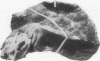
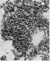
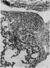
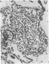
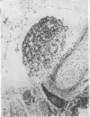
A quantitative study has been made of standardized sections taken from the right middle lobe of 316 children's lungs. Lungs showing no pathological state showed that lymph nodes are present at birth and these increase in prominence if not in numbers throughout the first year after birth. Lymphoreticular aggregates are not present among the alveoli of the normal infant lung at birth. These first appear around a week after birth and progressively increase in number throughout infancy and early childhood, being almost universally present by the age of 5 years. The development of peripheral lymphoreticular aggregates in the lungs of infants appears to be entirely environmentally and antigenically stimulated.
Full text
PDF


Images in this article


Selected References
These references are in PubMed. This may not be the complete list of references from this article.
- Bastianini A. Osservazioni sulla morfologia microscopica e l'istotopografia dei vasi linfatici del polmone umano. Boll Soc Ital Biol Sper. 1967 Nov 30;43(22):1567–1570. [PubMed] [Google Scholar]
- Bryant B. J., Shifrine M. Histogenesis of lymph nodes during development of the dog. J Reticuloendothel Soc. 1972 Jul;12(1):96–107. [PubMed] [Google Scholar]
- COLLET A., POLICARD A. [Attempted infra-structural localization in the lung of elements of the reticuloendothelial system]. C R Seances Soc Biol Fil. 1962;156:991–995. [PubMed] [Google Scholar]
- Emery J. L., Wilcock P. F. The post-natal development of the lung. Acta Anat (Basel) 1966;65(1):10–29. doi: 10.1159/000142865. [DOI] [PubMed] [Google Scholar]
- Jericho K. W., Austwick P. K., Hodges R. T., Dixon J. B. Intrapulmonary lymphoid tissue of pigs exposed to aerosols of carbon particles, of Salmonella oranienburg, of Mycoplasma granularum, and to an oral inoculum of larvae of Metastrongylus apri. J Comp Pathol. 1971 Jan;81(1):13–21. doi: 10.1016/0021-9975(71)90050-8. [DOI] [PubMed] [Google Scholar]
- Jericho K. W., Derbyshire J. B., Jones J. E. Intrapulmonary lymphoid tissue of pigs exposed to aerosols of haemolytic streptococcus group L and porcine adenovirus. J Comp Pathol. 1971 Jan;81(1):1–11. doi: 10.1016/0021-9975(71)90049-1. [DOI] [PubMed] [Google Scholar]
- Jericho K. W. Intrapulmonary lymphoid tissue of healthy pigs. Res Vet Sci. 1970 Nov;11(6):548–552. [PubMed] [Google Scholar]
- Lauweryns J. M. Stereomicroscopic funnel-like architecture of pulmonary lymphatic valves. Lymphology. 1971 Dec;4(4):125–132. [PubMed] [Google Scholar]
- TOBIN C. E. Human pulmonic lymphatics; an anatomic study. Anat Rec. 1957 Mar;127(3):611–633. doi: 10.1002/ar.1091270309. [DOI] [PubMed] [Google Scholar]