

Abstract
Background:
Marking pencils which are frequently used in orthodontics may cause microbial contamination. The purpose of this study was to evaluate and compare the effectiveness of three disinfection and sterilization methods (autoclave, glutaraldehyde solution, and Deconex spray) on orthodontic markers.
Materials and Methods:
One hundred and twenty orthodontic markers were divided into four groups each 30 pencils: One control group and three groups for three different disinfection/sterilization methods. To evaluate the effectiveness of these methods, pencils were initially contaminated by common pathogen by immersing the pencils in a suspension containing 1.5 × 10⁸ CFU/ml organisms. Then, the pencils were subjected to corresponding disinfection/sterilization methods, and the number of remaining microorganisms was calculated and compared with control group.
Results:
In the control group, the mean number of Escherichia coli was significantly higher than the other two microorganisms (P = 0.01, P = 0.031). However, the mean numbers of Staphylococcus aureus and Candida albicans were not significantly different (P = 0.1). After sterilization with autoclave and glutaraldehyde, no microbial growth was observed, whereas after disinfection with Deconx spray some colonies of microorganisms still could be observed.
Conclusion:
Autoclaving and glutaraldehyde solution are the best methods for disinfecting orthodontic markers.
Keywords: Infection control, microbial contamination, orthodontics marker
INTRODUCTION
The purpose of the infection control is to minimize the risk of transmitting the disease from the patient to dentist, the dentist to the patient, from patient to patient and dental personnel, family, and finally to society. The nature of many dental procedures is so that the specific methods must be taken for preventing transmission of infection among dental personnel and patients. Since all infected patients cannot identified according to disease history, clinical examination and laboratory tests, all patients should be considered infectious, and avoidance contact with blood and body fluids from all patients should be seriously implemented. Orthodontics is the branch of dentistry that compared with other fields has minimal contact with the blood, however, when placing and removal of fixed appliances and forming wires or replacing of chains, ligatures, springs and modules, contact with the saliva of the patient is mandatory.[1] In practice, orthodontists generally focus their attention on the sterilization of pliers, headpieces, and other instruments.[2,3,4,5] Orthodontic marking pencils are not usually considered as a possible vector in the chain of infection.[6] These pencils are frequently used to mark on the archwires in the oral cavity when orthodontist decides to place a bend or attach a hook on them. Therefore, these markers may contaminate with saliva and transmit pathogens between patients. In this regard, several studies have been performed.[7,8,9] However, few reports have been published on the permanent marker (PM) pens and pencils. Ascencio et al. stated that marking pencils can transfer bacteria from contaminated archwires, and they used gas sterilization which is effective in killing bacteria but is also costly and difficult, making it impractical for orthodontic clinics.[6] Tadiparthi et al. also demonstrated that marker pens can act as fomites for nosocomial infection. Furthermore, it was proved that dry whiteboard markers and PM pens carry a significant risk of transmitting infection among patients, and they suggested using disposable markers for immunocompromised patients to prevent cross infection.[10] Thomas et al. concluded that marking pens may transmit pathogens from one patient to others.[8]
According to this documentation, the aim of this study was to evaluate and compare the effectiveness of three disinfection and sterilization methods (autoclave, glutaraldehyde solution, and Deconex spray) on orthodontic markers.
MATERIALS AND METHODS
Three common pathogen - a Gram-negative rod (Escherichia coli), a Gram-positive cocci (Staphylococcus aureus), and a fungus (Candida albicans) were grown to logarithmic phase in trypticase soy broth. One hundred and twenty marking pencils (White Wax Marker, Dentaurum, Germany) were classified into four different groups including a control group and three test groups (autoclave, glutaraldehyde 2%, and Deconex spray), so each group consisted of 30 pencils. Initially, suspensions with a concentration of 1.5 × 10⁸ CFU/ml of organisms were prepared in liquid medium equal to 0.5 McFarland standard (Remel™, Thermoscientific, Lenexa, KS, USA). The optical density was 0.132 at 600 nm wavelength.[2]
Control Group
Within each 10 sterile test tubes, 4 ml of each microbial suspension poured pencils were immersed in bacterial suspensions. The pencils were air dried then the bacteria were washed and harvested by placing these pencils in 4 ml of sterile normal saline and by vigorous agitation. Ten microliters of this normal saline were inoculated on culture media and incubated at 37°C. After 24 h, the colonies were counted, and the numbers of remaining organisms were calculated per ml.
Autoclave Test Group
Similar to the control group, contamination step was performed and after 5 min, pencils air dried and autoclaved for 15 min at 121°C. Then pencils were placed in 4 ml of sterile saline and bacteria were washed and gathered by vigorous agitation. The numbers of bacteria were calculated by culturing it culture media.
Deconex Test Group
Similarly, contamination steps were performed and after 5 min, pencils were sprayed by Deconex (Borer, Switzerland) and after 15 min pencils were washed in 4 ml of sterile saline and bacteria harvested by vigorous agitation, and the number of remaining bacteria was counted.
Glutaraldehyde 2% Test Group
Similar to above steps the test procedure were performed, but the pencils were sprayed by glutaraldehyde 2% (Behsa, Iran) and after 30 min the pencils were washed in 4 ml of sterile saline and bacteria harvested after vigorous agitation. The number of bacteria was calculated similarly.
RESULTS
In this study, three different methods of sterilization/disinfection (autoclave, Deconex solution, glutaraldehyde) for marking pencils were evaluated. Three type of microorganism (E. coli, S. aureus, C. albicans) were used in our four groups (one control and three experimental groups).
In two groups (autoclave and glutaraldehyde), no microorganism was remained after sterilization/disinfection procedure. Therefore, these groups were considered complete and fully sufficient.
Control Group
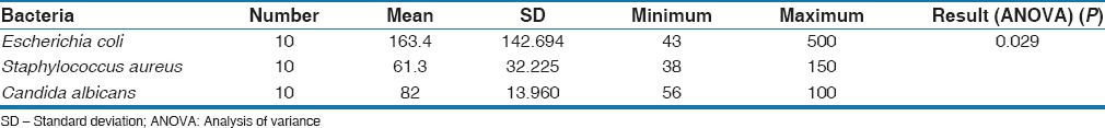
In the control group, the mean population of bacteria and C. albicans which settled after contamination was significantly different. Tukey test showed that the mean number of E. coli was significantly greater than C. albicans and S. aureus (P = 0.01, P = 0.031, respectively) There was no significant difference between the population of S. aureus and C. albicans (P = 0.1) [Table 1]. This shows that E. coli could contaminate markers much greater than Gram-positive bacteria and fungi.
Table 1.
The number of bacteria in control group
Deconex Group
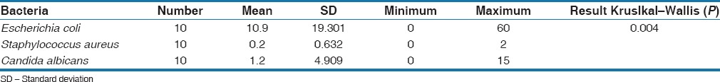
In this group, similarly Mann–Whitney U-test confirmed that the mean number of E. coli which settled on pencils was significantly greater than the mean number of C. albicans and S. aureus (P = 0.026, P = 0.003) and there was no significant difference between the number of S. aureus and C. albicans (P = 0.125) [Table 2].
Table 2.
The number of bacteria in Deconex group
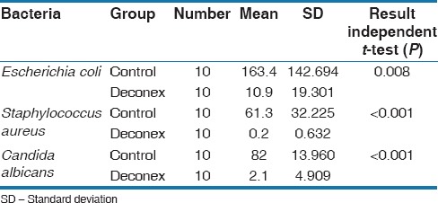
As it could be seen in Table 3, the numbers of bacteria and C. albicans after treatment with Deconex were reduced compared to control group.
Table 3.
The comparison of bacteria between Deconex and control group
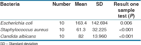
As the ideal sterilization/disinfection method should reduce the number of bacteria to zero. In the control group, the numbers of three microorganisms were significantly greater than zero [Table 4].
Table 4.
The comparison of bacteria in control group with zero
In Deconex group, the numbers of three microorganisms were not significantly greater than zero [Table 5]. Hence, using Deconex is not an appropriate method for disinfection markers. Figure 1 showed the mean number of microorganisms in groups.
Table 5.
The comparison of bacteria in Deconex group with zero
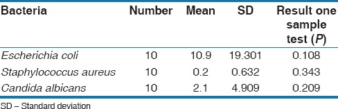
Figure 1.
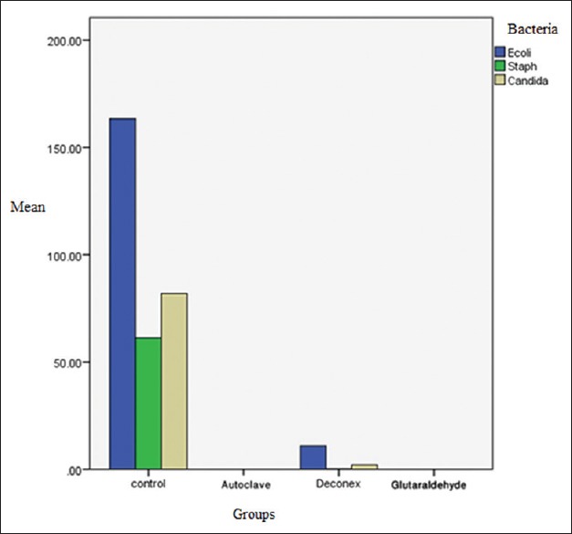
The number of bacteria in each groups
DISCUSSION
This study compared the efficacy of three disinfection/sterilization procedures on marking pencils contaminated with E. coli, S. aureus, and C. albicans. The results of the study showed that after disinfection of marking pencil by autoclave and glutaraldehyde solution all the microorganisms were killed while in disinfection with Deconex all three microorganisms had a little growth that was not significant. Based on the result of our study, all three procedures were acceptable for disinfecting of marking pencils in orthodontic offices.
Woo et al. evaluated the compliance with infection control procedures among California orthodontists and they concluded that orthodontists still need improvement in all aspects of their infection control procedures.[11] Ascencio et al. evaluated the effect of different disinfection procedures on contaminated marking pencils. The tip of pencils was wiped with either sterile gauze or gauze treated with IodoFive disinfectant. They concluded that only gas sterilization completely killed bacteria which are an expensive procedure. However, the pencils were used in this study were not autoclavable.[6] In our study, glutaraldehyde solution completely destroyed microorganisms, and this disinfectant can be used instead of gas sterilization. Terzic et al. evaluated the efficacy of autoclave in the sterilization of surgical marking pencils and they reported that no microorganisms were cultured after using autoclave which was in agreement with the results of our study.[12] Venkatasubramanian et al. compared the efficacy of four different disinfection methods on endodontic files. They used Bacillus stearothermophilus. They reported that autoclave and CO2 laser carried out sterilization completely while glutaraldehyde was not able to sterilize endodontic files completely. In our study, glutaraldehyde showed acceptable disinfection characteristics and this difference could be as a result of different types of bacteria, different solutions, and different subjects who were evaluated.[13] Camilla et al. evaluated the effect of different disinfection methods on orthodontic pliers. They reported that glutaraldehyde was an acceptable disinfectant agent which it was in agreement with our study.[14]
Parnia et al. examined the effect of different disinfecting agents on contaminated impression materials. They reported that Deconex was an acceptable disinfecting agent which was similar to our study.[15] It should be noted that every patient should be considered infectious. Therefore, this study was performed on contaminated markers to examine which infection control procedures are the best for marking pens and pencils in orthodontic clinics. However, the results do not reflect accurately the extent to which the sterilizing and disinfecting methods are reliable methods to be applied in-clinic.
CONCLUSION
Based on our result, autoclave and glutaraldehyde solution were the best methods for disinfection of orthodontic marking pencils. The remained bacterial contamination after disinfecting by Deconex solution was no significant, and therefore, this agent could be a proper substitute if the two aforementioned methods were not available.
Financial Support and Sponsorship
Nil.
Conflicts of Interest
There are no conflicts of interest.
REFERENCES
- 1.Aksoy A, Kılıç G, Hussein E, Aboukhalil D. Sterilization and Disinfection in Orthodontics. In: Naretto S, editor. Principles in Contemporary Orthodontics. Chapter 6. InTech. Rijeka, Croatia: 2011. p. 113. [Google Scholar]
- 2.Wichelhaus A, Bader F, Sander FG, Krieger D, Mertens T. Effective disinfection of orthodontic pliers. J Orofac Orthop. 2006;67:316–36. doi: 10.1007/s00056-006-0622-9. [DOI] [PubMed] [Google Scholar]
- 3.Cash RG. Trends in sterilization and disinfection procedures in orthodontic offices. Am J Orthod Dentofacial Orthop. 1990;98:292–9. doi: 10.1016/S0889-5406(05)81486-6. [DOI] [PubMed] [Google Scholar]
- 4.McCarthy GM, Koval JJ, MacDonald JK. Compliance with recommended infection control procedures among Canadian dentists: Results of a national survey. Am J Infect Control. 1999;27:377–84. doi: 10.1016/s0196-6553(99)70001-5. [DOI] [PubMed] [Google Scholar]
- 5.Mulick JF. Upgrading sterilization in the orthodontic practice. Am J Orthod. 1986;89:346–51. doi: 10.1016/0002-9416(86)90059-x. [DOI] [PubMed] [Google Scholar]
- 6.Ascencio F, Langkamp HH, Agarwal S, Petrone JA, Piesco NP. Orthodontic marking pencils: A potential source of cross-contamination. J Clin Orthod. 1998;32:307–10. [PubMed] [Google Scholar]
- 7.Kane TP, Greig M, Grover ML. Can the tips of marker pens act as a source of cross-infection? Ann R Coll Surg Engl. 2003;85:73. [PMC free article] [PubMed] [Google Scholar]
- 8.Thomas RJ, Goodbourne C, Goldie B. The transmission of MRSA via orthopaedic marking pens – Fact or fiction. Ann R Coll Surg Engl. 2004;86:51–2. doi: 10.1308/003588404772614731. [DOI] [PMC free article] [PubMed] [Google Scholar]
- 9.Wilson J, Tate D. Can pre-operative skin marking transfer methicillin-resistant Staphylococcus aureus between patients? A laboratory experiment. J Bone Joint Surg Br. 2006;88:541–2. doi: 10.1302/0301-620X.88B4.17454. [DOI] [PubMed] [Google Scholar]
- 10.Tadiparthi S, Shokrollahi K, Juma A, Croall J. Using marker pens on patients: A potential source of cross infection with MRSA. Ann R Coll Surg Engl. 2007;89:661–4. doi: 10.1308/003588407X209419. [DOI] [PMC free article] [PubMed] [Google Scholar]
- 11.Woo J, Anderson R, Maguire B, Gerbert B. Compliance with infection control procedures among California orthodontists. Am J Orthod Dentofacial Orthop. 1992;102:68–75. doi: 10.1016/0889-5406(92)70016-4. [DOI] [PubMed] [Google Scholar]
- 12.Terzic A, Dharan S, Pittet D, Scolozzi P. Autoclave sterilizable pencils in maxillofacial surgery. Plast Reconstr Surg. 2010;126:255e–7e. doi: 10.1097/PRS.0b013e3181ef81ed. [DOI] [PubMed] [Google Scholar]
- 13.Venkatasubramanian R, Jayanthi, Das UM, Bhatnagar S. Comparison of the effectiveness of sterilizing endodontic files by 4 different methods: An in vitro study. J Indian Soc Pedod Prev Dent. 2010;28:2–5. doi: 10.4103/0970-4388.60478. [DOI] [PubMed] [Google Scholar]
- 14.Camilla M, Adriana S, Danilo A. Evaluation of disinfection methods of orthodontic pliers. Dental Press J Orthod. 2012;17:105. [Google Scholar]
- 15.Parnia F, Hafezeqoran A, Moslehifard E, Mahboub F, Nahaei M, Akbari Dibavar M. Effect of different disinfectants on Staphylococcus aureus and Candida albicans transferred to alginate and polyvinylsiloxane impression materials. J Dent Res Dent Clin Dent Prospects. 2009;3:122–5. doi: 10.5681/joddd.2009.030. [DOI] [PMC free article] [PubMed] [Google Scholar]