

Summary:
The authors describe a case of retained sterile matrix in a 38-year-old Hispanic man with a history of remote trauma and soft-tissue coverage with a groin flap 13 years before presentation. The patient presented with a slowly enlarging, vertically growing dorsal thumb mass with occasional drainage. The patient had minimal functional impairment, and radiographic imaging showed a radiolucent mass projecting dorsally over the distal phalanx. Surgical exploration revealed an approximately 2 × 2 cm keratinized mass attached to a retained nail bed. The keratinized nail plate was removed, along with an ellipse of soft tissue around the draining tract. To the authors’ knowledge, this case is the largest reported vertically growing, retained, and cornified nail bed with an unusual size and shape. Physicians should consider the possibility of retained nail plates in patients who present with unusual large growths after trauma or surgery.
The hand is the most commonly injured part of the body. Among the injuries, most of them occur in the nail beds. These injuries can cause significant pain, long-term discomfort, and an aesthetically displeasing nail plate. Traumatic nail injuries may require avulsion as a therapeutic adjunct.1 Although complications of nail-unit surgery are rare, the most common complication is permanent dystrophy of the nail plate (eg, nail splitting, ridging, and pterygium formation), which results from damage of the nail matrix.2 Nail dystrophy occurs more frequently when the injury is located within the proximal nail matrix.2 We describe a case of the largest reported vertically growing, retained, and cornified nail bed with unusual size and shape.
CASE REPORT
A 38-year-old man presented with exacerbated swelling and pain in his right thumb (Fig. 1). He had sustained a crush injury 13 years before, with extensive damage to the soft tissue and bone, requiring a groin flap and nail-plate removal. After the injury, he had gradual swelling of the dorsum of his thumb with occasional bouts of drainage. The patient maintained function, with minimal impairment of flexion at the interphalangeal joint. Surgical exploration was performed for the condition that was considered to be a simple nail ablation and to explore the possibility of removal of the soft-tissue mass to alleviate the chronic drainage (Fig. 2). Intraoperative fluoroscopy allowed for better visualization of the nail plate and revealed that the mass was not a result of malunion fracture (Fig. 3). The large dorsal deformity was discovered to be a walnut-sized vertically growing nail plate (2 × 2 cm) completely covered by skin. The nail plate with nail-bed ablation was excised (Fig. 4). Histopathologic findings confirmed that the specimen was fragments of the nail plate and matrix with foreign-body giant-cell reaction in the surrounding area, and no bone was identified. The patient tolerated the procedure well with no adverse sequelae.
Fig. 1.

Preoperative image of a retained, entrapped nail plate resembling a large mass in a 38-year-old man.
Fig. 2.

Dorsal excision showing retained, entrapped, and cornified nail bed.
Fig. 3.
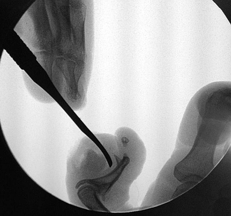
Intraoperative fluoroscopic image of the nail plate.
Fig. 4.

Excised walnut-sized nail plate (2 × 2 cm).
DISCUSSION
Nails are ectodermal appendages, formed in the germinal matrix. Loss or deformity of this germinal part results in permanent loss or permanent deformity of the nail.2,3 Our patient presented with entrapped nail bed after prior trauma. Retained nail bed is a common problem after traumatic injury to the distal phalanx, and it can lead to pain, swelling, drainage, and infection. A retained nail bed with the size and shape and with the vertical growth pattern described in this case is very uncommon.
Goikoetxea et al4 reported a case with a similar presentation as a mass: it was the case of an 8-year-old boy with entrapped and protruded ectopic nail in the dorsal aspect of the right middle phalanx of the fifth finger. The patient had a history of trauma and nail avulsion without fracture of the distal phalanx. He had episodes of soft-tissue infection of the site. By means of surgical excision and histopathologic examination, the presence of ectopic nail was confirmed.4
Vertical growth of the nail bed has been described previously. In one case, a 32-year-old man presented with a posttraumatic, vertically growing ectopic nail, which projected out of the dorsal aspect of the middle phalanx of the third finger.5 In another case, a 26-year-old man with a history of a vegetable thorn prick 8 years back presented with a slow-growing, nontender, hard projection in the dorsoulnar aspect of his left thumb. Excision and histopathologic findings confirmed that the projection was an ectopic nail with a nail matrix and nail bed.6
Radiographic findings were normal in our case, which was consistent with the findings in previous reports. In one report, magnetic resonance imaging was performed to identify the nail abnormality, but the ectopic nail was not well defined, probably because of its small size.7
With a growth rate of 1 mm/week for avulsed nails, complete regrowth of the nail typically takes 4–5 months.1 Separation of the nail plate from the nail bed decreases nail blood flow and may explain the slow growth of the nail in the present case. Histopathologic findings revealed fragments of the nail plate and matrix with no bone.
Retained nail plates can be confused with other conditions; physicians should ensure that any unusual growth or deformity in this area is not immediately ruled out as a soft-tissue mass or joint deformity. More aggressive preoperative imaging could help in guiding better operative planning as well.
CONCLUSIONS
The present case featured a nail with unusual size and shape with slow vertical growth after remote trauma and nail avulsion, which resembled a large mass and deformity of the finger with normal radiographic findings. Recognizing that unusual large growths after trauma or surgery can, in fact, be retained nail plates is important for treatment planning.
Footnotes
Disclosure: The authors have no financial interest to declare in relation to the content of this article. The Article Processing Charge was paid for by the authors.
REFERENCES
- 1.Pandhi D, Verma P. Nail avulsion: indications and methods (surgical nail avulsion). Indian J Dermatol Venereol Leprol. 2012;78:299–308. doi: 10.4103/0378-6323.95444. [DOI] [PubMed] [Google Scholar]
- 2.Moossavi M, Scher RK. Complications of nail surgery: a review of the literature. Dermatol Surg. 2001;27:225–228. [PubMed] [Google Scholar]
- 3.Haneke E. Surgical anatomy of the nail apparatus. Dermatol Clin. 2006;24:291–296. doi: 10.1016/j.det.2006.03.007. [DOI] [PubMed] [Google Scholar]
- 4.Goikoetxea X, Etxebarria I, Careaga M. Posttraumatic ectopic nail: case report. J Hand Surg Am. 2006;31:819–821. doi: 10.1016/j.jhsa.2005.12.025. [DOI] [PubMed] [Google Scholar]
- 5.Sasmaz S, Coban YK, Gumusalan Y, et al. Posttraumatic ectopic nail. J Am Acad Dermatol. 2004;50:323–324. doi: 10.1016/j.jaad.2003.07.020. [DOI] [PubMed] [Google Scholar]
- 6.Chatterjee K, Chaudhuri A, Chatterjee G. Onychoheterotopia: a unique case. Indian J Dermatol. 2013;58:150–151. doi: 10.4103/0019-5154.108064. [DOI] [PMC free article] [PubMed] [Google Scholar]
- 7.Kusano T, Hayashi M, Hosaka Y. Posttraumatic ectopic nail of the toe. Eur J Plast Surg. 2009;32:305–307. [Google Scholar]