Abstract
The Food and Drug Administration recently cleared silver diamine fluoride for reducing tooth sensitivity. Clinical trials document arrest and prevention of dental caries by silver diamine fluoride; this off-label use is now permissible and appropriate under U.S. law. A CDT code was approved for caries arresting medicaments for 2016 to facilitate documentation and billing. We present a systematic review, clinical indications, clinical protocol, and consent procedure to guide application for caries arrest treatment.
Introduction
Until now, no option for the treatment of dental caries in the United States besides restorative dentistry has shown substantial efficacy.1 Silver diamine fluoride is an inexpensive topical medicament used extensively in other countries to treat dental caries across the age spectrum. No other intervention approaches the ease of application and efficacy. Multiple randomized clinical trials – with hundreds of patients each – support use for caries treatment, thus substantiating an intervention that addresses an unmet need in American dentistry. In August 2014 the Food and Drug Administration (FDA) cleared the first silver diamine fluoride product for market, and as of April 2015 that product is available.
Since approval in Japan over 80 years ago,2 more than two million containers have been sold. The silver acts as an antimicrobial, the fluoride promotes remineralization, and the ammonia stabilizes high concentrations in solution.3
Because silver diamine fluoride is new to American dentistry and dental education, there is a need for a standardized guideline, protocol, and consent. The UCSF School of Dentistry Paradigm Shift Committee assembled a subcommittee with the following goals: Use available evidence to: 1. Develop a list of clinical indications; 2. Define a protocol that maximized safety and efficacy, and minimized inadvertent staining of clinical facilities; and 3. Build an informed consent document at the 8th grade reading level. We conducted a systematic review, inquired of authors of published clinical and in vitro studies about details and considerations in their protocols, and consulted experts in cariology and materials chemistry where evidence was lacking.
Methods
A literature review was designed by a medical librarian to search PubMed and the International Association of Dental Research abstract archive with the following search terms: ‘“33040-28-7” OR “1Z00ZK3E66” OR “silver diamine fluoride” OR “silver fluoride” OR “silver diammine fluoride”; OR “diammine silver fluoride” OR “ammonical silver fluoride” or “ammoniacal silver fluoride”. Differences in nomenclature have led to confusion around this material. Another review was completed with the terms: (“dental” or “caries”) and “silver nitrate” and “clinical”.
Material
Silver diamine fluoride (38% w/v Ag(NH3)2F, 30% w/w) is a colorless topical agent comprised of 24.4-28.8% (w/v) silver and 5.0-5.9% fluoride, at pH 10,4 and marketed as Advantage Arrest™ by Elevate Oral Care, LLC (West Palm Beach, FL). Other companies may market silver diamine fluoride in the future, following determination of substantial equivalence and FDA clearance.
Mechanisms
Silver diamine fluoride is used for caries arrest and treatment of dentin hypersensitivity. In treatment of exposed sensitive dentin surfaces, topical application results in development of a squamous layer on the exposed dentin, partially plugging the dentinal tubules.5 High concentration aqueous silver has been long known to form this protective layer.6 Decreased sensitivity in treated patients7,8 is consistent with the hydrodynamic theory of dentin hypersensitivity.9
Dental caries is a complex progression involving dietary sugars, bacterial metabolism, demineralization, and organic degradation. The collagenous organic matrix is exposed once a dentin surface is demineralized and destroyed by native and bacterial proteases to enable a lesion to enlarge.10 Upon application of silver diamine fluoride to a decayed surface, the squamous layer of silver-protein conjugates forms, increasing resistance to acid dissolution and enzymatic digestion.11 Hydroxyapatite and fluoroapatite form on the exposed organic matrix, along with the presence of silver chloride and metallic silver.5 The treated lesion increases in mineral density and hardness while the lesion depth decreases.5 Meanwhile, silver diamine fluoride specifically inhibits the proteins that break down the exposed dentin organic matrix: matrix metalloproteinases;11 cathepsins;12 and bacterial collagenases.5 Silver ions act directly against bacteria in lesions by breaking membranes, denaturing proteins, and inhibiting DNA replication.13,14 Ionic silver deactivates nearly any macromolecule. Silver diamine fluoride outperforms other anti-caries medicaments in killing cariogenic bacteria in dentinal tubules.15
Silver and fluoride ions penetrate ~25 microns into enamel,16 and 50-200 microns into dentin.17 Fluoride promotes remineralization, and silver is available for antimicrobial action upon release by re-acidification.18 Silver diamine fluoride arrested lesions are 150 microns thick.19
Artificial lesions treated with silver diamine fluoride are resistant to biofilm formation and further cavity formation, presumably due to remnant ionic silver.20,21 More silver and fluoride is deposited in demineralized than non-demineralized dentin; correspondingly, treated demineralized dentin is more resistant to caries bacteria than treated sound dentin.22 When bacteria killed by silver ions are added to living bacteria, the silver is re-activated, so that effectively the dead bacteria kill the living bacteria in a “zombie effect.”23 This reservoir effect helps explain why silver deposited on bacteria and dentin proteins within a cavity has sustained antimicrobial effects.
Clinical evidence
Silver nitrate + fluoride varnish
Before the FDA cleared silver diamine fluoride, some American dentists sequentially applied silver nitrate then fluoride varnish to dentinal decay as the only available non-invasive option for caries treatment. Duffin rediscovered silver nitrate from the early literature,24 which had been lost to modern cariology. Surprisingly, there is no mention of silver nitrate in either of the American Dental Association Council on Scientific Affairs reports on Nonfluoride Caries-preventive Agents,25 or Managing Xerostomia and Salivary Gland Hypofunction,26 and it is not part of standard dental school curricula. Case series of carious lesions arrested by silver nitrate date to the 1800s, for example in 1891 87 of 142 treated lesions were arrested.27 Percy Howe, then director of the Forsyth Institute in Boston, added ammonia to silver nitrate, making it more stable and effective as an antimicrobial for application to any infected tooth structure: from early cavitated lesions to infected root canals.28 Duffin added the application of fluoride varnish following silver nitrate, simulating silver diamine fluoride. While his clinic doubled in patients, cases needing general anesthesia disappeared. His review of randomly selected charts showed only 7 of 578 treated lesions progressed within 2.5 years to the point that extractions were needed.24 Thus, with the exception of Duffin’s and one other report, attention to silver nitrate largely disappeared by the 1950s. The lore is that use and teaching of this intervention were lost with the introduction of effective local anesthetic to enable painless restorations, and fluoride for caries prevention. Because no high quality clinical trials have been performed, we did not include the silver nitrate plus fluoride varnish regimen in our recommendation.
Silver diamine fluoride
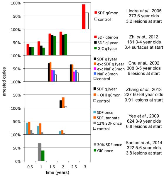
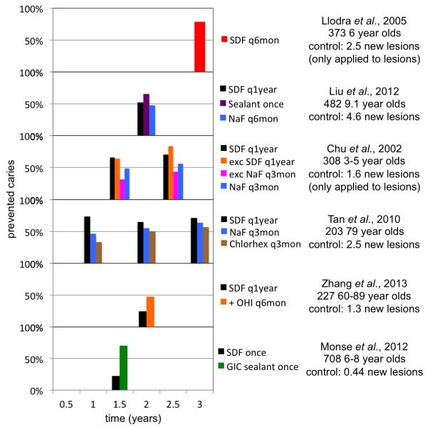
We found 9 published randomized clinical trials evaluating silver diamine fluoride for caries arrest and/or prevention, of at least 1 year in duration. These studies have each involved hundreds of children aged 3-9 or adults aged 60-89 (Figures 1 and 2). Most participants had low (<0.3 ppm) fluoride in the environmental water, and reported using fluoride toothpaste (e.g. 73%).29 Silver diamine fluoride was applied with cotton isolation. Lesions were detected with mirror and explorer only. All studies were registered and meet the Consolidated Standards of Reporting Trials requirements. Clinical cases and studies not meeting these criteria can be found elsewhere.30
Figure 1.
Graphic summary of randomized controlled trials demonstrating caries arrest after topical treatment with 38% silver diamine fluoride (SDF). Studies are arranged vertically by frequency of silver diamine fluoride application. Caries arrest is defined as the fraction of initially active carious lesions that became inactive and firm to a dental explorer. SDF (38% unless noted otherwise); q6mon, every six months; q1year, every year; q3mon, every three months; GIC, glass ionomer cement; NaF, 5% sodium fluoride varnish; + OHI q6mon, SDF every year and oral hygiene instructions every six months.
Figure 2.
Graphic summary of randomized controlled trials demonstrating caries prevention after topical treatment of carious lesions with 38% silver diamine fluoride. Prevented caries is defined as the fraction of new carious lesions in treatment groups as compared to those in the placebo or no treatment control group. Chlorhex, 1% chlorhexidine varnish.
Caries arrest
Caries arrest increased dramatically after re-application from 1 year post-treatment31-33 to 1.5 years,31,34 and increasingly to 2-3 years (Figure 1).29,31,35 Single application without repeat lost effect over time in elders.32 Twice per year application resulted in more arrest than once per year.31,35 12% silver diamine fluoride was markedly less effective.32
Darkening of the entire lesion indicated success at follow-up and is suggested to facilitate diagnosis of caries arrest status by non-dentists. A longitudinal study reported that color activation of silver diamine fluoride with 10% stannous fluoride resulted in less first molar caries.36 Tea extract was used in one group to activate color change for improved follow-up diagnosis; no differences in arrest were seen.32 Indeed, when stannous fluoride was used to activate color change, a break in the black color within a lesion at 6 months was highly sensitive and specific for active caries.37
Silver diamine fluoride greatly outperformed fluoride varnish for caries arrest29 and was equivalent or better than glass ionomer cement (GIC; Figure 1).31,33 The addition of semi-annual intensive oral health education with the application of silver diamine fluoride in elders increased arrest of root caries (Figure 1).38
Caries Prevention
When silver diamine fluoride was applied only to carious lesions, impressive prevention was seen for other tooth surfaces.29,35 Fluoride releasing GIC can have this effect but it is limited to surfaces adjacent to the treated surface and of short duration. Direct application to healthy surfaces in children also helps prevent caries (Figure 2).29,35,39 Two studies show great difference in the level of prevention in elders;38,40 the difference is hard to reconcile. As seen for arrest, prevention is less after 1 year without repeat application.41
Annual application of silver diamine fluoride prevented many more carious lesions than 4 times per year fluoride varnish in both children29 and elders.40 Prevention was roughly equivalent to twice per year varnish in one study (Figure 2).39 The addition of semi-annual intensive oral health education in a study of elders increased prevention.38 Although many fell out, GIC or resin sealants outperformed silver diamine fluoride in preventing caries in the first molars of children,39,41 though the cost was ~20 times more.
Ongoing trials
Unpublished reports of clinical studies unanimously confirm better caries arrest and/or prevention by silver diamine fluoride over control or other materials. A 1 year report of a study of elders demonstrated that the addition of a saturated solution of potassium iodide (SSKI) to decrease discoloration did not significantly alter caries arrest or prevention.42 This was confirmed in the 2 year examinations (personal communication, Edward Lo). A 1 year report of a study in children showed that the application once per week for 3 consecutive weeks, once per year, was more effective than that of single annual application.43 Other studies have recently begun to evaluate the ability of silver diamine fluoride to arrest interproximal carious lesions, to compare the relative efficacy of silver diamine fluoride to the combination of silver nitrate plus fluoride varnish, and to compare the effects on populations with or without access to fluoridated water. Final reports from these studies will follow in the coming years.
Recommendations from the Literature on Clinical Efficacy
These studies show that 38% silver diamine fluoride is effective and efficient in arresting and preventing carious lesions. Application only to lesions appears to be similarly effective in preventing cavities in other teeth and surfaces as applying directly. Single application appears insufficient for sustained effects, while annual re-application results in remarkable success, and even greater effects with semi-annual application. From these data we recommend twice per year application, only to carious lesions, without excavation, for at least the first 2 years.
For any patient with active caries, we recommend considering replacement of fluoride varnish as the primary means to prevent new lesions, with application of silver diamine fluoride to the active lesions only. For patients without access to both sealants and monitoring, silver diamine fluoride is the agent of choice for prevention of caries in permanent molars – particularly as there is no margin to leak and thereby facilitate deep caries, and it does not stain sound enamel.
Longer studies are needed to determine whether caries arrest and prevention can be maintained with decreased application after 2-3 years, and whether more frequent use would enhance efficacy. Traditional or non-traditional restorative approaches such as the Atraumatic Restorative Technique (ART)44 and Hall crowns45 should be performed as dictated by the response of the patient, disease progression, and the nature of individual lesions.
Safety
Maximum dose and Safety margin
The margin of safety for dosing is of paramount concern. In gaining clearance by the FDA, female and male rat and mouse studies were conducted to determine the lethal dose (LD50) of silver diamine fluoride by oral and subcutaneous administration. Average LD50 by oral administration was 520 mg/kg, and by subcutaneous administration was 380 mg/kg. The subcutaneous route is taken here as a worst-case scenario. One drop (25 μL) is ample material to treat 5 teeth, and contains 9.5 mg silver diamine fluoride. Assuming the smallest child with caries would be in the range of 10 kg, the dose would be 0.95 mg / kg child. Thus the relative safety margin of using an entire drop on a 10kg child is: 380 mg/kg LD50 / 0.95 mg / kg dose = 400-fold safety margin. Actual dose is likely to be much smaller, for example 2.37 mg total for 3 teeth was the largest dose measured in 6 patients.46 The most frequent application monitored in a clinical trial was weekly for 3 weeks, annually.43 Thus we set our recommended limit as 1 drop (25 μL) per 10 kg per treatment visit, with weekly intervals at most. This dose is commensurate with the EPA’s allowable short-term exposure of 1.142 mg silver per liter of drinking water for 1-10 days (ATSDR, 1990).
Cumulative exposure from lower level acute or chronic silver intake has no real physiologic disease importance, but the bluing of skin in argyria should obviously be avoided. The Environmental Protection Agency set the lifetime exposure conservatively at 1 gram to safely avoid argyria. The highest applied dose for 3 teeth measured in the pharmacokinetic study, 2.37 mg, would enable >400 applications.46 Silver nitrate (typically a 25% solution) has been used for more than 100 years in the United States without incident, including acceptance by the ADA, and in other countries for arresting dental caries.3
Adverse effects
Not a single adverse event has been reported to the Japanese authorities since they approved silver diamine fluoride (Saforide™, Toyo Seiyaku Kasei Co. Ltd., Osaka, JP) over 80 years ago.47 The manufacturer estimates that more than 2 million multi-use containers have been sold, including >41,000 units in each of the last three reporting years.
In the 9 randomized clinical trials in which silver diamine fluoride was applied to multiple teeth to arrest or prevent dental caries, the only side effect noted was for 3 of 1,493 children or elders monitored for 1-3 years who experienced “a small, mildly painful white lesion in the mucosa, which disappeared at 48 [hours] without treatment.”29,31-33,35,38,40,41,48 The occurrence of reversible localized changes to the oral mucosa was predicted in the first reports of longitudinal studies.49 No adverse pulpal response was observed.
Gingival responses have been minimal. In a pharmacokinetic study of silver diamine fluoride application to 3 teeth in each of 6 48-82 year olds, no erythema, bleeding, white changes, ulceration, or pigmentation was found after 24 hours. Serum fluoride hardly went up from baseline, while serum silver increased about 10-fold and stayed high past the 4 hours of measurement.46 In a 2 site hypersensitivity trial of 126 patients in Peru, at baseline 9% of patients presented redness scores of 2 (1 being normal, 2 being mild to moderate redness, and 3 being severe); and after 1 day 13% in silver diamine fluoride treated patients versus 4% in controls; all redness was gone at 7 days. Meanwhile, gingival index improved slightly in silver diamine fluoride treated patients.7 Nonetheless, gingival contact should be minimized. In our experience it has been adequate to coat the nearby gingiva with petroleum jelly, use the smallest available microsponge, and dab the side of the dappen dish to remove excess liquid before application.
Concerns for fluoride safety are most relevant to chronic exposure,50 whereas this is an acute exposure. Chronically high systemic fluoride results in dental fluorosis. The ubiquitous use of fluoride-based gas general anesthetics has shown that the first acute response is transient renal holding, and is rare.51 Concerns have been raised of poorly controlled silver diamine fluoride concentrations,52 and fluorosis appearing in treated rats.53 However, silver and fluoride levels are closely monitored for the U.S. product, and the Health Department of Western Australia conducted a study that found no evidence of fluorosis resulting from long-term proper use of silver diamine fluoride.54 Therefore, we have concluded that the development fluorosis after application of the U.S. approved product is not a clinically significant risk.
Silver allergy is a contraindication. Relative contraindications include any significant desquamative gingivitis or mucositis that disrupts the protective barrier formed by stratified squamous epithelium. Increased absorption and pain would be expected with contact. Heightened caution and use of a protective gingival coating may suffice.
SSKI (discussed below) is contraindicated in pregnant women and during the first six months of breastfeeding due to concern of overloading the developing thyroid with iodide; thyroid specialists suggested a pregnancy test prior to use in women of childbearing age uncertain of their status.
Non-medical side effects
Silver diamine fluoride darkens carious lesions. At least for children, many parents have seen the color changes as a positive indication that the treatment was effective.29 Application of a saturated solution of potassium iodide (SSKI) immediately following silver diamine fluoride treatment is thought to decrease staining (patent US6461161). This is an off-label use; potassium iodide is approved as an over the counter drug to facilitate mucus release to breathe more easily with chronic lung problems, and to protect the thyroid from radioactive iodine in radiation emergencies. In our clinical experience, SSKI helps but does not dramatically effect stain; arrested lesions normally darken. Most stain remains at the dentin-enamel or cementum-enamel junction. However, SSKI maintains resistance to biofilm formation or activity in laboratory studies.20 Also, SSKI maintained caries arrest efficacy in the early results of an ongoing clinical trial.42 Meanwhile, silver diamine fluoride-treated lesions can also be covered with GIC or composite (see below for discussion on bonding).
Patients note a transient metallic or bitter taste. In our experience with judicious use the taste and texture response is more favorable than the response to fluoride varnish.
Even a small amount of silver diamine fluoride can cause a “temporary tattoo” to skin (on the patient or provider), like a silver nitrate stain or henna tattoo, and does no harm. Stain on the skin resolves with the natural exfoliation of skin, in 2-14 days. Universal precautions prevent most exposures. Long-term mucosal stain: local argyria akin to an amalgam tattoo has been observed when applying silver nitrate to intra-oral wounds; we anticipate similar stains with submucosal exposure to silver diamine fluoride.
Silver diamine fluoride stains clinic surfaces and clothes. The stain does not come out once it sets. Spills can be cleaned up immediately with copious water, ethanol, or bleach. High pH solvents such as ammonia may be more successful. Secondary containers and plastic liners for surfaces are adequate preventives.
Effects on bonding
Using a contemporary bonding system, silver diamine fluoride had no effect on composite bonding to noncarious dentin using either self-etch or full etch systems.55 In one study, simply rinsing after silver diamine fluoride application avoided a 50% decrease in bond strength for GIC.56 In another study, increased dentin bond strength to GIC was observed.57 Silver diamine fluoride decreased dentin bonding strength of resin-based crown cement by ~1/3.58 Thus, rinsing will suffice for direct restorations, while excavation of the silver diamine fluoride-treated superficial dentin is appropriate for cementing crowns.
Indications
Countless patients would benefit from conservative treatment of non-symptomatic active carious lesions. We discuss five indications.
First, extreme caries risk is defined as patients with salivary dysfunction, usually secondary to cancer treatment, Sjogren syndrome, polypharmacy, aging, or methamphetamine abuse. For these patients, frequent prevention visits and traditional restorations fail to stop disease progression. Similar disease recurrence occurs in Severe Early Childhood Caries.
Second, some patients cannot tolerate standard treatment for medical or psychological reasons. These include the pre-cooperative child, the frail elder, and those with severe cognitive or physical disabilities, and dental phobias. Various forms of immunocompromise mean that these same patients have a much higher risk of systemic infection arising from untreated dental caries. Many only receive restorative care with general anesthesia or sedation and others are not good candidates for general anesthesia due to frailty or other medical complexity. The CDC estimates 1.4 million people in the U.S. live in nursing homes and 1.2 million live in hospice.59 These individuals tend to have medical, behavioral, physical, and financial limitations that beg a reasonable option.
Third, some patients have more lesions than can be treated in one visit, such that new lesions arise or existing lesions become symptomatic while awaiting completion of treatment. This is particularly relevant to the dental school setting where treatment is slow. American dentistry has been desperately lacking an efficient instrument to be used at the diagnostic visit to provide a step towards controlling the disease.
Fourth, some lesions are just difficult to treat. Recurrent caries at a crown margin, root caries in a furcation, or the occlusal of a partially erupted wisdom tooth pose a challenge to access, isolation, and cleansability necessary for restorative success.
Following the above considerations, we developed four indications for treatment of dental caries with silver diamine fluoride:
Extreme caries risk (Xerostomia or Severe Early Childhood Caries).
Treatment challenged by behavioral or medical management.
Patients with carious lesions that may not all be treated in one visit.
Difficult to treat dental carious lesions.
Finally, These indications are for our school clinics. They do not address access to care. The U.S. Department of Health and Human Services estimates 108 million Americans without dental insurance, and 4,230 shortage areas with 49 million people without access to a dental health professional.60 Unlike fillings, failure of silver diamine fluoride treatment does not appear to create an environment that promotes caries, and thus need to be monitored. Thus, a final important indication is:
-
5.
Patients without access to dental care.
Clinical application
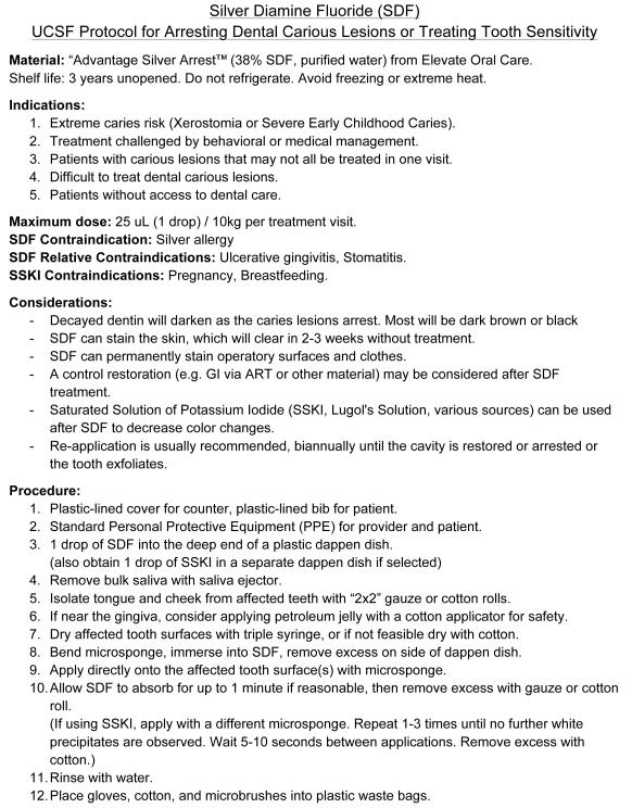
We considered practical strategies to maximize safety and effectiveness in the design of a clinical protocol for the UCSF dental clinics (Figure 3).
Figure 3.
Clinical protocol for the UCSF dental clinics.
The key factor is repeat application over multiple years. We believe that dryness of the lesion during application is also important. Isolation with gauze and/or cotton rolls is sufficient, while air-drying prior to application is thought to improve effectiveness. Allowing 1-3 minutes for the silver diamine fluoride to soak into and react with a lesion is thought to effect success. Allowing only a few seconds to soak in due to the cooperation limits of very young patients commonly results in arrest. Application time in clinical studies does not correlate to outcome. However, our committee decided to be cautious in our recommendations for initial use. Longer absorption time also decreases concerns about removing silver diamine fluoride with a post-treatment rinse. Removing any excess material with the same cotton used to isolate is routine to minimize systemic absorption.
Many clinicians place silver diamine fluoride at the diagnostic visit, then at 1 and/or 3 month follow ups, then at semi-annual recall visits (6, 12, 18, 24 months). Whether application needs to continue after 2 or 3 years to maintain caries arrest is not known. Another approach is simply to substitute silver diamine fluoride for any application of fluoride varnish to a patient with untreated carious lesions. Increased frequency with higher disease burden follows the Caries Management by Risk Assessment (CAMBRA) principles.61 It is relevant to take photographs to track lesions over time.
Efforts to improve the penetration of silver diamine fluoride into affected dentin by chemical cavity preparation have not been studied but are being explored clinically. Pretreatment with EDTA to remove superficial hydroxyapatite in affected dentin may open the dentinal tubules to further silver diamine fluoride penetration. Pretreatment with hypochlorite (bleach) may help breakdown bacteria and exposed dentin proteins, but this may be redundant to the action of the silver. Hypochlorite to decrease discoloration after silver diamine fluoride treatment is not recommended, as the color comes from silver that cannot be broken down like organic chromophores, and might break down dentin proteins stabilized against the effects of bacteria and acid by interactions with silver.
Experience with the combination of silver nitrate plus fluoride varnish (see above) has many practitioners asking about a topical varnish after silver diamine fluoride placement to prevent silver diamine fluoride taste and keep the silver diamine fluoride in the lesion. We see no evidence that varnish would help achieve either goal. Varnish does not seal. Rather, allowing more time for residence and diffusion of silver diamine fluoride to react with and dry into the lesion is more likely to improve effectiveness. Also, in our experience silver diamine fluoride results in less aversive taste and texture responses than to fluoride varnish.
Decreased darkening of lesions in the esthetic zone improves acceptance. SSKI is an option if the patient is not pregnant, though significant darkening should still be expected. SSKI and silver diamine fluoride are not to be combined prior to application: SSKI can be placed after drying the silver diamine fluoride-treated tooth. Silver diamine fluoride does not prevent restoration of a lesion, thus it does not prevent esthetic options. While silver diamine fluoride has been shown to be more effective than the atraumatic restorative technique (ART, often called IRT),33 the two are compatible and can be combined across 1 or more visits.
The California Business and Professions Code permits dental hygienists and assistants to apply silver diamine fluoride for the control of caries because they are topical fluorides (Section 1910.(b)). Physicians, nurses, and their assistants are permitted to apply fluorides in California and in many other states and federal programs. The recent decision of the Oregon Dental Board to allow dental hygienists and assistants to place silver diamine fluoride under existing rules for topical fluoride medicaments sets a precedent. Dental hygienists and assistants in Oregon were barred from providing silver nitrate in a previous decision. All providers need to be trained. Applications should be tracked if applied to the same patient by multiple clinics.
Documentation and Billing
A new code, D1354, for “interim caries arresting medication application” was approved by the Code on Dental Procedures and Nomenclature (CDT) Code Maintenance Commission for 2016. The code definition is “Conservative treatment of an active, non-symptomatic carious lesion by topical application of a caries arresting or inhibiting medicament and without mechanical removal of sound tooth structure”. The CDT Code is the U.S. HIPAA standard code set and is required for billing. The Commission includes representatives from the major insurers, Medicaid, ADA, AGD and specialty organizations. Insurers are in the process of evaluating coverage for this treatment.
Legal considerations
Silver diamine fluoride is cleared by the FDA for marketing as a Class II medical device to treat tooth sensitivity. We are discussing off label use as a drug to treat and prevent dental caries. This is a parallel situation to fluoride varnish, which has the same device clearance but is ubiquitously used off label by dentists and physicians as a drug to prevent caries. The same public health dentists who achieved the FDA device clearance are now applying for a dental caries indication. However, this is a more complicated process, normally only carried out by large pharmaceutical companies, and is likely to take longer.
Consent
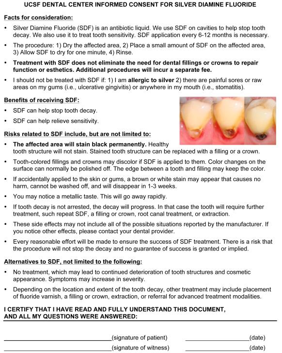
Because silver diamine fluoride is new in the U.S., it is important to communicate effectively. In the UCSF clinics, we are using a special consent form (Figure 4) as a way to inform patients, parents, and caregivers, and to standardize procedures because we have so many inexperienced student clinicians. All practices have established procedures for consent and an extra form may not be needed in the community. The normal elements of informed consent apply: we sought to ensure awareness of the expected change in color of the dentin as the decay arrests, likelihood of reapplication, and contraindications in the presence of silver allergy and stomatitis. Note the importance of distinguishing between allergy to nickel and other trace metals rather than silver allergy, which is rare. We used readability measurements to guide intelligibility, and included a progressively discoloring lesion to show stain of a lesion but not healthy enamel.
Figure 4.
UCSF special consent form.
Conclusion
Silver diamine fluoride is a safe, effective treatment for dental caries across the age spectrum. At UCSF it is indicated for patients with extreme caries risk, those who cannot tolerate conventional care, patients who must be stabilized so they can be restored over time, patients who are medically compromised or too frail to be treated conventionally, and those in disparity populations with little access to care.
Application twice-per-year outperforms all minimally invasive options including the atraumatic restorative technique – with which it is compatible but 20 times less expensive. It approaches the success of dental fillings after 2 or more years, and again, prevents future caries – while fillings do not. Silver diamine fluoride is more effective as a primary preventative than any other available material, with the exception of dental sealants which are >10 times more expensive and need to be monitored.
Saliva may play a role in caries arrest by silver diamine fluoride. Lower rates of arrest are seen in geriatric patients.38 Elders tend to have less abundant and less functional saliva, which generally explains their higher caries rate. In pediatric patients, higher rates of arrest are noted for buccal or lingual smooth surfaces, and anterior teeth.31 These surfaces bathe more directly in saliva than others. It is surprising that silver chloride is the main precipitant in treated dentin, as chloride is not a common component of dentin or silver diamine fluoride, so may come from the saliva.
Traditional approaches often provide only temporary benefit, given the highest rates of recurrent caries are in patients with the worst disease burden. The advent of a treatment for non-symptomatic caries not requiring general anesthesia or sedation addresses long-standing concerns about the expense, danger, and practical complexity of these services.
Experience suggests that dryness prior to application enhances effectiveness; good patient management is still profoundly relevant to the very young and otherwise challenged patients, though this 1 minute intervention is more tolerable than other options. Silver diamine fluoride can readily replace fluoride varnish for the prevention of caries in patients that have active caries. This as a powerful new tool in the fight against dental caries, particularly suited for those who suffer most from this disease.
Clinical evidence supports continued application 1-2 times per year until the tooth is restored or exfoliates, and otherwise perhaps indefinitely. Some treated lesions keep growing, particularly those in the inner third of dentin. It is unclear what will happen if treatment is stopped after 2-3 years and research is needed.
Acknowledgements
The UCSF Paradigm Shift Committee subcommittee on Silver Caries Arrest included Drs. Sean Mong, Spomenka Djordjevic, Paul Atkinson, George Taylor, Ling Zhan, Natalie Heaivilin, John Featherstone, Hellene Ellenikiotis, and Jeremy Horst. Thanks to Linda Milgrom for designing the PubMed search. Thanks to Chad Zillich for help with literature review. Thanks to study authors, particularly Drs. Edward Lo and Geoff Knight, for helpful discussions. JAH’s work on this manuscript was supported in part by NIH/NIDCR grant T32-DE007306.
References
- 1.Clarkson BH, Exterkate RAM. Noninvasive dentistry: a dream or reality? Caries Res. 2015;49(Suppl 1(1)):11–17. doi: 10.1159/000380887. [DOI] [PubMed] [Google Scholar]
- 2.Yamaga R, Yokomizo I. Arrestment of caries of deciduous teeth with diamine silver fluoride. Dental Outlook. 1969;33:1007–1013. [Google Scholar]
- 3.Rosenblatt A, Stamford TCM, Niederman R. Silver diamine fluoride: a caries “silver-fluoride bullet”. Journal of Dental Research. 2009;88(2):116–125. doi: 10.1177/0022034508329406. [DOI] [PubMed] [Google Scholar]
- 4.Mei ML, Chu CH, Lo ECM, Samaranayake LP. Fluoride and silver concentrations of silver diammine fluoride solutions for dental use. Int J Paediatr Dent. 2013;23(4):279–285. doi: 10.1111/ipd.12005. [DOI] [PubMed] [Google Scholar]
- 5.Mei ML, Ito L, Cao Y, Li QL, Lo ECM, Chu CH. Inhibitory effect of silver diamine fluoride on dentine demineralisation and collagen degradation. Journal of Dentistry. 2013;41(9):809–817. doi: 10.1016/j.jdent.2013.06.009. [DOI] [PubMed] [Google Scholar]
- 6.Hill TJ, Arnold FA. The effect of silver nitrate in the prevention of dental caries. I. The effect of silver nitrate upon the decalcification of enamel. Journal of Dental Research. 1937;16:23–28. [Google Scholar]
- 7.Castillo JL, Rivera S, Aparicio T, et al. The short-term effects of diammine silver fluoride on tooth sensitivity: a randomized controlled trial. Journal of Dental Research. 2011;90(2):203–208. doi: 10.1177/0022034510388516. [DOI] [PMC free article] [PubMed] [Google Scholar]
- 8.Craig GG, Knight GM, McIntyre JM. Clinical evaluation of diamine silver fluoride/potassium iodide as a dentine desensitizing agent. A pilot study. Australian Dental Journal. 2012;57(3):308–311. doi: 10.1111/j.1834-7819.2012.01700.x. [DOI] [PubMed] [Google Scholar]
- 9.Markowitz K, Pashley DH. Discovering new treatments for sensitive teeth: the long path from biology to therapy. J Oral Rehabil. 2008;35(4):300–315. doi: 10.1111/j.1365-2842.2007.01798.x. [DOI] [PubMed] [Google Scholar]
- 10.Featherstone JDB. The continuum of dental caries--evidence for a dynamic disease process. Journal of Dental Research. 2004;83(Spec No C):C39–C42. doi: 10.1177/154405910408301s08. [DOI] [PubMed] [Google Scholar]
- 11.Mei ML, Li QL, Chu CH, Yiu CKY, Lo ECM. The inhibitory effects of silver diamine fluoride at different concentrations on matrix metalloproteinases. Dental Materials. 2012;28(8):903–908. doi: 10.1016/j.dental.2012.04.011. [DOI] [PubMed] [Google Scholar]
- 12.Mei ML, Ito L, Cao Y, Li QL, Chu CH, Lo ECM. The inhibitory effects of silver diamine fluorides on cysteine cathepsins. Journal of Dentistry. 2014;42(3):329–335. doi: 10.1016/j.jdent.2013.11.018. [DOI] [PubMed] [Google Scholar]
- 13.Klasen HJ. A Historical Review of the Use of Silver in the Treatment of Burns. II. Renewed Interest for Silver. 2000:131–138. doi: 10.1016/s0305-4179(99)00116-3. [DOI] [PubMed] [Google Scholar]
- 14.Youravong N, Carlen A, Teanpaisan R, Dahlén G. Metal-ion susceptibility of oral bacterial species. Lett Appl Microbiol. 2011;53(3):324–328. doi: 10.1111/j.1472-765X.2011.03110.x. [DOI] [PubMed] [Google Scholar]
- 15.Hamama HH, Yiu CK, Burrow MF. Effect of silver diamine fluoride and potassium iodide on residual bacteria in dentinal tubules. Australian Dental Journal. 2015;60(1):80–87. doi: 10.1111/adj.12276. [DOI] [PubMed] [Google Scholar]
- 16.Suzuki T, Nishida M, Sobue S, Moriwaki Y. Effects of diammine silver fluoride on tooth enamel. J Osaka Univ Dent Sch. 1974;14:61–72. [PubMed] [Google Scholar]
- 17.Chu CH, Lo ECM. Microhardness of dentine in primary teeth after topical fluoride applications. Journal of Dentistry. 2008;36(6):387–391. doi: 10.1016/j.jdent.2008.02.013. [DOI] [PubMed] [Google Scholar]
- 18.ENGLANDER HR, JAMES VE, MASSLER M. Histologic effects of silver nitrate of human dentin and pulp. J Am Dent Assoc. 1958;57(5):621–630. doi: 10.14219/jada.archive.1958.0258. [DOI] [PubMed] [Google Scholar]
- 19.Mei ML, Ito L, Cao Y, Lo ECM, Li QL, Chu CH. An ex vivo study of arrested primary teeth caries with silver diamine fluoride therapy. Journal of Dentistry. 2014;42(4):395–402. doi: 10.1016/j.jdent.2013.12.007. [DOI] [PubMed] [Google Scholar]
- 20.Knight GM, McIntyre JM, Craig GG, Mulyani, Zilm PS, Gully NJ. Inability to form a biofilm of Streptococcus mutans on silver fluoride- and potassium iodide-treated demineralized dentin. Quintessence Int. 2009;40(2):155–161. [PubMed] [Google Scholar]
- 21.Knight GM, McIntyre JM, Craig GG, Mulyani, Zilm PS, Gully NJ. An in vitro model to measure the effect of a silver fluoride and potassium iodide treatment on the permeability of demineralized dentine to Streptococcus mutans. Australian Dental Journal. 2005;50(4):242–245. doi: 10.1111/j.1834-7819.2005.tb00367.x. [DOI] [PubMed] [Google Scholar]
- 22.Knight GM, McIntyre JM, Craig GG, Mulyani, Zilm PS, Gully NJ. Differences between normal and demineralized dentine pretreated with silver fluoride and potassium iodide after an in vitro challenge by Streptococcus mutans. Australian Dental Journal. 2007;52(1):16–21. doi: 10.1111/j.1834-7819.2007.tb00460.x. [DOI] [PubMed] [Google Scholar]
- 23.Wakshlak RB-K, Pedahzur R, Avnir D. Antibacterial activity of silver-killed bacteria: the “zombies” effect. Sci Rep. 2015;5:9555. doi: 10.1038/srep09555. [DOI] [PMC free article] [PubMed] [Google Scholar]
- 24.Duffin S. Back to the future: the medical management of caries introduction. J Calif Dent Assoc. 2012;40(11):852–858. [PubMed] [Google Scholar]
- 25.Rethman MP, Beltrán-Aguilar ED, Billings RJ, et al. Nonfluoride caries-preventive agents: executive summary of evidence-based clinical recommendations. J Am Dent Assoc. 2011;142(9):1065–1071. doi: 10.14219/jada.archive.2011.0329. [DOI] [PubMed] [Google Scholar]
- 26.Plemons JM, Al-Hashimi I, Marek CL. Managing xerostomia and salivary gland hypofunction. The Journal of the American Dental Association. 2014;145(8):867–873. doi: 10.14219/jada.2014.44. [DOI] [PubMed] [Google Scholar]
- 27.Stebbins EA. What value has argenti nitras as a therapeutic agent in dentistry? Int Dent J. 1891;12:661–670. [PMC free article] [PubMed] [Google Scholar]
- 28.PR H. A method of sterilizing and at the same time impregnating with a metal affected dentinal tissue. Dent Cosmos. 1917;59:891–904. [Google Scholar]
- 29.Chu CH, Lo ECM, Lin HC. Effectiveness of silver diamine fluoride and sodium fluoride varnish in arresting dentin caries in Chinese pre-school children. Journal of Dental Research. 2002;81(11):767–770. doi: 10.1177/0810767. [DOI] [PubMed] [Google Scholar]
- 30.Shah S, Bhaskar V, Venkataraghavan K, Choudhary P, Ganesh M, Trivedi K. Efficacy of silver diamine fluoride as an antibacterial as well as antiplaque agent compared to fluoride varnish and acidulated phosphate fluoride gel: an in vivo study. Indian J Dent Res. 2013;24(5):575–581. doi: 10.4103/0970-9290.123374. [DOI] [PubMed] [Google Scholar]
- 31.Zhi QH, Lo ECM, Lin HC. Randomized clinical trial on effectiveness of silver diamine fluoride and glass ionomer in arresting dentine caries in preschool children. Journal of Dentistry. 2012;40(11):962–967. doi: 10.1016/j.jdent.2012.08.002. [DOI] [PubMed] [Google Scholar]
- 32.Yee R, Holmgren C, Mulder J, Lama D, Walker D, van Palenstein Helderman W. Efficacy of silver diamine fluoride for Arresting Caries Treatment. Journal of Dental Research. 2009;88(7):644–647. doi: 10.1177/0022034509338671. [DOI] [PubMed] [Google Scholar]
- 33.Santos Dos VE, de Vasconcelos FMN, Ribeiro AG, Rosenblatt A. Paradigm shift in the effective treatment of caries in schoolchildren at risk. International Dental Journal. 2012;62(1):47–51. doi: 10.1111/j.1875-595X.2011.00088.x. [DOI] [PMC free article] [PubMed] [Google Scholar]
- 34.Lo EC, Chu CH, Lin HC. A community-based caries control program for pre-school children using topical fluorides: 18-month results. Journal of Dental Research. 2001;80(12):2071–2074. doi: 10.1177/00220345010800120901. [DOI] [PubMed] [Google Scholar]
- 35.Llodra JC, Rodriguez A, Ferrer B, Menardia V, Ramos T, Morato M. Efficacy of silver diamine fluoride for caries reduction in primary teeth and first permanent molars of schoolchildren: 36-month clinical trial. Journal of Dental Research. 2005;84(8):721–724. doi: 10.1177/154405910508400807. [DOI] [PubMed] [Google Scholar]
- 36.Green E. A clinical evaluation of two methods of caries prevention in newly-erupted first permanent molars. Australian Dental Journal. 1989;34(5):407–409. doi: 10.1111/j.1834-7819.1989.tb00696.x. [DOI] [PubMed] [Google Scholar]
- 37.Craig GG, Powell KR, Price CA. Clinical evaluation of a modified silver fluoride application technique designed to facilitate lesion assessment in outreach programs. BMC Oral Health. 2013;13(1):73. doi: 10.1186/1472-6831-13-73. [DOI] [PMC free article] [PubMed] [Google Scholar]
- 38.Zhang W, McGrath C, Lo ECM, Li JY. Silver diamine fluoride and education to prevent and arrest root caries among community-dwelling elders. Caries Res. 2013;47(4):284–290. doi: 10.1159/000346620. [DOI] [PubMed] [Google Scholar]
- 39.Liu BY, Lo ECM, Chu CH, Lin HC. Randomized trial on fluorides and sealants for fissure caries prevention. Journal of Dental Research. 2012;91(8):753–758. doi: 10.1177/0022034512452278. [DOI] [PubMed] [Google Scholar]
- 40.Tan HP, Lo ECM, Dyson JE, Luo Y, Corbet EF. A randomized trial on root caries prevention in elders. Journal of Dental Research. 2010;89(10):1086–1090. doi: 10.1177/0022034510375825. [DOI] [PubMed] [Google Scholar]
- 41.Monse B, Heinrich-Weltzien R, Mulder J, Holmgren C, van Palenstein Helderman WH. Caries preventive efficacy of silver diammine fluoride (SDF) and ART sealants in a school-based daily fluoride toothbrushing program in the Philippines. BMC Oral Health. 2012;12(1):52. doi: 10.1186/1472-6831-12-52. [DOI] [PMC free article] [PubMed] [Google Scholar]
- 42.Li R, Lo ECM, Chu CH, Liu BY. Preventing and arresting root caries through silver diammine fluoride applications. 2014 doi: 10.1016/j.jdent.2016.05.005. https://iadr.confex.com/iadr/14iags/webprogram/Paper189080.html. [DOI] [PubMed]
- 43.Duangthip D, Lo ECM, Chu CH. Arrest of dentin caries in preschool children by topical fluorides. 2014 https://iadr.confex.com/iadr/14iags/webprogram/Paper189031.html.
- 44.Frencken JE, Pilot T, Songpaisan Y, Phantumvanit P. Atraumatic Restorative Treatment (ART): Rationale, Technique, and Development. J Public Health Dent. 1996;56(3):135–140. doi: 10.1111/j.1752-7325.1996.tb02423.x. [DOI] [PubMed] [Google Scholar]
- 45.Innes NP, Evans DJ, Stirrups DR. The Hall Technique; a randomized controlled clinical trial of a novel method of managing carious primary molars in general dental practice: acceptability of the technique and outcomes at 23 months. BMC Oral Health. 2007;7(1):18. doi: 10.1186/1472-6831-7-18. [DOI] [PMC free article] [PubMed] [Google Scholar]
- 46.Vasquez E, Zegarra G, Chirinos E, et al. Short term serum pharmacokinetics of diammine silver fluoride after oral application. BMC Oral Health. 2012;12(1):60. doi: 10.1186/1472-6831-12-60. [DOI] [PMC free article] [PubMed] [Google Scholar]
- 47.Chu CH, Lo ECM. Promoting caries arrest in children with silver diamine fluoride: a review. Oral Health Prev Dent. 2008;6(4):315–321. [PubMed] [Google Scholar]
- 48.Liu Y, Zhai W, Du F. Clinical observation of treatment of tooth hypersensitiveness with silver ammonia fluoride and potassium nitrate solution. Zhonghua Kou Qiang Yi Xue Za Zhi. 1995;30(6):352–354. [PubMed] [Google Scholar]
- 49.Yamaga R, Nishino M, Yoshida S, Yokomizo I. Diammine silver fluoride and its clinical application. J Osaka Univ Dent Sch. 1972;(12):1–20. [PubMed] [Google Scholar]
- 50.Milgrom P, Taves DM, Kim AS, Watson GE, Horst JA. Pharmacokinetics of fluoride in toddlers after application of 5% sodium fluoride dental varnish. Pediatrics. 2014;134(3):e870–e874. doi: 10.1542/peds.2013-3501. [DOI] [PMC free article] [PubMed] [Google Scholar]
- 51.Goldberg ME, Cantillo J, Larijani GE, Torjman M, Vekeman D, Schieren H. Sevoflurane versus isoflurane for maintenance of anesthesia: are serum inorganic fluoride ion concentrations of concern? Anesth Analg. 1996;82(6):1268–1272. doi: 10.1097/00000539-199606000-00029. [DOI] [PubMed] [Google Scholar]
- 52.Gotjamanos T, Afonso F. Unacceptably high levels of fluoride in commercial preparations of silver fluoride. Australian Dental Journal. 1997;42(1):52–53. doi: 10.1111/j.1834-7819.1997.tb00097.x. [DOI] [PubMed] [Google Scholar]
- 53.Gotjamanos T, Ma P. Potential of 4 per cent silver fluoride to induce fluorosis in rats: clinical implications. Australian Dental Journal. 2000;45(3):187–192. doi: 10.1111/j.1834-7819.2000.tb00555.x. [DOI] [PubMed] [Google Scholar]
- 54.Neesham DC. Fluoride concentration in AgF and dental fluorosis. Australian Dental Journal. 1997;42(4):268–269. [PubMed] [Google Scholar]
- 55.Quock RL, Barros JA, Yang SW, Patel SA. Effect of Silver Diamine Fluoride on Microtensile Bond Strength to Dentin. Operative Dentistry. 2012;37(6):610–616. doi: 10.2341/11-344-L. [DOI] [PubMed] [Google Scholar]
- 56.Knight GM, McIntyre JM, Mulyani The effect of silver fluoride and potassium iodide on the bond strength of auto cure glass ionomer cement to dentine. Australian Dental Journal. 2006;51(1):42–45. doi: 10.1111/j.1834-7819.2006.tb00399.x. [DOI] [PubMed] [Google Scholar]
- 57.Yamaga M, Koide T, Hieda T. Adhesiveness of glass ionomer cement containing tannin-fluoride preparation (HY agent) to dentin--an evaluation of adding various ratios of HY agent and combination with application diammine silver fluoride. Dent Mater J. 1993;12(1):36–44. doi: 10.4012/dmj.12.36. [DOI] [PubMed] [Google Scholar]
- 58.Soeno K, Taira Y, Matsumura H, Atsuta M. Effect of desensitizers on bond strength of adhesive luting agents to dentin. J Oral Rehabil. 2001;28(12):1122–1128. doi: 10.1046/j.1365-2842.2001.00756.x. [DOI] [PubMed] [Google Scholar]
- 59.Centers for Disease Control and Prevention Long-Term Care Services in the United States: 2013 Overview, table 4. http://www.cdc.gov/nchs/data/nsltcp/long_term_care_services_2013.pdf.
- 60.U.S. Department of Health and Human Services [Accessed June 5, 2015];Integration of Oral Health and Primary Care Practice. http://www.hrsa.gov/publichealth/clinical/oralhealth/
- 61.Featherstone JDB, Adair SM, Anderson MH, et al. Caries management by risk assessment: consensus statement, April 2002. J Calif Dent Assoc. 2003;31:257–269. [PubMed] [Google Scholar]


