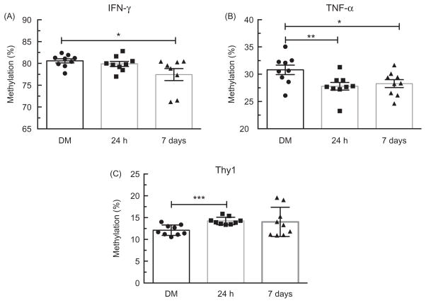
Figure 6.
Promoter methylation status in lung tissue of C57BL/6 mice. Change in percent methylation 24 h and 7 days after MWCNT exposure for three genes. (A) IFN-γ at 7 days post-MWCNT exposure was significantly hypomethylated (p<0.05) compared to the DM controls. (B) TNF-α promoter was statistically hypomethylaed at 24 h (p<0.01) and 7 days (p<0.05) post-MWCNT exposure compared to the DM controls. (C) Thy-1 was significantly hypermethylated within the promoter region 24 h post-MWCNT exposure compared with the DM controls (p<0.05). Data expressed as mean ± SEM percent methylation. Asterisks indicate significance at **p<0.01, *p<0.05 compared to the DM controls. n = 9 per condition.

