Abstract
The artery of Percheron (AOP) is a rare vascular variant in which a single dominant thalamoperforating artery arises from the P1 segment and bifurcates to supply both paramedian thalami. Occlusion of this uncommon vessel results in a characteristic pattern of bilateral paramedian thalamic infarcts with or without mesencephalic infarctions. We report a case of a 37-year-old man with acute bilateral thalamic infarcts. The scans revealed symmetric bilateral hyperintense paramedian thalamic lesions consistent with an acute ischemic event. The posterior circulation was patent including the tip of the basilar artery and both posterior cerebral arteries, making the case compatible with occlusion of the AOP. This type of infarct is associated with embolic phenomena, and further evaluation revealed a patent foramen ovale as the source of emboli in the cerebrovascular circulation. The occlusion of the AOP is a rare cause of coma in young patients, and early recognition of this rare disease entity may lead to more favorable outcomes.
Keywords: Artery of Percheron, bilateral thalamic infarcts, patent foramen ovale
INTRODUCTION
The artery of Percheron (AOP) is a rare vascular variant in which a single dominant thalamoperforating artery arises from the P1 segment and bifurcates to supply both paramedian thalami.[1] Occlusion of this uncommon vessel results in a characteristic pattern of bilateral paramedian thalamic infarcts with or without mesencephalic infarctions. This type of infarct might be associated with embolic phenomena; nevertheless, all the workup for embolic cause was negative including cardiac arrhythmias, and the finding of the patent foramen ovale (PFO) was incidental without an evidence of a source of paradoxical embolization such as venous thromboembolism (VTE), also the possibility of underlying thrombophilia was excluded in regard of arterial thrombosis as a possible underlying etiology.[2,3]
CASE REPORT
A 37-year-old man with no medical history presented to the Emergency Department (ED) after being found unresponsive in bed. He was last seen normal approximately 6 h before presentation. There was no recent history of fever, headache, seizure or trauma, and no known toxic substance exposure. On arrival at the ED, his vital signs were normal. On neurological examination, he was comatose with a Glasgow Coma Scale (GCS) of 6. His neck was supple, and his pupils were anisocoric, with a 4 mm right pupil and a 6 mm left pupil. The pupillary light reflex was absent in both eyes. The vertical oculocephalic reflex was absent, and his left eye did not show adduction with turning of the head to the left. His limbs moved in response to painful stimuli. The rest of his examination was unremarkable.
Laboratory data including blood glucose, complete blood count, electrolytes, liver and renal function tests, lipid profile, thyroid function tests, arterial blood gas, and ammonia were unremarkable. Urine drug screen and alcohol level were negative. Electrocardiogram showed a normal sinus rhythm, and the 72 h telemetry monitoring did not reveal any arrhythmias. Anti-beta-2 glycoprotein I antibodies, anticardiolipin (immunoglobulin G and M), antiphospholipid, lupus anticoagulant, activated partial thromboplastin time, prothrombin gene mutation, venereal disease research laboratory, Factor V Leiden, activated protein C, and S tests were all negative.
Emergent head computed tomography (CT) revealed chronic/subacute left posterior frontal focal infarct and small right occipital lobe infarct, which did not fully explain his findings. Unseen acute ischemic stroke (AIS) was suspected. Oral aspirin was administered, and he was admitted to the Intensive Care Unit. The patient was intubated with a GCS of 7/15 for airway protection, improved after 2 days, and was extubated.
Magnetic resonance imaging (MRI) performed on the next day demonstrated areas of abnormal signal with mildly restricted diffusion within the medial thalami bilaterally and mid brain suggestive of acute/subacute infarcts, secondary to AIS in the AOP territory. There were additional small acute/subacute infarcts in both cerebellar hemispheres as well as chronic infarcts in the right cerebellum and left frontal lobe [Figures 1–5].
Figure 1.
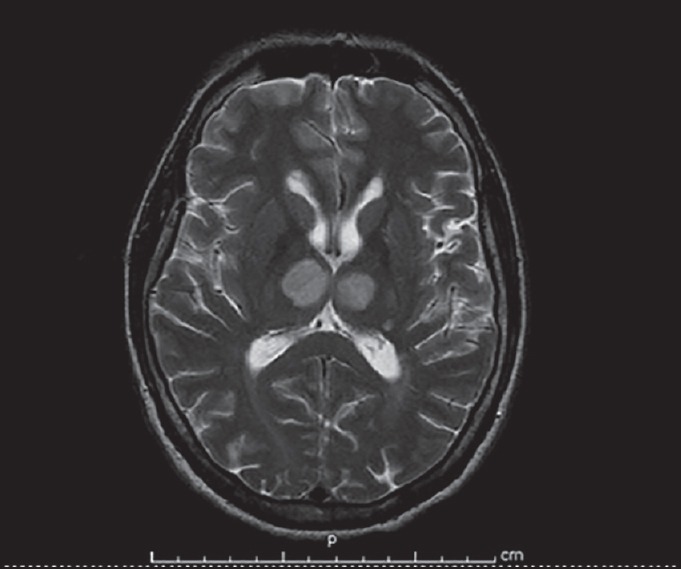
Magnetic resonance image revealing bilateral thalamic infarct
Figure 5.
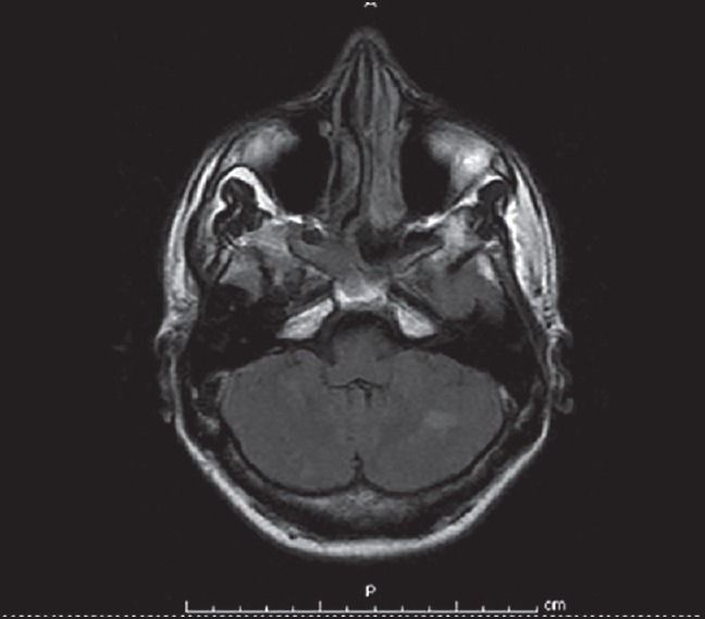
Magnetic resonance imaging flair image showing bilateral cerebellar infarct
Figure 2.
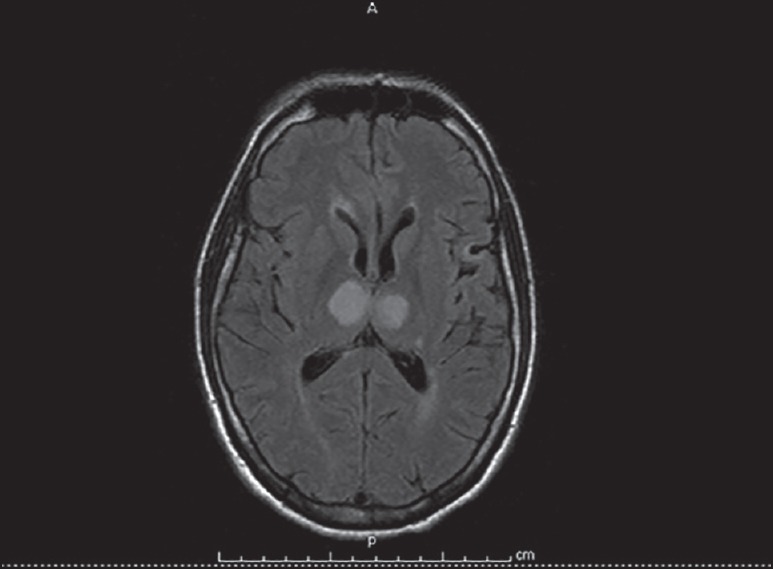
MRI revealed acute/subacute infarcts in both cerebellar hemispheres
Figure 3.
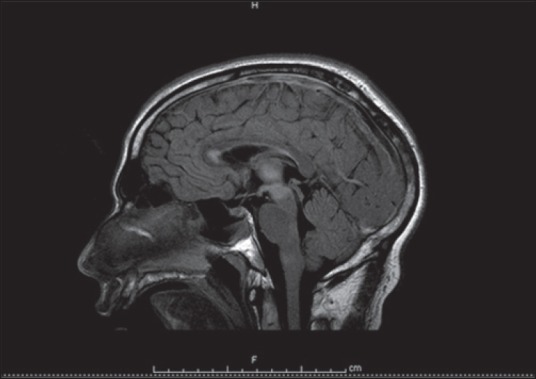
MRI revealed acute/subacute infarcts in the right cerebellum and left frontal lobe
Figure 4.
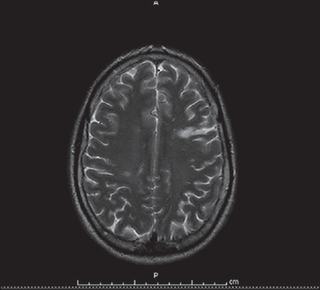
Magnetic resonance imaging T2 image showing left frontal infarct
MR angiography demonstrated patent posterior circulation including the tip of the basilar artery and both posterior cerebral arteries [Figures 6 and 7]. Further evaluation with an aim of defining the etiology of multiple strokes in a young patient without risk factors revealed a PFO on transesophageal echocardiography with spontaneous passage of contrast bubbles from the right auricle to the left cavities, though VTE was excluded as a possible source of paradoxical embolization, still there might be unrevealed underlying venous thrombosis (VT). Thus, the patient was started on anticoagulation despite the midbrain involvement.
Figure 6.
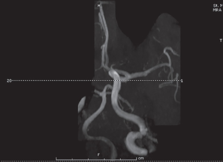
Magnetic resonance angiography showing patent posterior circulation
Figure 7.
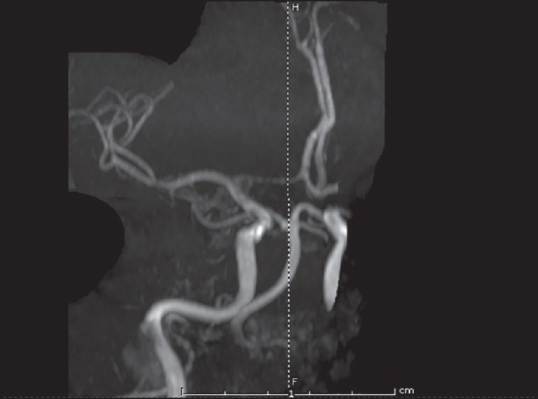
MR angiography revealed patency of both the tip of the basilar artery and both posterior cerebral arteries
During his stay, he gradually regained consciousness during the hospitalization days with GCS of 10/15. Upon discharge to acute care facility, his eye signs remained along with persistent memory impairment.
DISCUSSION
The thalamus has four major vascular territories, which are anterior, paramedian, inferolateral, and posterior. AOP is an uncommon anatomic variant of the paramedian arteries in which a single dominant thalamoperforating artery supplies bilateral paramedian thalami with variable contribution to the rostral midbrain. Occlusion of this artery results in symmetrical bilateral paramedian thalamic infarcts with or without midbrain involvement.[3] This may result in an acutely ill and severely impaired patient, having problems of disorientation, confusion, hypersomnolence, deep coma, akinetic mutism, and severe memory impairment with perseveration, confabulation, and abnormal eye movements.[4]
Our case illustrates the importance of considering ischemic stroke in the AOP territory in the differential diagnosis of acute disturbance of consciousness in young patients. The clinical presentation can mimic nonconvulsive status epilepticus, subarachnoid hemorrhage, metabolic or toxic encephalopathy, and encephalitis. The low sensitivity of CT makes AOP infarction diagnosis difficult and diagnosis is usually delayed. Diffusion-weighted MRI is the imaging modality of choice.[5] It is important to note, AOP is rarely visualized on MR angiography, and lack of visualization does not exclude its presence.[5,6] AOP prevalence remains unknown. In a recent study, AOP infarction was found in 0.4% patients with a first-ever stroke in their stroke registry.[6] Small artery disease and cardioembolism were the most frequent stroke mechanisms.[6] These patients must be differentiated from those with “top of the basilar artery” syndrome.[3,4,5,6,7] However, “top of the basilar artery” syndrome also tends to involve the superior cerebellar artery and posterior cerebral artery territories. The lack of associated lesions and no thrombus seen on MR angiogram in our patient excluded this diagnosis. Deep cerebral VT (DCVT) may also be confused with AOP infarction because bilateral thalamic infarcts can also result from DCVT.[7,8] However, DCVT produces simultaneous bilateral thalamic and basal ganglia lesions on MRI that do not respect typical arterial delineations. In addition, the MRI pattern usually suggests vasogenic edema rather than arterial infarcts; this is discordant with our patient's findings. Other differential diagnoses considered in our case were Wernicke's encephalopathy, osmotic demyelination, and Japanese encephalitis. These diagnoses were excluded according to clinical characteristics and imaging features.[8] Successful tissue plasminogen activator therapy for AOP occlusion is reported in literature, but our patient was outside the treatment time window on initial presentation.[9,10] Li et al. suggested that patients with AOP occlusion without mesencephalic involvement should receive long-term anticoagulant therapy and rehabilitation. These patients also have a better prognosis with many having full or partial recovery over a 6 months period.[11] Because of the rare occurrence of AOP occlusion, further clinical studies are needed to identify optimal treatment options and prognosis.
CONCLUSION
Occlusion of the AOP is a rare cause of coma in young patients. Diffusion-weighted MRI is the imaging modality of choice for early diagnosis and should not be delayed for altered mentation, which is a hallmark of this condition since early recognition of AOP occlusion may lead to more favorable outcomes.
Financial support and sponsorship
Nil.
Conflicts of interest
There are no conflicts of interest.
REFERENCES
- 1.López-Serna R, González-Carmona P, López-Martínez M. Bilateral thalamic stroke due to occlusion of the artery of Percheron in a patient with patent foramen ovale: A case report. J Med Case Rep. 2009;3:7392. doi: 10.4076/1752-1947-3-7392. [DOI] [PMC free article] [PubMed] [Google Scholar]
- 2.Jiménez Caballero PE. Bilateral paramedian thalamic artery infarcts: Report of 10 cases. J Stroke Cerebrovasc Dis. 2010;19:283–9. doi: 10.1016/j.jstrokecerebrovasdis.2009.07.003. [DOI] [PubMed] [Google Scholar]
- 3.Lazzaro NA, Wright B, Castillo M, Fischbein NJ, Glastonbury CM, Hildenbrand PG, et al. Artery of Percheron infarction: Imaging patterns and clinical spectrum. AJNR Am J Neuroradiol. 2010;31:1283–9. doi: 10.3174/ajnr.A2044. [DOI] [PMC free article] [PubMed] [Google Scholar]
- 4.Reilly M, Connolly S, Stack J, Martin EA, Hutchinson M. Bilateral paramedian thalamic infarction: A distinct but poorly recognized stroke syndrome. Q J Med. 1992;82:63–70. [PubMed] [Google Scholar]
- 5.Matheus MG, Castillo M. Imaging of acute bilateral paramedian thalamic and mesencephalic infarcts. AJNR Am J Neuroradiol. 2003;24:2005–8. [PMC free article] [PubMed] [Google Scholar]
- 6.Arauz A, Patiño-Rodríguez HM, Vargas-González JC, Arguelles-Morales N, Silos H, Ruiz-Franco A, et al. Clinical spectrum of artery of Percheron infarct: Clinical-radiological correlations. J Stroke Cerebrovasc Dis. 2014;23:1083–8. doi: 10.1016/j.jstrokecerebrovasdis.2013.09.011. [DOI] [PubMed] [Google Scholar]
- 7.Ameridou I, Spilioti M, Amoiridis G. Bithalamic infarcts: Embolism of the top of basilar artery or deep cerebral venous thrombosis? Clin Neurol Neurosurg. 2004;106:345–7. doi: 10.1016/j.clineuro.2004.01.004. [DOI] [PubMed] [Google Scholar]
- 8.Brami-Zylberberg F, Méary E, Oppenheim C, Gobin-Metteil MP, Delvat D, De Montauzan-Rivière I, et al. Abnormalities of the basal ganglia and thalami in adults. J Radiol. 2005;86:281–93. doi: 10.1016/s0221-0363(05)81357-5. [DOI] [PubMed] [Google Scholar]
- 9.Kostanian V, Cramer SC. Artery of Percheron thrombolysis. AJNR Am J Neuroradiol. 2007;28:870–1. [PMC free article] [PubMed] [Google Scholar]
- 10.Cao W, Dong Q, Li L, Dong Y. Bilateral thalamic infarction and DSA demonstrated AOP after thrombosis. Acta radiologica short reports. 2012;1:5. doi: 10.1258/arsr.2012.110004. [DOI] [PMC free article] [PubMed] [Google Scholar]
- 11.Li X, Agarwal N, Hansberry DR, Prestigiacomo CJ, Gandhi CD. Contemporary therapeutic strategies for occlusion of the artery of Percheron: A review of the literature. J Neurointerv Surg. 2015;7:95–8. doi: 10.1136/neurintsurg-2013-010913. [DOI] [PubMed] [Google Scholar]


