

Abstract
Rhinosporidiosis is a chronic localized granulomatous disease caused by Rhinosporidium seeberi, an aquatic protistan parasite belonging to a clade, Mesomycetozoea. Infestation of Rhinosporidiosis to the eye and adnexa is termed oculosporidiosis, in such cases, conjunctival mucosa is mostly involved; however in our case, it involved only the lacrimal sac and deeper periorbital tissue and presented as a case of orbital swelling. Surgical excision of the lesion was done, postoperatively dapsone therapy was given for 6 months, and the patient responded very well with no recurrence till date.
Keywords: Dapsone, lacrimal sac, oculosporidiosis
INTRODUCTION
Rhinosporidiosis is a chronic localized granulomatous disease caused by Rhinosporidium seeberi, an aquatic protistan parasite belonging to a new clade, Mesomycetozoea.[1] The disease is endemic in different parts of India, especially coastal areas.[2] Most common site of involvement is nasal mucosa, followed by ocular involvement; other natural orifices such as penis, urethra, anus, vagina, skin[3] may also be involved; few cases of disseminated rhinosporidiosis have also been reported.[4,5] Rhinosporidiosis of the eye and adnexa is termed oculosporidiosis. Here, we present an interesting case of oculosporidiosis, how the patient was diagnosed, treated; along with the recent literature review.
CASE REPORT
A 16-year-old female patient presented with a swelling over left lower orbital area which was present for last 3 years. On examination, the swelling was pinkish in color with a deep “T” shaped scar over it due to previous intervention [Figure 1]. On palpation, the swelling was soft and nontender, seemed to be fixed with the deeper tissue.
Figure 1.

Patient on initial presentation with lower orbital swelling with a scar on it
Two years ago, she was treated as a case of dacryocystitis with osteomyelitis of lower orbital rim (left maxillary bone) for which incision and drainage was done by an ENT surgeon in her local area; it took around one month for the wound to heal. Though there was an initial reduction of the size of the swelling for few months, the swelling started to grow again for last 8 months, for which she came to us.
We then referred the patient to the department of ophthalmology for detailed cheek up of the eye and lacrimal system. On examination, her visual acuity was found to be normal; conjunctiva was normal; syringing through both puncta of her left upper and lower eyelid proved the patency of lacrimal system.
Contrast-enhanced computed tomography scan of paranasal sinuses (PNS) and orbit showed an ill-defined, enhancing, soft tissue density mass in the left lacrimal sac region extending up to superior aspect of right maxillary antrum; disruption of medial wall of left orbit was noted; adjacent fat planes were blurred [Figure 2]. The impression of the radiologist was an inflammatory lesion of the lacrimal sac with pyocele. Her routine blood investigation along with erythrocyte sedimentation rate was normal. The patient was tested negative for HIV, and she did not have any other immunodeficiency. Her X-ray chest was done for routine preanesthesia check-up, which was found to be normal.
Figure 2.

Contrast-enhanced computed tomography showing the extent of the swelling
Hence, with a preoperative diagnosis of orbital swelling due to lacrimal sac pyocele, we decided to explore the area surgically in presence of an ophthalmologist. The operation was done under general anesthesia. An incision was placed over the old scar extending medially up to the medial canthal ligament. On exploration, we found an ill-defined soft tissue mass adherent with the surrounding soft tissues; meticulous dissection was done and the swelling was found to be extending deep into the orbit. As the lacrimal sac was intimately adherent with it, it was removed. There were also some erosion of medial part the bony orbit [Figure 3]. Extra-ocular muscles were not involved and intraoperative tests were done to check their functions. Electro-cauterization of the wall of the cavity was done and gelfoam was packed into the cavity to achieve hemostasis; the wound was then closed.
Figure 3.
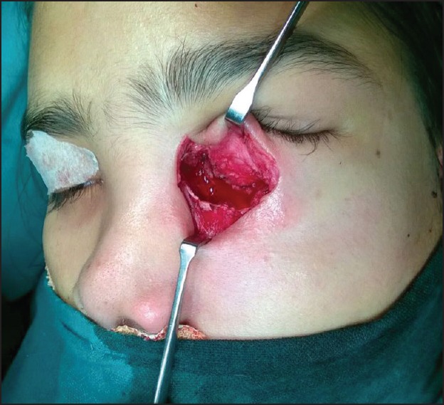
Intraoperative picture showing the cavity after removal of the mass
Histopathological examination of the specimen was done. Sections from the orbital cyst and lacrimal sac showed tissue lined partially by stratified squamous epithelium and partially by columnar epithelium. Sub-epithelium showed sporangia with endospores. Surrounding tissues showed chronic inflammatory cell infiltrate [Figure 4]. Thus, a diagnosis of orbital rhinosporidiosis was made.
Figure 4.

Histopathology showing the typical sporangia containing numerous spores
The patient made a good recovery and was discharged on the 5th postoperative day on oral antimicrobial therapy (dapsone 100 mg/day, for 6 days a week, for 6 months). Before discharge, a through ENT cheek-up was done with nasal endoscope and fiberoptic laryngoscope to look for any such lesion in the upper respiratory passage, but no such lesion indicating infestation by Rhinosporidium in that area was found. There was no sign of recurrence in last 6 months follow-up [Figure 5].
Figure 5.

Patient on 6 months follow-up
DISCUSSION
Oculosporidiosis is found among 10-15% of cases of rhinosporidiosis, and among oculosporidiosis, conjunctiva is the most commonly affected tissue (77.6%), next is lacrimal sac (26%) with or without conjunctiva involvement. Clinical features of oculosporidiosis of the lacrimal sac were well described by Kuriakose as it may present with a swelling on the lower eyelid; which is painless; soft and fluctuating to the touch; there may be history of bleeding from the nose; and the lacrimal passage is seldom completely obstructed.[6]
In our case, though there was no bleeding from nose, other features were present. But still we missed the diagnosis because isolated oculosporidiosis of the lacrimal sac is very rare,[7] so it becomes very difficult to suspect an isolated lacrimal sac swelling to be due to infestation by Rhinosporidium. Such also happened to Mukherjee et al.[8] They described a patient with recurrent painful swelling of right lower eyelid, who was diagnosed elsewhere as a case of chronic dacryocystitis and they diagnosed it as a case of idiopathic orbital inflammatory disease. Final diagnosis was made only after tissue biopsy from the lacrimal sac area during aconchoplasty by the otolaryngologist that the lacrimal sac swelling was due to Rhinosporidium infestation.
A computed tomography scan of the orbit and PNS may show an enlarged lacrimal sac, along with polyps inside the air sinuses, thus helping to suspect rhinosporidiosis. However, in our case the sinuses were normal; only the lacrimal sac was enlarged, giving enhancement same as abscess. Thus she was diagnosed as a case of lacrimal sac pyocele. However, in suspected cases confirmation of this disease can be done by histopathological examination, which shows characteristic features as numerous sporocysts in various stages of maturation.
Management of lacrimal oculosporidiosis is primarily surgical excision;[4] but there is a very high chance of recurrence as reported by two large case series.[2,6] Ghorpade et al.[9] and Sood and Badhu[10] suspect that the recurrence may be due to incomplete removal of the infested sac and they recommended more aggressive approach that includes complete and meticulous surgical excision with electric cautery[10,11] to ensure complete eradication of the disease.
During surgery, dissection of involved lacrimal sac becomes very difficult, because of the risk of severe bleeding,[2] in our case also we experienced the same and the bleeding was controlled by gel foam application. After excision, copious irrigation with 5% providine iodine solution and 5 mg/ml amphotericin-B have also been tried with good results.[9] However, as our case was not suspected or diagnosed as a case of oculosporidiosis, we have not done any irrigation during surgery.
Postoperatively antimicrobial dapsone therapy as an additional measure to reduce the possibility of recurrence as it has been found to arrest the maturation of spores and promote fibrosis in the stroma;[9,10] though some study suggests that dapsone therapy may not be very effective.[12,13]
CONCLUSION
Isolated oculosporidiosis involving the lacrimal sac and presenting as an orbital swelling is a rare clinical finding, which can initially be misdiagnosed, like in this case it was wrongly diagnosed as a pyocele. The lesion, in such cases, must be surgically excised. Postoperatively dapsone therapy may be an additional measure to reduce the possibility of recurrence.
Declaration of patient consent
The authors certify that they have obtained all appropriate patient consent forms. In the form the patient(s) has/have given his/her/their consent for his/her/their images and other clinical information to be reported in the journal. The patients understand that their names and initials will not be published and due efforts will be made to conceal their identity, but anonymity cannot be guaranteed.
Financial support and sponsorship
Nil.
Conflicts of interest
There are no conflicts of interest.
REFERENCES
- 1.Herr RA, Ajello L, Taylor JW, Arseculeratne SN, Mendoza L. Phylogenetic analysis of Rhinosporidium seeberi's 18S small-subunit ribosomal DNA groups this pathogen among members of the protoctistan Mesomycetozoa clade. J Clin Microbiol. 1999;37:2750–4. doi: 10.1128/jcm.37.9.2750-2754.1999. [DOI] [PMC free article] [PubMed] [Google Scholar]
- 2.Chowdhury RK, Behera S, Bhuyan D, Das G. Oculosporidiosis in a tertiary care hospital of western Orissa, India: A case series. Indian J Ophthalmol. 2007;55:299–301. doi: 10.4103/0301-4738.33045. [DOI] [PubMed] [Google Scholar]
- 3.Ahluwalia KB, Maheshwari N, Deka RC. Rhinosporidiosis: A study that resolves etiologic controversies. Am J Rhinol. 1997;11:479–83. doi: 10.2500/105065897780914938. [DOI] [PubMed] [Google Scholar]
- 4.Amritanand R, Nithyananth M, Cherian VM, Venkatesh K, Shah A. Disseminated rhinosporidiosis destroying the talus: A case report. J Orthop Surg (Hong Kong) 2008;16:99–101. doi: 10.1177/230949900801600123. [DOI] [PubMed] [Google Scholar]
- 5.Sood N, Agarwal MC, Gugnani HC. Ocular rhinosporidiosis: A case report from Delhi. J Infect Dev Ctries. 2012;6:825–7. doi: 10.3855/jidc.2397. [DOI] [PubMed] [Google Scholar]
- 6.Kuriakose ET. Oculosporidiosis: Rhinosporidiosis of the eye. Br J Ophthalmol. 1963;47:346–9. doi: 10.1136/bjo.47.6.346. [DOI] [PMC free article] [PubMed] [Google Scholar]
- 7.Shrestha SP, Hennig A, Parija SC. Prevalence of rhinosporidiosis of the eye and its adnexa in Nepal. Am J Trop Med Hyg. 1998;59:231–4. doi: 10.4269/ajtmh.1998.59.231. [DOI] [PubMed] [Google Scholar]
- 8.Mukherjee B, Mohan A, Sumathi V, Biswas J. Infestation of the lacrimal sac by Rhinosporidium seeberi: A clinicopathological case report. Indian J Ophthalmol. 2013;61:588–90. doi: 10.4103/0301-4738.121084. [DOI] [PMC free article] [PubMed] [Google Scholar]
- 9.Ghorpade A, Gurumurthy J, Banerjee PK, Banerjee AK, Bhalla M, Ravindranath M. Oculosporidiosis presenting as an under-eye swelling. Indian J Dermatol Venereol Leprol. 2007;73:196–7. doi: 10.4103/0378-6323.32749. [DOI] [PubMed] [Google Scholar]
- 10.Sood A, Badhu B. Oculosporidiosis in a tertiary care hospital of Western Orissa, India. Indian J Ophthalmol. 2008;56:165. doi: 10.4103/0301-4738.39130. [DOI] [PMC free article] [PubMed] [Google Scholar]
- 11.John SS, Mohandas SG. Conjunctival oculosporidiosis with scleral thinning and staphyloma formation. Indian J Ophthalmol. 2005;53:272–4. doi: 10.4103/0301-4738.18912. [DOI] [PubMed] [Google Scholar]
- 12.Job A, Venkateswaran S, Mathan M, Krishnaswami H, Raman R. Medical therapy of rhinosporidiosis with dapsone. J Laryngol Otol. 1993;107:809–12. doi: 10.1017/s002221510012448x. [DOI] [PubMed] [Google Scholar]
- 13.Venkateswaran S, Date A, Job A, Mathan M. Light and electron microscopic findings in rhinosporidiosis after dapsone therapy. Trop Med Int Health. 1997;2:1128–32. doi: 10.1046/j.1365-3156.1997.d01-212.x. [DOI] [PubMed] [Google Scholar]