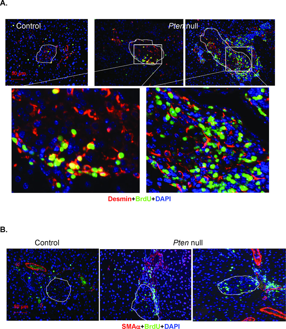
Figure 5. PTEN loss leads to expansion of surrounding mesenchymes and pancreatic stellate cells.
(A) STZ-treated control and Pten null pancreas sections were costained with Desmin (red) and BrdU (green). Bottom row, the magnified images of the boxed areas. (B) STZ-treated control and Pten null pancreas sections were costained with smooth muscle actin α (SMAα, red) and BrdU (green). Islets are designated in dashed circles. Bar, 50µM. Blue, DAPI.

