Abstract
Therapy for large symptomatic keloids is often plagued with complicated reconstruction manner and recurrence. This article reports a rare treatment combination for a chest keloid with internal mammary artery perforator flap reconstruction and radiation therapy. We excised the keloid and covered the defect with an internal mammary artery perforator flap. Immediate electron-beam irradiation therapy was applied on the second postoperative day. There was no sign of recurrence over the follow-up period of 18 months. The combination of internal mammary artery perforator flap and immediate radiation therapy is useful when faced with chest keloids of similar magnitude and intractability.
Keywords: Keloid, Perforator Flap, Radiotherapy
INTRODUCTION
Keloids are common, benign, hyperproliferative growths of dermal collagen of unknown etiology. Due to pain, hyperesthesia, pruritus, and significant psychosocial impact on quality of life, strategic approaches should be taken in keloid management. Many treatment modalities, including surgery, intralesional corticosteroid injection, pressure therapy, radiation, cryotherapy, silicone gel application, lasers, photodynamic therapy, have yielded limited success for large keloids.1 The lesions are frequently located at areas of stretching tension such as the anterior chest, shoulder and upper back. According to previous studies, the presence of tension seems to be one of the determinant factors for keloid formation.2 Here we relate a case of chest keloid, involving the combined treatments of internal mammary artery perforator (IMAP) flap reconstruction and radiation therapy. Recommendation of their use is underpinned by the core concepts of tension-free reconstruction manner and rational multimodality therapies focused on prevention of recurrence.
CASE REPORT
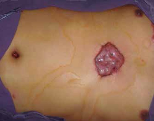
A 45-year-old man was diagnosed with a recurrent major keloid over his chest, which he had had for over 13 years. Past history consisted of repeated failed excisions and intralesional steroid injections. The patient suffered from localized pain and itching as well as progressive enlargement of the keloid. On physical examination, a large 6(W) (×) 8(L) (×) 1.5cm (thick) epidermal mass was seen over the midline of his chest below the cartilago ensiformis (Figure 1). The patient was concerned about the risk of recurrence. Therefore, a radical excision with flap coverage was planned, followed by electron-beam irradiation therapy.
Figure 1.

Preoperative view of reluctant chest keloid
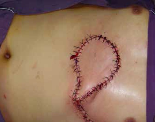
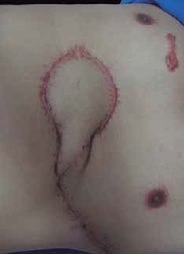
The patient underwent complete resection of the keloid (Figure 2). An IMAP flap of 9×7cm skin dimension was harvested on the lateral side of his chest. The flap was based on the perforators in the fifth intercostal space and rotated 180°along the vascular axis to the chest wall defect. After suturing the flap completely without tension, a percutaneous drain was placed. The donor site was closed directly, which releasing tension by subcutaneous and fascial sutures (Figure 3).Histological analysis revealed a typical keloid feature.
Figure 2.

IMAP Flap design during the excision
Figure 3.

IMAP Flap was rotated 180° with primary closure of the donor site
To achieve tight control in preventing recurrence, our patient initiated adjuvant electron-beam irradiation therapy on the second postoperative day. He received 13 Gy over four fractions in total. After being discharged, the patient continued to heal well and the flap showed stable circulation without limited necrosis or other conditions. At his last follow-up (18 months later), he was asymptomatic and demonstrated no sign of keloids at the donor or recipient site (Figure 4). The patient was highly satisfied with his result, and was rated "good" by the surgeons.
Figure 4.

Follow up after postoperative radiotherapy
DISCUSSION
This patient is one of few reported cases of flaps for keloid reconstruction in the English literature. Treatment of a large sternum keloid was reported, associated with bilateral mammaplasty, from which the remaining skin was used for the sternum defect.3 There are two recorded cases of microvascular free tissue transfer for keloid reconstruction using transverse rectus abdominis myocutaneous flap and free myocutaneous left lateral arm flap.4Furthermore, clinical trials revealed that skin flaps, combined with postoperative radiotherapy, can be more effective in keloid treatment.5
As a general rule, closing the wound should be performed with minimal tension, therefore, Z-plasties, W-plasties and advancement local flaps may indeed be indicated.5In our case, the IMAP flap was chosen for his chest keloid. IMAP flap is an island flap with perforators from the internal mammary artery. Previous studies have shown that this flap is a reliable and less invasive option for localized reconstruction of the chest wall.6 Its advantages lie in the flap's mobility range (90-180° to reach the opposite chest wall), minimal morbidity at the donor site, the cosmetic match to the reception site, as well as its relatively thin tissue.
Keloid resection and defect reconstruction were only the first step. The second challenge was postoperative keloid prevention. Keloids in the sternum area are frequently complicated by high recurrence. Surgical excision alone leads to recurrence rates ranging from 80% to 100%.1 The use of radiotherapy as an adjunct treatment has shown success rates of between 52.4% and 91%.7 It has been suggested that radiation may inhibit fibroblasts and neovascular buds.4 Assessed among other existing adjunct modalities, flapscans provide bulk in addition to cover. Hence, they provide better radiation resistance than skin grafts and other coverage with poor vascularity. By applying the IMAP flap in our patient, postoperative radiation may be considered the primary treatment option.
For scar management recommendations in Asian, low-dose fractionated radiotherapy (12 Gy in three fractions) remains an effective adjuvant option.8 Although scar sites with high stretch tension could be treated with higher doses, up to 32 Gy in the literature, we opted for a normal dose, as the IMAP flap had ensured maximum tension relief.7 Moreover, for low dose therapy, the risk of carcinogenesis attributed to radiation therapy is very low when the surrounding tissues are adequately protected.8
The limitation of our treatment combination lies in the surgical needs of a donor site and the essential radiation protection. Diverse adjuvant methods have been associated with recurrence rates of less than 10%.1 Novel immune modulators, such as topical imiquimod or intralesional interferon-alpha and 5-fluorouracil, used in adjunction with surgery and the latest available technology in dermal replacement, could yield better alternatives.5For future undertakings, it may be useful to enroll a series of patients,observe multiple adjuvant therapies and ensure comprehensive documentation of results.
Funding Statement
Financial support: The National Natural Science Foundation of China (No. 81301654) and the Foundation of Education Department of Zhejiang Province (No. 491020-G21393) provided funding for this work.
Footnotes
Conflict of Interest: None.
Financial support: The National Natural Science Foundation of China (No. 81301654) and the Foundation of Education Department of Zhejiang Province (No. 491020-G21393) provided funding for this work.
Work performed at the Department of Plasticand Reconstruction Surgery The Second Affiliated Hospital of Zhejiang University - Zhejiang, China.
REFERENCES
- 1.Ud-Din S, Bayat A. Strategic management of keloid disease in ethnic skin: a structured approach supported by the emerging literature. Br J Dermatol. 2013;169:71–81. doi: 10.1111/bjd.12588. [DOI] [PubMed] [Google Scholar]
- 2.Park TH, Seo SW, Kim JK, Chang CH. Management of chest keloids. J Cardiothorac Surg. 2011;6:49–49. doi: 10.1186/1749-8090-6-49. [DOI] [PMC free article] [PubMed] [Google Scholar]
- 3.de Almeida OM, Braz JC, Jorgetti C, de Lemos RG, Neves RI. Treatment of extensive sternum keloid scar with surgical removal and immediate reconstruction with skin flaps from a combined mammaplasty. Plast Reconstr Surg. 2000;105:2452–2454. doi: 10.1097/00006534-200006000-00023. [DOI] [PubMed] [Google Scholar]
- 4.Chen Y, Chhabra N, Liu YC, Zender CA. Lateral arm microvascular free tissue reconstruction of a large neck keloid. Am J Otolaryngol. 2014;35:514–516. doi: 10.1016/j.amjoto.2014.01.008. [DOI] [PubMed] [Google Scholar]
- 5.Arno AI, Gauglitz GG, Barret JP, Jeschke MG. Up-to-date approach to manage keloids and hypertrophic scars: A useful guide. Burns. 2014;40:1255–1266. doi: 10.1016/j.burns.2014.02.011. [DOI] [PMC free article] [PubMed] [Google Scholar]
- 6.Takeuchi M, Sakurai H. Internal mammary artery perforator flap for reconstruction of the chest wall. J Plast Surg Hand Surg. 2013;47:328–330. doi: 10.3109/2000656X.2012.718893. [DOI] [PubMed] [Google Scholar]
- 7.Oliveira B, Junior, Schellini AS, Lastória JC, Carvalho LR, Stolf HO, Oliveira ALP. Keloid treatment using electron beams with postoperative radiotherapy: a randomized comparative study of two methods. Surg Cosmet Dermatol. 2013;5:16–26. [Google Scholar]
- 8.Kim S, Choi TH, Liu W, Ogawa R, Suh JS, Mustoe TA. Update on scar management: guidelines for treating Asian patients. Plast Reconstr Surg. 2013;132:1580–1589. doi: 10.1097/PRS.0b013e3182a8070c. [DOI] [PubMed] [Google Scholar]


