Abstract
The protein kinase D (PKD) family consists of three serine/threonine protein kinases involved in the regulation of fundamental biological processes in response to their activation and intracellular redistribution. Although a substantial amount of information is available describing the mechanisms regulating the activation and intracellular distribution of the PKD isozymes during interphase, nothing is known of their activation status, localization and role during mitosis. The results presented in this study indicate that during mitosis, PKD3 and PKD are phosphorylated at Ser731 and Ser744 within their activation loop by a mechanism that requires protein kinase C. Mitosis-associated PKD3 Ser731 and PKD Ser744 phosphorylation is related to the catalytic activation of these kinases as evidenced by in vivo phosphorylation of histone deacetylase 5, a substrate of PKD and PKD3. Activation loop-phosphorylated PKD3 and PKD, as well as PKD2, associate with centrosomes, spindles and midbody suggesting that these activated kinases establish dynamic interactions with the mitotic apparatus. Thus, this study reveals a connection between the PKD isozymes and cell division, suggesting a novel role for this family of serine/threonine kinases.
Keywords: PKD, PKD2, PKD3, Mitosis, Centrosomes, Spindles, GPCR, PKC, HDAC5, done
Introduction
The protein kinase D (PKD) family consists of three serine/threonine protein kinases termed PKD, PKD2 and PKD3 with different structural, enzymological and regulatory properties from the protein kinase C (PKC) family [1–5]. The N-terminal regulatory portion of the PKDs contains a cysteine-rich domain, which is comprised of a tandem repeat of cysteine-rich, zinc finger like motifs termed cys1 and cys2 that confer high affinity binding to phorbol esters and diacylglycerol (DAG), and a pleckstrin homology domain that regulates catalytic activity [6–9]. The C-terminal region of the PKDs contains their catalytic domain, which is distantly related to the Ca2+-regulated kinases [1].
The PKD isozymes can be activated in intact cells by pharmacological agents, including biologically active phorbol esters and physiological stimuli mediated by G protein-coupled receptor (GPCR) agonists, growth factors and antigen-receptor engagement [10]. In many cases, activation involves a PKC-dependent phosphorylation of two serine residues within the activation loop of the PKDs [5,11,12].
Reversible protein phosphorylation has emerged as a fundamental mechanism for transducing growth-promoting signals in quiescent cells and for controlling subsequent checkpoints throughout the cell cycle. Previous studies have indicated that PKDs play a critical role in facilitating early events in growth stimulation, including prolongation of ERK signaling and entry into the S phase of the cell cycle [13,14]. Recently, PKD has been shown to mediate cardiac growth and hypertrophy in response to Gq-coupled agonists [15].
In contrast to these studies implicating PKD in early stages of cell cycle control, nothing is known of any PKD isozyme regarding activation status, localization and role during mitosis. The results presented here show that PKD, PKD2 and PKD3 are activated and recruited to centrosomes, spindles and midbody during mitosis.
Materials and methods
Cell culture
Rat intestinal epithelial IEC-18 cells were maintained as previously described [16]. Human embryonic kidney HEK-293 cells were maintained in DMEM supplemented with 10% fetal bovine serum (FBS), 1000 U/ml penicillin, 1 mg/ml streptomycin (complete medium), and incubated in a humidified atmosphere containing 90% air and 10% CO2 at 37 °C [17]. The nontransformed human colonic epithelial cell line NCM460 was obtained from INCELL (San Antonio, TX). Cultures of these cells were maintained as previously described in a humidified atmosphere containing 5% CO2 and 95% air at 37 °C. T-Rex cells (HEK-T) (Invitrogen), a cell line derived from HEK-293 cells that constitutively express the tetracycline (Tet) repressor, were grown in a humidified atmosphere of 90% air and 10% CO2 at 37 °C in DMEM supplemented with 10% FBS, 1000 U/ml penicillin, 1 mg/ml streptomycin, and 5 µg/ml blasticidin. HEK-T and IEC-18 cells predominantly express PKD over PKD2 [18]. Both cell lines express PKD3. PKD2 is the only detectable member of the PKD family in NCM460 cells [17].
Construction of plasmids for tetracycline-regulated expression of PKD3 and PKD
The cDNA for PKD3was amplified from pGFP-PKD3 [5] by PCR with Taq DNA Polymerase High Fidelity (Invitrogen) using the forward primer 5-GGGGTACCGCCACCATGTCTGCAAATAATTCCCCTCC-3 and reverse primer 5-TTATTAAGGATCTTCTTCCATATCATCTGGATTAGG-3. The PCR product was digested with KpnI and resolved in a 1% low-melting agarose gel-1× TAE buffer. The band with the predicted size for PKD3 was eluted from the gel and subcloned into pENTR-2B (Invitrogen) previously digested with KpnI and EcoRV. The cDNA for PKD was obtained by EcoRI digestion of pGFP-PKD [19], gel purified and subcloned into pENTR-2B previously digested with EcoRI and treated with alkaline phosphatase. pENTR-2B-PKD3 and pENTR-2B-PKD were used as donor vectors to recombine the coding sequences for PKD3 and PKD into the destination vector pLenti4/TO/V5-Dest (Invitrogen). The recombinant constructs pDEST-PKD3 and pDEST-PKD cDNA sequences were confirmed by DNA sequence analysis and the products of their expression analyzed by Western blot using antibodies against PKD3 or PKD.
Transfection and selection of stable transformant cells expressing PKD3 and PKD
To generate cell lines stably expressing PKD3 or PKD under the control of an inducible promoter, exponentially growing HEK-293 cells constitutively expressing a Tet repressor (HEK-T) were transfected with plasmids encoding PKD3 or PKD (pDEST-PKD3 or pDEST-PKD) under the control of a Tet-inducible promoter using Lipofectamine Plus (Invitrogen). After 6 h, the media was replaced with complete medium supplemented with 5 µg/ml blasticidin and the cells further incubated at 37 °C. After 48 h, 250 µg/ml Zeocin (Invitrogen) was added for selection of the transfected cells. Growth was monitored for two weeks, with the culture medium refreshed at 48 h intervals. Colonies were analyzed separately for PKD3 or PKD expression by Tet induction (100 ng/ml for 16 h) using Western blot and indirect immunofluorescence.
Cell synchronization
G1/S synchronized HEK-293 derived cultures were obtained using previously described protocols [20–24]. In brief, cells plated in 10 cm dishes at 3.5×10⁵ cells/dish in complete medium were rinsed (3×) 24 h after plating with pre-warmed (37 °C) DMEM minus serum. The cells were then incubated for 36 h with DMEM supplemented with 0.2% FBS, 1000 U/ml penicillin and 1 mg/ml streptomycin followed by 18 h incubation in complete medium supplemented with 5µg/ml aphidicolin. For HEK-T, HEK-TPKD3 and HEK-TPKD, 100 ng/ml Tet in conjunction with aphidicolin was added to induce expression of PKD3 or PKD. After 18 h, the cells were rapidly rinsed twice with pre-warmed DMEM with a final 10 min incubation/wash with complete medium at 37 °C. After aspirating the final wash, the cells were incubated in fresh medium with 10% FBS, supplemented or not, with 100 ng/ml Tet. Mitotic cells were collected at different times after aphidicolin release by shake-off [20]. The collected cells were pelleted at 700 g at 4 °C, washed once with ice-cold PBS, pelleted again and lysed in 2× SDS-protein sample buffer [25]. Control cells were kept at all times with 5 µg/ml aphidicolin. Briefly, after the 18 h aphidicolin incubation, the cultures were rapidly rinsed twice with pre-warmed DMEM with a final 10 min incubation/wash with complete medium at 37 °C in the presence of aphidicolin. After aspirating the final wash, the cells were incubated in fresh medium with 10% FBS, supplemented or not, with 100 ng/ml Tet plus aphidicolin and cell lysates obtained at the indicated times by lysing the monolayers in 2× SDS-protein sample buffer. A synchronization protocol previously described [20] was used to obtain G2/M HEK-293 derived cultures [26–28]. Briefly, cells plated in 60-mm dishes at 1.5×10⁵ cells/dish in complete medium were incubated for 18–20 h at 37 °C before being rinsed 3× with DMEM and incubated in DMEM supplemented with 0.2% FBS, plus or minus Tet (100 ng/ml), for 36 h at 37 °C. Subsequently, these cells were incubated in complete medium, i. e. DMEM plus 10% FBS, without or with Tet (100 ng/ml) and nocodazole (40 ng/ml) for 12 h at 37 °C. Mitotic cells were collected as described above. The efficiency of the aphidicolin and nocodazole treatments was determined by examining the percentage of cells undergoing mitosis by immunofluorescence analysis of DAPI-stained cells. In agreement with published results, we found that the number of HEK-T, HEK-TPKD and HEK-TPKD3 cells undergoing mitosis reached a peak (20–30%) between 12–14 h after aphidicolin removal [22,23] and 50–70% 12 h after nocodazole incubation [26–28]. IEC-18 cells were synchronized at G1/S using the procedure described above for HEK-293 cells.
Western blot analysis
Cell lysates directly solubilized by boiling in 2× SDS-PAGE sample buffer were resolved in SDS-4.5–15% or 12.5%-PAGE, transferred to Immobilon-P membranes (Millipore Corp.) and processed for Western blot as described [19] using horseradish peroxidase-conjugated IgG and enhanced chemiluminescence ECL reagent (GE Healthcare). Autoradiograms were scanned using a Calibrated Densitometer GS800 (BioRad) and the intensity of the detected bands was quantified using the BioRad Quantity One 4.6 software. The Western blots displayed in the appropriate figures were representative of at least three independent experiments.
Immunocytochemistry and cell imaging
Cells fixed in 4% paraformaldehyde in phosphate-buffered saline (PBS) and permeabilized with a solution of 0.4% Triton X-100 in PBS for 5 min at 25 °C were processed as described [19]. The samples were examined with a laser scanning confocal microscope (LSCM) 5 Pascal (Carl Zeiss MicroImaging) and images collected in multi-track mode using the same image acquisition settings as previously described [29]. In some cases, the cells were examined with an epifluorescence microscope (Zeiss Axioskop, Carl Zeiss MicroImaging) and images captured as previously described [19]. Fifty cells were analyzed per experiment and each experiment was performed at least five times. The selected mitotic cells displayed in the appropriate figures were representative of 80% of the population of dividing cells.
Materials
Primary antibodies were obtained from: Cell Signaling Technology (rabbit anti-PKD/PKCµ Ser744/Ser748 (pS744) recognizes predominantly phosphorylated Ser731 in PKD3, Ser744 in PKD and Ser706 in PKD2; rabbit anti-PKD/PKCµ Ser916 (pS916) that recognizes phosphorylated Ser916 in PKD and Ser876 in PKD2); Upstate (rabbit anti-human PKD2; rabbit anti-human PKD2 Ser876 (pS876) that recognizes predominantly phosphorylated Ser876 in PKD2 and Ser916 in PKD); Abcam Inc. (mouse monoclonal [DM1A] against α-tubulin, rabbit anti-histone deacetylase 5 phospho-Ser498); EMD Biosciences (rabbit anti-PKD3, the epitope recognized by this antibody maps to a region between amino acid residues 840 and 890 in the C-terminus of human PKD3). The rabbit anti-PKD3 N17, which was raised against a peptide corresponding to the human PKD3 N-terminal residues 1-SANNSPPSAQKSVLPTA-18, was previously described [5]. The antibodies against PKD3 and PKD used in this study do not cross react as determined by Western blot analysis of transiently overexpressed GFP-tagged PKD3 and PKD (data not shown). The rabbit anti-histone 3 phospho-Ser10 (H3pS10) and rabbit anti-PKCµ/PKD C20 antibodies were obtained from Santa Cruz Biotechnology. Alexa Fluor-conjugated secondary antibodies were obtained from Molecular Probes. Horseradish peroxidase-conjugated secondary antibodies were from GE Healthcare DAPI (4′, 6-Diamidino-2-phenylindole), aphidicolin, tetracycline, and Gö6983 were obtained from Sigma Chemical Company. GF 109203X, Ro 31-8220, H-89 and Toxin B from Clostridium difficile were obtained from Calbiochem. ZM-447439 was purchased from Tocris Bioscience. All other reagents were of the highest grade commercially available.
Results and discussion
Mitosis-associated phosphorylation of PKD isozymes
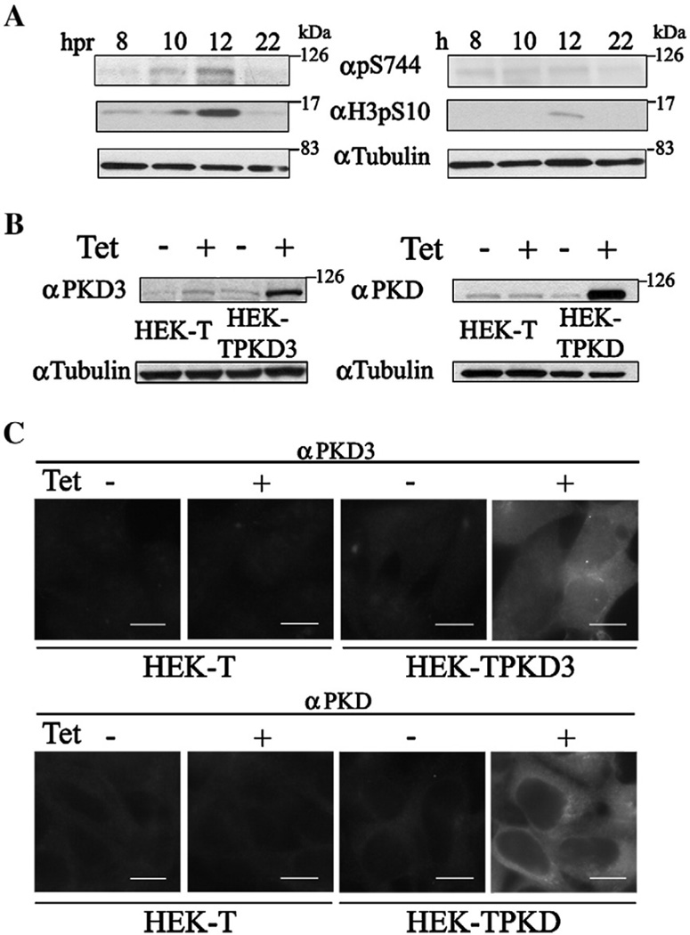
The rapid activation of PKD isozymes in response to a variety of stimuli occurs via phosphorylation of two conserved serine residues within their activation loop [10]. To determine whether the PKDs are regulated during mitosis, HEK-293 cells were incubated with aphidicolin, a reversible DNA polymerases inhibitor [30–32] extensively used to induce G1/S synchronization in different cell types [20], including HEK-293 cells [21–24]. The phosphorylation of PKD3 and PKD was examined at various time-points after aphidicolin removal. In these studies, we used an antibody that predominantly recognizes the phosphorylated state of the first serine residue in the activation loop of all the PKD isoforms (Ser744 in PKD, Ser706 in PKD2 and Ser731 in PKD3) [33]. Cell lysates obtained from parallel cultures kept in the presence of aphidicolin were used as controls.
As shown in Fig. 1A, the pS744 antibody revealed a prominent band with a molecular mass of approximately 100 kDa 12 h after the aphidicolin removal. Because the predicted molecular masses of PKD3 and PKD are very similar, 100.5 and 102 kDa respectively, this band could correspond to phosphorylated PKD3 and/or PKD. This time point coincided with the phosphorylation of Ser10 in the N-terminus of histone H3, a widely used mitosis marker [34–38], and with published results indicating that HEK-293 cells reach mitosis between 12–14 h after aphidicolin removal [22,23]. In contrast, no proteins were detected with the pS744 antibody in lysates obtained from the cells that were kept in the presence of aphidicolin. Accordingly, only a very faint band was revealed with the H3pS10 antibody at 12 h, indicating that most of the cells did not reach mitosis (Fig. 1A). Isoform-specific antibodies reveal no changes in the expression level of endogenous PKD3 or PKD in HEK-293 cells during aphidicolin incubation and subsequent release (data not shown). These results suggest that members of the PKD family are phosphorylated at a critical residue of the activation loop in cells traversing the mitotic phase of the cell cycle.
Fig. 1.
PKD3/PKD activation loop phosphorylation after cell cycle synchronization (A). HEK-293 cells synchronized at G1/S by aphidicolin treatment were obtained at the indicated times (hpr) after aphidicolin removal (left panels) or not (right panels). Equal amounts of the lysates were resolved in a 4.5–15% (PKDs/α-tubulin) or 12.5% (histone H3) SDS-PAGE and proteins transferred to PVDF membranes. The membranes were incubated with primary rabbit polyclonal antibody (pS744) that recognizes the phosphorylation of Ser731 and Ser744 within the activation loop of PKD3 and PKD, respectively, or with the primary rabbit polyclonal antibody H3pS10 that recognizes the phosphorylation of Ser10 in histone H3. Additionally, the membranes were incubated with a murine monoclonal antibody against α-tubulin. Signals were detected as described under Materials and methods. Analysis of PKD3 and PKD expression in stable cell lines (B). Cell lines stably transfected with plasmids encoding PKD3 (HEK-TPKD3) or PKD (HEK-TPKD) under the control of a Tet-inducible promoter were incubated for 16 h without or with Tet (100 ng/ml). Equal amounts of cell lysates, including control HEK-T cells, were resolved in a 4.5–15% SDS-PAGE and proteins transferred to PVDF membranes. The membranes were incubated with primary antibodies against PKD3, PKD or α-tubulin and signals detected as described under Materials and methods. PKD3 and PKD intracellular distribution in stable cell lines (C). HEK-T (control), HEK-TPKD3 and HEK-TPKD cells incubated without or with Tet (100 ng/ml) for 16 h were processed for indirect immunofluorescence as described under Material and methods using primary antibodies against PKD3 or PKD and Alexa 488-conjugated anti-rabbit IgG as secondary antibody. Bar: 10 µm, hpr: hours post aphidicolin release, h: hours.
Ser731 and Ser744 in the activation loop of PKD3 and PKD are phosphorylated during mitosis
Because the amino acid sequence of the activation loop of all the mammalian PKD isozymes is identical [10], there is no isoform-specific activation loop phospho-antibody. Furthermore, since the molecular masses of PKD3 and PKD are almost the same, the identity of the phosphorylated PKD during mitosis as revealed by the pS744 antibody could not be determined. Accordingly, we established HEK-293-derived cell lines overexpressing PKD3 (HEK-TPKD3) or PKD (HEK-TPKD) under the control of an inducible tetracycline (Tet) promoter to clarify which of these kinases becomes phosphorylated during mitosis. As shown in Fig. 1B, HEK-TPKD3 cells showed a marked increase in PKD3 expression after 16 h of Tet induction (4.7±0.5-fold increase; n=3). Similarly, HEK-TPKD cells also exhibited a marked increase in PKD expression in response to Tet exposure (8.6±1.5-fold increase; n=3). The intracellular distribution of the overexpressed proteins at interphase was identical to endogenous PKD3 and PKD [5,19]. Specifically, PKD3 was present in the cytoplasm and nucleus and PKD was found in the cytoplasm of HEK-TPKD3 and HEK-TPKD cells, respectively (Fig. 1C).
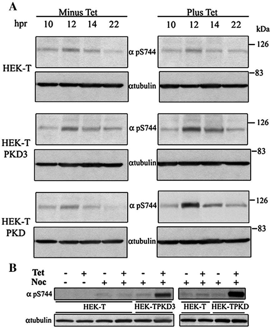
To determine whether PKD3 and/or PKD are phosphorylated in their activation loop during mitosis, HEK-TPKD3 and HEK-TPKD cells were incubated with aphidicolin and the phosphorylation of Ser731 in PKD3 and Ser744 in PKD was examined at various time-points after removing the aphidicolin from the medium. In agreement with the results presented in Fig.1A, the pS744 antibody revealed a distinct band 12 h after aphidicolin removal in Tet-treated or untreated HEK-T cells (Fig. 2A). The same antibody revealed prominent bands at 12 h and 14 h after aphidicolin removal in Tet-treated HEK-TPKD3 and HEK-TPKD cells, demonstrating that both PKD3 and PKD become phosphorylated in their activation loop during mitosis (Fig. 2A).
Fig. 2.
PKD3 Ser731 and PKD Ser744 phosphorylation during mitosis. HEK-T (control), HEK-TPKD3 and HEK-TPKD cells, without or with Tet (100 ng/ml) synchronized at G1/S by aphidicolin treatment (A), were collected at the indicated times after aphidicolin removal. Alternatively, the cultures, without or with Tet (100 ng/ml), were synchronized at G2/M by nocodazole treatment (B) for 12 h. Equal amounts of cell lysates obtained from aphidicolin or nocodazole-treated cells were resolved in a 4.5–15% SDS-PAGE. Proteins were transferred to PVDF membranes and the membranes incubated with pS744 or α-tubulin antibodies. Signals were detected as described under Materials and methods.
To independently validate these results, we also examined cells arrested at the G2/M transition. Growing HEK-T, HEK-TPKD3 and HEK-TPKD cells were incubated for 12 h with nocodazole, an inhibitor of tubulin polymerization extensively employed to induce G2/M synchronization in different cell types [20], including HEK-293 cells [24,26,27], and the phosphorylation of PKD3 and PKD was examined using Western blots. A very prominent band became evident with the pS744 antibody after Tet-induced PKD3 and PKD expression in nocodazole-treated cells (Fig. 2B), reinforcing the concept that PKD3 and PKD are phosphorylated at Ser731 and Ser744 in their activation loop during mitosis. No protein was detected by the pS744 antibody in non-synchronized HEK-T cells (Fig. 2B). Because PKD3 and PKD were phosphorylated during nocodazole-mediated G2/M arrest [20], our results suggest that Ser731 and Ser744 phosphorylation occurs during early mitosis.
Mitosis-dependent phosphorylation of PKD3 Ser731 and PKD Ser744 requires PKC activity
The members of the PKC family are central components in pathways regulating a wide variety of cellular processes [39]. For example, conventional and novel PKCs are involved in G2/M transition and cytokinesis [40–47]. Furthermore, PKC activity is indispensable for the rapid phosphorylation of Ser744 in the activation loop of PKD [33]. In view of these observations and the results presented in Figs. 2A, B indicating that PKD3 Ser731 and PKD Ser744 are phosphorylated during mitosis, we examined whether PKC inhibitors would prevent the phosphorylation of these serine residues.
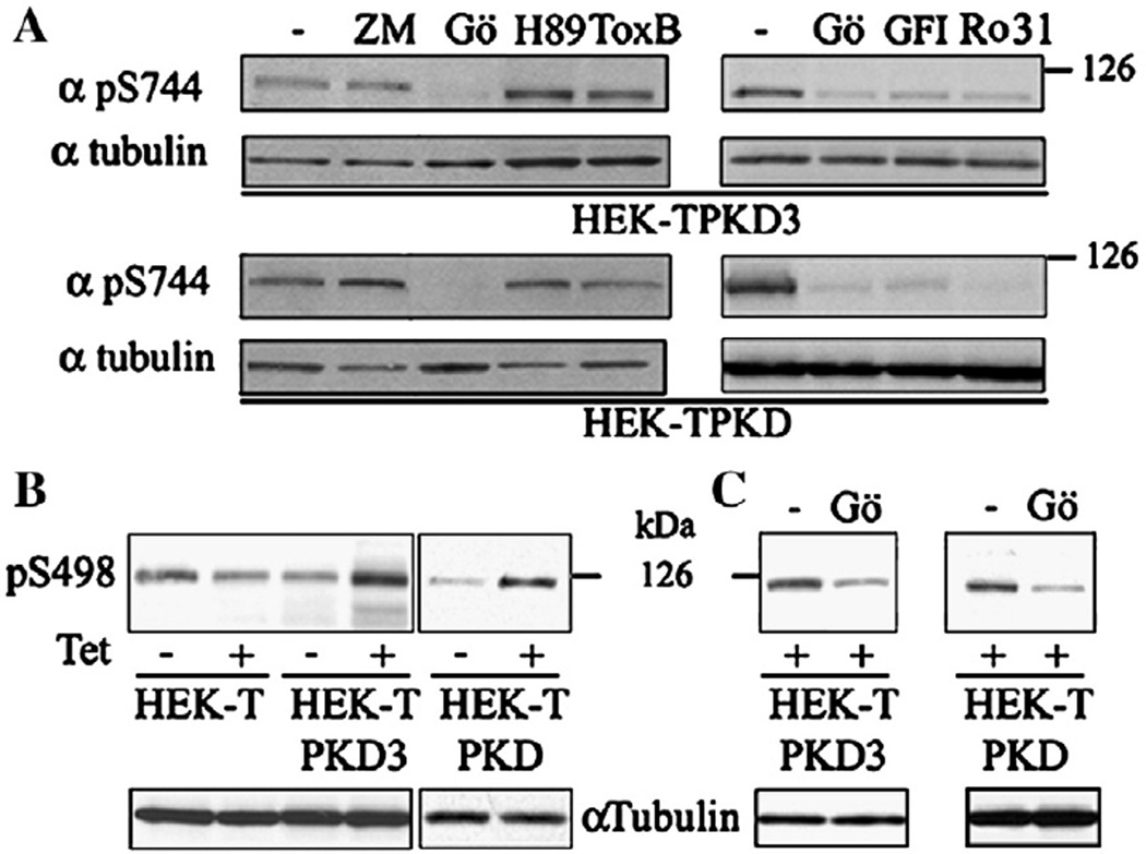
HEK-TPKD3 and HEK-TPKD cells were synchronized at G1/S by exposure to aphidicolin for 18 h. After aphidicolin removal, the cells were further incubated for 10 h before being treated with the selective PKC inhibitor Gö6983 [48] and the phosphorylation of PKD3 and PKD was determined 2 h later by Western blot using the pS744 antibody. We also examined the phosphorylation of Ser731 in PKD3 and Ser744 in PKD after inhibiting the activity of Aurora Kinase A and B [49,50], protein kinase A (PKA) [51–53] and Rho GTPases [54,55], which are signaling molecules that regulate cell cycle progression from G2 through cytokinesis. Our results show that exposure to the PKC inhibitor Gö6983 blocked the phosphorylation of Ser731 in PKD3 and Ser744 in PKD (Fig. 3A, left panel). In contrast, the treatment with the Aurora Kinase A and B inhibitor ZM-447439 [56], the PKA inhibitor H-89 [57] or the Rho GTPases inhibitor Toxin B from C. difficile [58,59] did not prevent the phosphorylation of Ser731 or Ser744 (Fig. 3A, left panels). In agreement with the results obtained with Gö6983 and published data [33], other selective PKC inhibitors, including GF 109203X (also known as bisindolylmaleimide I) or Ro 31-8220 [60,61], also prevented the phosphorylation of Ser731 and Ser744 (Fig. 3A, right panels). Thus, these results indicate that the phosphorylation of PKD3 Ser731 and PKD Ser744 during mitosis is mediated by a mechanism that requires PKC activity.
Fig. 3.
Effect of PKC inhibitors on mitosis-dependent PKD3 Ser731 and PKD Ser744 phosphorylation (A). HEK-TPKD3 and HEK-TPKD cells synchronized at G1/S by aphidicolin in the presence of Tet (100 ng/ml) were treated 10 h after aphidicolin removal with inhibitors of Aurora Kinase A and B (5 µM ZM-447439), PKA (10 µM H-89), Rho GTPases (Tox B 40 ng/ml) or PKC (2.5 µM Gö 6983, 3.5 µM GF 109203X, 2.5 µM Ro 31-8220) and the phosphorylation of PKD3 and PKD examined 2 h later by Western blot using the pS744 antibody. PKD3 and PKD-mediated HDAC5 Ser498 phosphorylation during mitosis (B). Cell lysates obtained from HEK-T (control), HEK-TPKD3 and HEK-TPKD cells synchronized at G2/M by 12 h nocodazole treatment, plus or minus Tet (100 ng/ml), were resolved in a 4.5–15% SDS-PAGE. Proteins were transferred to PDVF membranes and the membranes incubated with pS498 or α-tubulin antibodies. Effect of PKC inhibition on mitosis-dependent HDAC5 Ser498 phosphorylation (C). HEK-TPKD3 and HEK-TPKD cells synchronized at G1/S by aphidicolin in the presence of Tet (100 ng/ml) were treated 10 h after aphidicolin removal with the selective PKC inhibitor Gö6983 (2.5 µM) and the phosphorylation of HADC5 Ser498 examined 2 h later by Western blot using the pS498 antibody. Signals were detected as described under Materials and methods.
Catalytic activation of PKD3 and PKD during mitosis
Histone deacetylase (HDAC) 5 and 7, two members of the class II of classical HDAC [62], are in vivo substrates of PKD3 and PKD [63]. In response to a variety of signals, including phorbol esters, T cell receptor engagement, vascular endothelial growth factor and angiotensin stimulation, the activity of HDAC5 and 7 are regulated by a mechanism that involves PKD3 and PKD-mediated phosphorylation of the highly conserved Ser259 and Ser498 residues that are located in N-terminus of class II HDACs [63–67]. To determine whether mitosis-mediated phosphorylation of Ser731 and Ser744 in the activation loop of PKD3 and PKD was associated with an increase in their catalytic activity, we examined the phosphorylation of HDAC5 Ser498 in G2/M synchronized and PKD3 or PKD overexpressing cells.
Lysates obtained from nocodazole-treated HEK-T, HEK-TPKD3 and HEK-TPKD cells were analyzed by Western blot using an antibody that recognizes the phosphorylated Ser498 in HDAC5. As shown in Fig. 3B, the pS498 antibody revealed a band migrating within the predicted molecular mass for HDAC5 (122 kDa). This immunoreactive band, which was detected in all the samples, showed a dramatic increase only in the cells overexpressing PKD3 or PKD. Thus, these results suggest that the phosphorylation of Ser731 and Ser744 in the activation loop of PKD3 and PKD that occurs during mitosis is associated with an increase in their catalytic activity. No changes in the expression level of HDAC5 were detected because of nocodazole treatment or PKDs overexpression (data not shown).
Our data indicate that PKD3 Ser731 and PKD Ser744 phosphorylation during mitosis is mediated by a mechanism that requires PKC activity (Fig. 3A). Accordingly, and to further reinforce the association between PKD3 and PKD activation during mitosis and downstream events, we examined whether the phosphorylation of HDAC5 Ser498 required PKC activity. HEK-TPKD3 and HEK-TPKD cells were synchronized at G1/S by exposure to aphidicolin for 18 h. After aphidicolin removal, the cells were further incubated for 10 h before being treated with the selective PKC inhibitor Gö6983 and the phosphorylation of HDAC5 Ser498 determined 2 h later by Western blot using the pS498 antibody. As shown in Fig. 3C, Gö6983 inhibited the phosphorylation of Ser498 further reinforcing the concept that during mitosis PKD3 and PKD become activated by a mechanism that requires PKC activity and that their activation is associated with the phosphorylation of downstream substrates. Similar results, i. e. Ser498 phosphorylation inhibition during mitosis, were obtained using GF 109203X (data not shown).
PKD3 and PKD associate with the mitotic apparatus
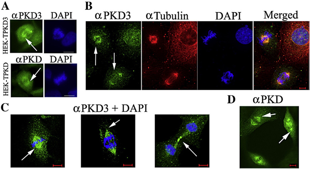
To characterize the intracellular distribution of PKD3 and PKD during mitosis, non-synchronized HEK-TPKD3 and HEK-TPKD cells treated with Tet for 16 h were fixed and processed for indirect immunofluorescence, as described under Materials and methods, using antibodies against PKD3 or PKD. As Fig. 4A shows, PKD3 and PKD were associated with structures resembling spindles (arrows) in cells undergoing mitosis, suggesting that both proteins are recruited to the mitotic apparatus during cell division. To confirm that this association was not due to PKD3 and PKD overexpression, we examined the intracellular distribution of endogenous PKD3 and PKD in intestinal epithelial IEC-18 cells.
Fig. 4.
Intracellular distribution of overexpressed PKD3 and PKD during mitosis (A). Unsynchronized HEK-TPKD3 and HEK-TPKD cells (A) incubated with Tet (100 ng/ml) for 16 h were processed for immunocytochemistry as described under Materials and methods using rabbit primary antibodies against PKD3 or PKD and Alexa 488-conjugated anti-rabbit IgG. The preparations were examined with an epifluorescence microscope and images acquired as previously described [19]. Intracellular distribution of endogenous PKD3 and PKD during mitosis (B–D). Unsynchronized IEC-18 cells were processed for indirect immunofluorescence and laser scanning confocal microscopy as described under Materials and methods using rabbit primary antibodies against PKD3 (B, C) or PKD (D), and Alexa 488-conjugated chicken anti-rabbit IgG. α-tubulin was detected using a murine monoclonal antibody and secondary Alexa 568-conjugated goat anti-mouse IgG. DNA was counterstained using 0.0005% DAPI. Bar: 10 µm.
In agreement with the results presented in Fig. 4A, immunocytochemistry and laser scanning confocal microscopy of non-synchronized IEC-18 cells undergoing mitosis showed that endogenous PKD3 associates with the mitotic spindles (Fig. 4B, arrows). This association was confirmed with another antibody (N17) generated in our laboratory [5] against the N-terminus of PKD3 (data not shown). The results also show that the association of PKD3 was not limited to the spindles. Specifically, PKD3 was present in centrosomes and midbody (Fig. 4C, arrows), indicating that this kinase undergoes a dynamic redistribution during mitosis. PKD3 did not colocalize with microtubules during interphase (Fig. 4B), suggesting that the interaction between PKD3 and the spindles is mediated by a protein(s) specifically present in these mitotic structures. In agreement with our published data [5] and results obtained with HEK-TPKD3 cells (see Fig. 1C), PKD3 was present in the nucleus and cytoplasm of IEC-18 cells during interphase. The localization of endogenous PKD3 to the mitotic apparatus was detected in different cell types, including human pancreatic cancer Panc-1 cells, murine Swiss 3T3 fibroblasts and human primary keratinocytes (data not shown). We also found that endogenous PKD is associated with the mitotic apparatus in non-synchronized IEC-18 cells undergoing mitosis (Fig. 4D, arrows), substantiating the results obtained with overexpressed PKD (Fig. 4A). Interestingly, we found that long term-overexpression (72 h) of PKD3 or PKD in HEK-TPKD3 and HEK-TPKD cells was associated with the appearance of multinucleated cells and mitotic bridges, both indicators of cytokinesis failure [68] (data not shown). Thus, the present results indicate that PKD3 and PKD are recruited to the mitotic apparatus during cell division.
Activation loop-phosphorylated PKDs are associated with the mitotic apparatus
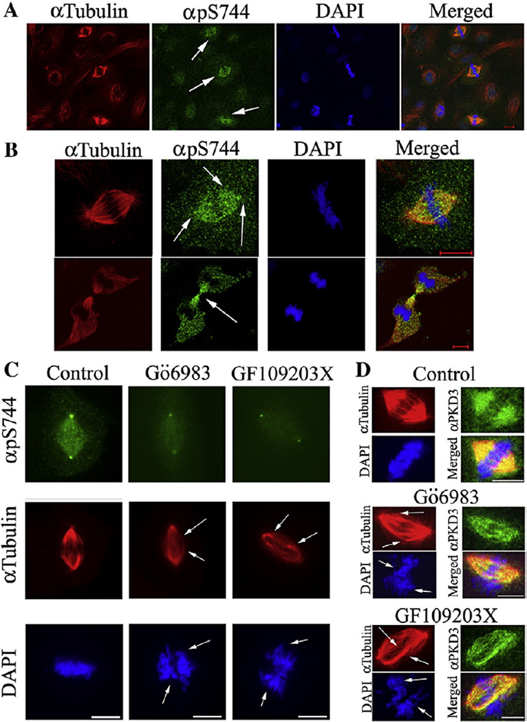
The association of PKD3 and PKD with the spindles, centrosomes and midbody could be important to regulate the function of these structures. Accordingly, we examined the distribution of activation loop-phosphorylated PKDs using the pS744 antibody. Immunocytochemistry and laser scanning confocal microscopy of non-synchronized IEC-18 cells undergoing mitosis revealed a strong reactivity of the pS744 antibody with cells at different mitotic stages (Figs. 5A, B). Specifically, the pS744 antibody reacted with spindles, centrosomes and midbody (Fig. 5B, arrows), a pattern that closely matches the distribution of endogenous PKD3 and PKD presented in Figs. 4B–D. In agreement with the results presented in Fig. 3A, the reactivity of pS744 with IEC-18 cells undergoing mitosis was reduced in response to the PKC inhibitors Gö6983 or GF 109203X (Fig. 5C), consistent with the notion that during mitosis a mechanism involving PKC mediates PKD3 and PKD activation loop phosphorylation. These PKC inhibitors did not interfere in the association of PKD3 (Fig. 5D) or PKD (data not shown) with the mitotic apparatus. However, atypical spindle morphology and organization, as well as abnormal chromosomal segregation, were common in cells treated with Gö6983 or GF 109203X (Figs. 5C and D, arrows), suggesting that the activity of PKC is involved in the regulation of spindle assembly and/or dynamics.
Fig. 5.
Intracellular distribution of activation loop-phosphorylated PKDs during mitosis. Unsynchronized IEC-18 cells were processed for indirect immunofluorescence and examined using a LSCM as described under Materials and methods using the rabbit phospho-specific antibody pS744 (A, B) and Alexa 488-conjugated chicken anti-rabbit IgG. α-tubulin was detected using a murine monoclonal antibody and secondary Alexa 568-conjugated goat anti-mouse IgG. DNA was counterstained using 0.0005% DAPI. Effect of PKC inhibition on PKDs mitosis-dependent activation loop phosphorylation. IEC-18 cells synchronized at G1/S by exposure to aphidicolin, following the same procedure described in Materials and methods for HEK-T derived cell lines, were further incubated for 4 h after aphidicolin removal before being treated for 2 h with the selective PKC inhibitors Gö6983 (2.5 µM) or GF 109203X (3.5 µM). After the treatment, the cells were fixed and processed for indirect immunofluorescence using the rabbit pS744 antibody and a murine monoclonal antibody against α-tubulin (C) or rabbit anti-PKD3 and a murine monoclonal antibody against α-tubulin (D). Alexa 488-conjugated chicken anti-rabbit IgG and Alexa 568-conjugated goat anti-mouse IgG were used as secondary antibodies. DNA was counterstained using 0.0005% DAPI. Preparations were examined using a LSCM (A, B, D) or an epifluorescence microscope (C). Bar: 10 µm.
Thus, these results obtained with non-synchronized and synchronized cells expressing endogenous PKD3 and PKD support the conclusion that PKD3 Ser731 and PKD Ser744 are phosphorylated during mitosis and that these phosphorylated kinases associate with centrosomes, spindles and midbody.
PKD has in its C-terminus an autophosphorylation site (Ser916) which is absent in PKD3 [10]. The phosphorylation of this serine residue has been extensively used to evaluate the catalytic activity of PKD using the phospho-specific pS916 antibody. As shown in Fig. 6A, the pS916 antibody reacted with the same mitotic structures as pS744, further demonstrating that catalytically active PKD is associated with the mitotic apparatus. To corroborate this observation, cell lysates obtained from G2/M arrested IEC-18 cells were examined by Western blot using the pS916 antibody. Lysates obtained from non-synchronized IEC-18 cells that were either left unstimulated or stimulated with the GPCR agonist vasopressin were used as negative and positive controls, respectively. As previously reported [18], vasopressin promotes a rapid phosphorylation of PKD Ser916 in IEC-18 cells (Fig. 6B). Importantly, a dramatic increase in the reactivity of the pS916 antibody equivalent to the phosphorylation observed after GPCR stimulation was also detected in nocodazole-treated IEC-18 cells, further demonstrating that PKD autophosphorylation is elevated during mitosis.
Fig. 6.
Intracellular distribution of activated PKD during mitosis (A). Unsynchronized IEC-18 cells were processed for indirect immunofluorescence and examined using a LSCM as described under Materials and methods using a rabbit phospho-specific antibody pS916 and Alexa 488-conjugated chicken anti-rabbit IgG. DNA was counterstained using 0.0005% DAPI. Bar: 10 µm. Western blot analysis of endogenous PKD Ser916 phosphorylation during mitosis (B). Unsynchronized IEC-18 cells were left unstimulated (−) or stimulated with 100 nM vasopressin (Vas) for 10 min at 37 °C and then lysed. Parallel cultures of IEC-18 cells synchronized at G2/M following the same procedure described in Materials and methods for HEK-T derived cell lines were also collected and lysed. Equal amounts of the different lysates were resolved in a 4.5–15% SDS-PAGE and proteins transferred to PVDF membranes. The membranes were incubated with pS916 or α-tubulin antibodies and signals detected as described in Materials and methods.
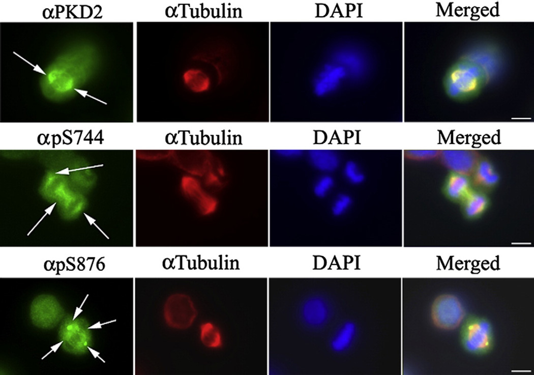
We next examined the intracellular distribution and phosphorylation of PKD2 during mitosis. We employed the nontransformed human colonic epithelial NCM460 cells because PKD2 is the only detectable PKD isoform in this cell line [17]. Non-synchronized NCM460 cells were processed for indirect immunofluorescence as described under Materials and methods and stained with a rabbit antibody highly specific for human PKD2 [17]. As shown in Fig. 7, top panels, this antibody revealed that PKD2 associates with the mitotic spindles (arrows) as well as centrosomes and midbody (data not shown). We also found a pronounced reactivity of the pS744 antibody with spindles and centrosomes (Fig. 7, middle panels, arrows), indicating that Ser706 in the activation loop of PKD2 is also phosphorylated during mitosis. PKD2 has also in its C-terminus an autophosphorylation site (Ser876) that is homologous to Ser916 present in PKD [10]. The phosphorylation of this serine residue has also been used to evaluate the catalytic activity of PKD2. As shown in Fig. 7, bottom panels, the phospho-specific pS876 antibody strongly reacted with centrosomes and spindles, demonstrating that catalytically active PKD2 is associated with the mitotic apparatus.
Fig. 7.
Intracellular distribution of activated PKD2 during mitosis. Unsynchronized NCM-460 cells were processed for indirect immunofluorescence and examined with an epifluorescence microscope as described under Materials and methods. Samples were stained with an antibody that recognizes PKD2 (top panels), with the phospho-specific antibody pS744 (middle panels) which recognizes phosphorylated Ser706 within the activation loop of PKD2, or with the phospho-specific antibody pS876 that recognizes PKD2 phosphorylated at Ser876 (bottom panels), a PKD2 autophosphorylation site. Alexa 488-conjugated chicken anti-rabbit IgG was used as secondary antibody. α-tubulin was detected using a murine monoclonal antibody and secondary Alexa 568-conjugated goat anti-mouse IgG. DNA was counterstained using 0.0005% DAPI. Bar: 10 µm.
In summary, the studies presented here reveal the existence of a cell cycle-regulated signaling pathway involving the activation of the PKD isozymes and their localization to centrosomes, spindles and midbody. Considering that the nuclear levels of polyunsaturated DAG species increase during G2/M [69] and that the PKD isozymes can be activated by DAG, it is tempting to speculate that during mitosis DAG mediates the activation of these kinases directly and/or indirectly via PKC stimulation.
The PKD isozymes have been implicated in the regulation of a variety of important cellular functions including DNA synthesis and cell proliferation, chromatin modification, Golgi organization and function, c-Jun signaling, NFκB-mediated gene expression and cell migration [10]. In addition to these processes, the results presented here raise the possibility that these kinases are implicated in the regulation of cell cycle events directly associated with the progression of mitosis.
Acknowledgments
This work was supported by a grant of the Margaret E. Early Medical Research Trust to O. R., a recipient of a NIH K22CA128883. R. T. W. and E. R. are recipients of NIH R21DK071783 and RO1DK55003 awards, respectively. Support from the Morphology/Imaging Core of the CURE Center Grant 5P30DK41301 is gratefully acknowledged.
REFERENCES
- 1.Valverde AM, Sinnett-Smith J, Van Lint J, Rozengurt E. Molecular cloning and characterization of protein kinase D: a target for diacylglycerol and phorbol esters with a distinctive catalytic domain. Proc. Natl. Acad. Sci. U. S. A. 1994;91:8572–8576. doi: 10.1073/pnas.91.18.8572. [DOI] [PMC free article] [PubMed] [Google Scholar]
- 2.Johannes FJ, Prestle J, Eis S, Oberhagemann P, Pfizenmaier K. PKCu is a novel, atypical member of the protein kinase C family. J. Biol. Chem. 1994;269:6140–6148. [PubMed] [Google Scholar]
- 3.Hayashi A, Seki N, Hattori A, Kozuma S, Saito T. PKCnu, a new member of the protein kinase C family, composes a fourth subfamily with PKCmu. Biochem. Biophys. Acta. 1999;1450:99–106. doi: 10.1016/s0167-4889(99)00040-3. [DOI] [PubMed] [Google Scholar]
- 4.Sturany S, Van Lint J, Muller F, Wilda M, Hameister H, Hocker M, Brey A, Gern U, Vandenheede J, Gress T, Adler G, Seufferlein T. Molecular cloning and characterization of the human protein kinase D2. A novel member of the protein kinase D family of serine threonine kinases. J. Biol. Chem. 2001;276:3310–3318. doi: 10.1074/jbc.M008719200. [DOI] [PubMed] [Google Scholar]
- 5.Rey O, Yuan J, Young SH, Rozengurt E. Protein kinase C nu/protein kinase D3 nuclear localization, catalytic activation, and intracellular redistribution in response to G protein-coupled receptor agonists. J. Biol. Chem. 2003;278:23773–23785. doi: 10.1074/jbc.M300226200. [DOI] [PubMed] [Google Scholar]
- 6.Rozengurt E, Sinnett-Smith J, Zugaza JL. Protein kinase D: a novel target for diacylglycerol and phorbol esters. Biochem. Soc. Trans. 1997;25:565–571. doi: 10.1042/bst0250565. [DOI] [PubMed] [Google Scholar]
- 7.Iglesias T, Matthews S, Rozengurt E. Dissimilar phorbol ester binding properties of the individual cysteine-rich motifs of protein kinase D. FEBS Lett. 1998;437:19–23. doi: 10.1016/s0014-5793(98)01189-2. [DOI] [PubMed] [Google Scholar]
- 8.Iglesias T, Rozengurt E. Protein kinase D activation by mutations within its pleckstrin homology domain. J. Biol. Chem. 1998;273:410–416. doi: 10.1074/jbc.273.1.410. [DOI] [PubMed] [Google Scholar]
- 9.Iglesias T, Rozengurt E. Protein kinase D activation by deletion of its cysteine-rich motifs. FEBS Lett. 1999;454:53–56. doi: 10.1016/s0014-5793(99)00772-3. [DOI] [PubMed] [Google Scholar]
- 10.Rozengurt E, Rey O, Waldron RT. Protein kinase D signaling. J. Biol. Chem. 2005;280:13205–13208. doi: 10.1074/jbc.R500002200. [DOI] [PubMed] [Google Scholar]
- 11.Waldron RT, Rey O, Iglesias T, Tugal T, Cantrell D, Rozengurt E. Activation loop Ser744 and Ser748 in protein kinase D are transphosphorylated in vivo. J. Biol. Chem. 2001;276:32606–32615. doi: 10.1074/jbc.M101648200. [DOI] [PubMed] [Google Scholar]
- 12.Sturany S, Van Lint J, Gilchrist A, Vandenheede JR, Adler G, Seufferlein T. Mechanism of activation of protein kinase D2 (PKD2) by the CCK(B)/gastrin receptor. J. Biol. Chem. 2002;277:29431–29436. doi: 10.1074/jbc.M200934200. [DOI] [PubMed] [Google Scholar]
- 13.Sinnett-Smith J, Zhukova E, Hsieh N, Jiang X, Rozengurt E. Protein kinase D potentiates DNA synthesis induced by Gq-coupled receptors by increasing the duration of ERK signaling in Swiss 3T3 cells. J. Biol. Chem. 2004;279:16883–16893. doi: 10.1074/jbc.M313225200. [DOI] [PubMed] [Google Scholar]
- 14.Sinnett-Smith J, Zhukova E, Rey O, Rozengurt E. Protein kinase D2 potentiates MEK/ERK/RSK signaling, c-Fos accumulation and DNA synthesis induced by bombesin in Swiss 3T3 cells. J. Cell. Physiol. 2007;211:781–790. doi: 10.1002/jcp.20984. [DOI] [PubMed] [Google Scholar]
- 15.Fielitz J, Kim MS, Shelton JM, Qi X, Hill JA, Richardson JA, Bassel-Duby R, Olson EN. Requirement of protein kinase D1 for pathological cardiac remodeling. Proc. Natl. Acad. Sci. U. S. A. 2008;105:3059–3063. doi: 10.1073/pnas.0712265105. [DOI] [PMC free article] [PubMed] [Google Scholar]
- 16.Rey O, Zhukova E, Sinnett-Smith J, Rozengurt E. Vasopressin-induced intracellular redistribution of protein kinase D in intestinal epithelial cells. J. Cell Physiol. 2003;196:483–492. doi: 10.1002/jcp.10323. [DOI] [PubMed] [Google Scholar]
- 17.Chiu TT, Leung WY, Moyer MP, Strieter RM, Rozengurt E. Protein kinase D2 mediates lysophosphatidic acid-induced interleukin 8 production in nontransformed human colonic epithelial cells through NF-kappaB. Am. J. Physiol. Cell Physiol. 2007;292:C767–C777. doi: 10.1152/ajpcell.00308.2006. [DOI] [PubMed] [Google Scholar]
- 18.Chiu T, Rozengurt E. PKD in intestinal epithelial cells: rapid activation by phorbol esters, LPA, and angiotensin through PKC. Am. J. Physiol. Cell Physiol. 2001;280:C929–C942. doi: 10.1152/ajpcell.2001.280.4.C929. [DOI] [PubMed] [Google Scholar]
- 19.Rey O, Young SH, Cantrell D, Rozengurt E. Rapid protein kinase D translocation in response to G protein-coupled receptor activation: dependence on protein kinase Ce. J. Biol. Chem. 2001;276:32616–32626. doi: 10.1074/jbc.M101649200. [DOI] [PubMed] [Google Scholar]
- 20.Krek W, DeCaprio JA. Cell synchronization. Methods Enzymol. 1995;254:114–124. doi: 10.1016/0076-6879(95)54009-1. [DOI] [PubMed] [Google Scholar]
- 21.Fatatis A, Miller RJ. Cell cycle control of PDGF-induced Ca(2+) signaling through modulation of sphingolipid metabolism. FASEB J. 1999;13:1291–1301. doi: 10.1096/fasebj.13.11.1291. [DOI] [PubMed] [Google Scholar]
- 22.Katayama K, Fujita N, Tsuruo T. Akt/protein kinase B-dependent phosphorylation and inactivation of WEE1Hu promote cell cycle progression at G2/M transition. Mol. Cell. Biol. 2005;25:5725–5737. doi: 10.1128/MCB.25.13.5725-5737.2005. [DOI] [PMC free article] [PubMed] [Google Scholar]
- 23.Schneikert J, Behrens J. Truncated APC is required for cell proliferation and DNA replication. Int. J. Cancer. 2006;119:74–79. doi: 10.1002/ijc.21826. [DOI] [PubMed] [Google Scholar]
- 24.Forester CM, Maddox J, Louis JV, Goris J, Virshup DM. Control of mitotic exit by PP2A regulation of Cdc25C and Cdk1. Proc. Natl. Acad. Sci. U. S. A. 2007;104:19867–19872. doi: 10.1073/pnas.0709879104. [DOI] [PMC free article] [PubMed] [Google Scholar]
- 25.Laemmli UK. Cleavage of structural proteins during the assembly of the head of bacteriophage T4. Nature. 1970;227:680–685. doi: 10.1038/227680a0. [DOI] [PubMed] [Google Scholar]
- 26.Cude K, Wang Y, Choi HJ, Hsuan SL, Zhang H, Wang CY, Xia Z. Regulation of the G2-M cell cycle progression by the ERK5-NFkappaB signaling pathway. J. Cell Biol. 2007;177:253–264. doi: 10.1083/jcb.200609166. [DOI] [PMC free article] [PubMed] [Google Scholar]
- 27.Khare L, Astrinidis A, Senapedis W, Adams PD, Henske EP. Expression of wild type and mutant TSC2, but not TSC1, causes an increase in the G1 fraction of the cell cycle in HEK293 cells. J. Med. Genet. 2002;39:676–680. doi: 10.1136/jmg.39.9.676. [DOI] [PMC free article] [PubMed] [Google Scholar]
- 28.Kani S, Nakayama E, Yoda A, Onishi N, Sougawa N, Hazaka Y, Umeda T, Takeda K, Ichijo H, Hamada Y, Minami Y. Chk2 kinase is required for methylglyoxal-induced G2/M cell-cycle checkpoint arrest: implication of cell-cycle checkpoint regulation in diabetic oxidative stress signaling. Genes Cells. 2007;12:919–928. doi: 10.1111/j.1365-2443.2007.01100.x. [DOI] [PubMed] [Google Scholar]
- 29.Rey O, Papazyan R, Waldron RT, Young SH, Lippincott-Schwartz J, Jacamo R, Rozengurt E. The nuclear import of protein kinase D3 requires its catalytic activity. J. Biol. Chem. 2006;281:5149–5157. doi: 10.1074/jbc.M508014200. [DOI] [PubMed] [Google Scholar]
- 30.Ikegami S, Taguchi T, Ohashi M, Oguro M, Nagano H, Mano Y. Aphidicolin prevents mitotic cell division by interfering with the activity of DNA polymerase-alpha. Nature. 1978;275:458–460. doi: 10.1038/275458a0. [DOI] [PubMed] [Google Scholar]
- 31.Hammond RA, Byrnes JJ, Miller MR. Identification of DNA polymerase delta in CV-1 cells: studies implicating both DNA polymerase delta and DNA polymerase alpha in DNA replication. Biochemistry. 1987;26:6817–6824. doi: 10.1021/bi00395a035. [DOI] [PubMed] [Google Scholar]
- 32.Cheng CH, Kuchta RD. DNA polymerase epsilon: aphidicolin inhibition and the relationship between polymerase and exonuclease activity. Biochemistry. 1993;32:8568–8574. doi: 10.1021/bi00084a025. [DOI] [PubMed] [Google Scholar]
- 33.Jacamo R, Sinnett-Smith J, Rey O, Waldron RT, Rozengurt E. Sequential PKC-dependent and PKC-independent protein kinase D catalytic activation via Gq-coupled receptors: differential regulation of activation loop Ser744 and Ser748 phosphorylation. J. Biol. Chem. 2008;283:12877–12887. doi: 10.1074/jbc.M800442200. [DOI] [PMC free article] [PubMed] [Google Scholar]
- 34.Ajiro K, Nishimoto T, Takahashi T. Histone H1 and H3 phosphorylation during premature chromosome condensation in a temperature-sensitive mutant (tsBN2) of baby hamster kidney cells. J. Biol. Chem. 1983;258:4534–4538. [PubMed] [Google Scholar]
- 35.Ajiro K, Nishimoto T. Specific site of histone H3 phosphorylation related to the maintenance of premature chromosome condensation. Evidence for catalytically induced interchange of the subunits. J. Biol. Chem. 1985;260:15379–15381. [PubMed] [Google Scholar]
- 36.Gong J, Traganos F, Darzynkiewicz Z. Growth imbalance and altered expression of cyclins B1, A, E, and D3 in MOLT-4 cells synchronized in the cell cycle by inhibitors of DNA replication. Cell Growth Differ. 1995;6:1485–1493. [PubMed] [Google Scholar]
- 37.Hendzel MJ, Wei Y, Mancini MA, Van Hooser A, Ranalli T, Brinkley BR, Bazett-Jones DP, Allis CD. Mitosis-specific phosphorylation of histone H3 initiates primarily within pericentromeric heterochromatin during G2 and spreads in an ordered fashion coincident with mitotic chromosome condensation. Chromosoma. 1997;106:348–360. doi: 10.1007/s004120050256. [DOI] [PubMed] [Google Scholar]
- 38.Li DW, Yang Q, Chen JT, Zhou H, Liu RM, Huang XT. Dynamic distribution of Ser-10 phosphorylated histone H3 in cytoplasm of MCF-7 and CHO cells during mitosis. Cell Res. 2005;15:120–126. doi: 10.1038/sj.cr.7290276. [DOI] [PubMed] [Google Scholar]
- 39.Mellor H, Parker PJ. The extended protein kinase C superfamily. Biochem. J. 1998;332(Pt 2):281–292. doi: 10.1042/bj3320281. [DOI] [PMC free article] [PubMed] [Google Scholar]
- 40.Watanabe T, Ono Y, Taniyama Y, Hazama K, Igarashi K, Ogita K, Kikkawa U, Nishizuka Y. Cell division arrest induced by phorbol ester in CHO cells overexpressing protein kinase C-delta subspecies. Proc. Natl. Acad. Sci. U. S. A. 1992;89:10159–10163. doi: 10.1073/pnas.89.21.10159. [DOI] [PMC free article] [PubMed] [Google Scholar]
- 41.Dai Z, Dulyaninova NG, Kumar S, Bresnick AR, Lawrence DS. Visual snapshots of intracellular kinase activity at the onset of mitosis. Chem. Biol. 2007;14:1254–1260. doi: 10.1016/j.chembiol.2007.10.007. [DOI] [PMC free article] [PubMed] [Google Scholar]
- 42.Chen D, Purohit A, Halilovic E, Doxsey SJ, Newton AC. Centrosomal anchoring of protein kinase C betaII by pericentrin controls microtubule organization, spindle function, and cytokinesis. J. Biol. Chem. 2004;279:4829–4839. doi: 10.1074/jbc.M311196200. [DOI] [PubMed] [Google Scholar]
- 43.Black JD. Protein kinase C-mediated regulation of the cell cycle. Front. Biosci. 2000;5:D406–D423. doi: 10.2741/black. [DOI] [PubMed] [Google Scholar]
- 44.Barboule N, Lafon C, Chadebech P, Vidal S, Valette A. Involvement of p21 in the PKC-induced regulation of the G2/M cell cycle transition. FEBS Lett. 1999;444:32–37. doi: 10.1016/s0014-5793(99)00022-8. [DOI] [PubMed] [Google Scholar]
- 45.Passalacqua M, Patrone M, Sparatore B, Pedrazzi M, Melloni E, Pontremoli S. Protein kinase C-theta is specifically activated in murine erythroleukaemia cells during mitosis. FEBS Lett. 1999;453:249–253. doi: 10.1016/s0014-5793(99)00729-2. [DOI] [PubMed] [Google Scholar]
- 46.Passalacqua M, Patrone M, Sparatore B, Melloni E, Pontremoli S. Protein kinase C-theta is specifically localized on centrosomes and kinetochores in mitotic cells. Biochem. J. 1999;337(Pt 1):113–118. [PMC free article] [PubMed] [Google Scholar]
- 47.Arita Y, Buffolino P, Coppock DL. Regulation of the cell cycle at the G2/M boundary in metastatic melanoma cells by 12-O-tetradecanoyl phorbol-13-acetate (TPA) by blocking p34cdc2 kinase activity. Exp. Cell Res. 1998;242:381–390. doi: 10.1006/excr.1997.3911. [DOI] [PubMed] [Google Scholar]
- 48.Gschwendt M, Dieterich S, Rennecke J, Kittstein W, Mueller HJ, Johannes FJ. Inhibition of protein kinase C mu by various inhibitors. Differentiation from protein kinase c isoenzymes. FEBS Lett. 1996;392:77–80. doi: 10.1016/0014-5793(96)00785-5. [DOI] [PubMed] [Google Scholar]
- 49.Marumoto T, Zhang D, Saya H. Aurora-A — a guardian of poles. Nat. Rev. Cancer. 2005;5:42–50. doi: 10.1038/nrc1526. [DOI] [PubMed] [Google Scholar]
- 50.Vader G, Medema RH, Lens SM. The chromosomal passenger complex: guiding Aurora-B through mitosis. J. Cell Biol. 2006;173:833–837. doi: 10.1083/jcb.200604032. [DOI] [PMC free article] [PubMed] [Google Scholar]
- 51.Grieco D, Porcellini A, Avvedimento EV, Gottesman ME. Requirement for cAMP-PKA pathway activation by M phase-promoting factor in the transition from mitosis to interphase. Science. 1996;271:1718–1723. doi: 10.1126/science.271.5256.1718. [DOI] [PubMed] [Google Scholar]
- 52.Kotani S, Tugendreich S, Fujii M, Jorgensen PM, Watanabe N, Hoog C, Hieter P, Todokoro K. PKA and MPF-activated polo-like kinase regulate anaphase-promoting complex activity and mitosis progression. Mol. Cell. 1998;1:371–380. doi: 10.1016/s1097-2765(00)80037-4. [DOI] [PubMed] [Google Scholar]
- 53.Shibuya EK. G2 cell cycle arrest—a direct link between PKA and Cdc25C. Cell Cycle. 2003;2:39–41. doi: 10.4161/cc.2.1.291. [DOI] [PubMed] [Google Scholar]
- 54.Schmidt A, Durgan J, Magalhaes A, Hall A. Rho GTPases regulate PRK2/PKN2 to control entry into mitosis and exit from cytokinesis. EMBO J. 2007;26:1624–1636. doi: 10.1038/sj.emboj.7601637. [DOI] [PMC free article] [PubMed] [Google Scholar]
- 55.Wolf A, Keil R, Gotzl O, Mun A, Schwarze K, Lederer M, Huttelmaier S, Hatzfeld M. The armadillo protein p0071 regulates Rho signalling during cytokinesis. Nat. Cell Biol. 2006;8:1432–1440. doi: 10.1038/ncb1504. [DOI] [PubMed] [Google Scholar]
- 56.Girdler F, Gascoigne KE, Eyers PA, Hartmuth S, Crafter C, Foote KM, Keen NJ, Taylor SS. Validating Aurora B as an anti-cancer drug target. J. Cell Sci. 2006;119:3664–3675. doi: 10.1242/jcs.03145. [DOI] [PubMed] [Google Scholar]
- 57.Davies SP, Reddy H, Caivano M, Cohen P. Specificity and mechanism of action of some commonly used protein kinase inhibitors. Biochem. J. 2000;351:95–105. doi: 10.1042/0264-6021:3510095. [DOI] [PMC free article] [PubMed] [Google Scholar]
- 58.Aktories K. Bacterial toxins that target Rho proteins. J. Invest. Clin. 1997;99:827–829. doi: 10.1172/JCI119245. [DOI] [PMC free article] [PubMed] [Google Scholar]
- 59.Just I, Wilm M, Selzer J, Rex G, von Eichel-Streiber C, Mann M, Aktories K. The enterotoxin from Clostridium difficile (ToxA) monoglucosylates the Rho proteins. J. Biol. Chem. 1995;270:13932–13936. doi: 10.1074/jbc.270.23.13932. [DOI] [PubMed] [Google Scholar]
- 60.Martiny-Baron G, Kazanietz MG, Mischak H, Blumberg PM, Kochs G, Hug H, Marme D, Schachtele C. Selective inhibition of protein kinase C isozymes by the indolocarbazole Go 6976. J. Biol. Chem. 1993;268:9194–9197. [PubMed] [Google Scholar]
- 61.Wilkinson SE, Parker PJ, Nixon JS. Isoenzyme specificity of bisindolylmaleimides, selective inhibitors of protein kinase C. Biochem. J. 1993;294:335–337. doi: 10.1042/bj2940335. [DOI] [PMC free article] [PubMed] [Google Scholar]
- 62.de Ruijter AJ, van Gennip AH, Caron HN, Kemp S, van Kuilenburg AB. Histone deacetylases (HDACs): characterization of the classical HDAC family. Biochem. J. 2003;370:737–749. doi: 10.1042/BJ20021321. [DOI] [PMC free article] [PubMed] [Google Scholar]
- 63.Matthews SA, Liu P, Spitaler M, Olson EN, McKinsey TA, Cantrell DA, Scharenberg AM. Essential role for protein kinase D family kinases in the regulation of class II histone deacetylases in B lymphocytes. Mol. Cell. Biol. 2006;26:1569–1577. doi: 10.1128/MCB.26.4.1569-1577.2006. [DOI] [PMC free article] [PubMed] [Google Scholar]
- 64.Vega RB, Harrison BC, Meadows E, Roberts CR, Papst PJ, Olson EN, McKinsey TA. Protein kinases C and D mediate agonist-dependent cardiac hypertrophy through nuclear export of histone deacetylase 5. Mol. Cell. Biol. 2004;24:8374–8385. doi: 10.1128/MCB.24.19.8374-8385.2004. [DOI] [PMC free article] [PubMed] [Google Scholar]
- 65.Dequiedt F, Van Lint J, Lecomte E, Van Duppen V, Seufferlein T, Vandenheede JR, Wattiez R, Kettmann R. Phosphorylation of histone deacetylase 7 by protein kinase D mediates T cell receptor-induced Nur77 expression and apoptosis. J. Exp. Med. 2005;201:793–804. doi: 10.1084/jem.20042034. [DOI] [PMC free article] [PubMed] [Google Scholar]
- 66.Ha CH, Wang W, Jhun BS, Wong C, Hausser A, Pfizenmaier K, McKinsey TA, Olson EN, Jin ZG. Protein kinase D-dependent phosphorylation and nuclear export of histone deacetylase 5 mediates vascular endothelial growth factor-induced gene expression and angiogenesis. J. Biol. Chem. 2008;283:14590–14599. doi: 10.1074/jbc.M800264200. [DOI] [PMC free article] [PubMed] [Google Scholar]
- 67.Xu X, Ha CH, Wong C, Wang W, Hausser A, Pfizenmaier K, Olson EN, McKinsey TA, Jin ZG. Angiotensin II stimulates protein kinase D-dependent histone deacetylase 5 phosphorylation and nuclear export leading to vascular smooth muscle cell hypertrophy. Arterioscler. Thromb. Vasc. Biol. 2007;27:2355–2362. doi: 10.1161/ATVBAHA.107.151704. [DOI] [PMC free article] [PubMed] [Google Scholar]
- 68.Shi Q, King RW. Chromosome nondisjunction yields tetraploid rather than aneuploid cells in human cell lines. Nature. 2005;437:1038–1042. doi: 10.1038/nature03958. [DOI] [PubMed] [Google Scholar]
- 69.Deacon EM, Pettitt TR, Webb P, Cross T, Chahal H, Wakelam MJ, Lord JM. Generation of diacylglycerol molecular species through the cell cycle: a role for 1-stearoyl, 2-arachidonyl glycerol in the activation of nuclear protein kinase C-betaII at G2/M. J, Cell Sci. 2002;115:983–989. doi: 10.1242/jcs.115.5.983. [DOI] [PubMed] [Google Scholar]


