Abstract
Recent evidence demonstrated that most fertilizing mouse sperm undergo acrosomal exocytosis (AE) before binding to the zona pellucida of the eggs. However, the sites where fertilizing sperm could initiate AE and what stimuli trigger it remain unknown. Therefore, the aim of this study was to determine physiological sites of AE by using double transgenic mouse sperm, which carried EGFP in the acrosome and DsRed2 fluorescence in mitochondria. Using live imaging of sperm during in vitro fertilization of cumulus-oocyte complexes, it was observed that most sperm did not undergo AE. Thus, the occurrence of AE within the female reproductive tract was evaluated in the physiological context where this process occurs. Most sperm in the lower segments of the oviduct were acrosome-intact; however, a significant number of sperm that reached the upper isthmus had undergone AE. In the ampulla, only 5% of the sperm were acrosome-intact. These results support our previous observations that most of mouse sperm do not initiate AE close to or on the ZP, and further demonstrate that a significant proportion of sperm initiate AE in the upper segments of the oviductal isthmus.
Keywords: Sperm, acrosomal exocytosis, oviduct, fertilization, acrosome
1. Introduction
Mammalian spermatozoa are not able to fertilize oocytes immediately after ejaculation; they must first undergo a complex process called capacitation in the female reproductive tract or in vitro (Austin, 1951; Chang, 1951). These changes include the development of hyperactivated motility and the ability to undergo acrosomal exocytosis (AE) in response to specific stimuli (Buffone et al., 2014; Suarez, 2008). AE is essential for fertilization. Mice and men that produce sperm lacking acrosomes are sterile (Dam et al., 2007; Kang-Decker et al., 2001; Lin et al., 2007). The occurrence of AE allows IZUMO1, a protein that is essential for sperm-egg fusion, to relocalize to the equatorial region of mouse sperm head (Sosnik et al., 2009).
Not long ago, it was broadly accepted that sperm undergo AE upon interaction with the zona pellucida (ZP) of the egg, and many of the advances in our knowledge of this process were derived from in vitro studies using solubilized ZP (Cherr et al., 1986; Florman and Storey, 1982; Storey et al., 1984). However, recent evidence acquired using transgenic mice that produce sperm carrying enhanced green fluorescent protein (EGFP) in the acrosome and Ds-Red2 red fluorescence in the mitochondria of the flagellar midpiece (Hasuwa et al., 2010) suggest that sperm binding to the ZP is not sufficient to induce AE. Real-time imaging of in vitro fertilization of cumulus-oocyte complexes (COCs) showed that most fertilizing sperm undergo AE before contacting the ZP (Jin et al., 2011). In fact, most acrosome-intact sperm in that study were seen to move away from the ZP without penetrating it. A subsequent study demonstrated that acrosome-reacted sperm recovered from the perivitelline space of oviductal CD9(−/−) oocytes, which cannot fuse with sperm, are able to fertilize other cumulus enclosed oocytes in vitro (Inoue et al., 2011; Kuzan et al., 1984). This confirmed an earlier report that rabbit sperm recovered from the perivitelline space can fertilize zona-intact oocytes (Kuzan et al., 1984). When taken together with the observations that AE of sperm is minimal or nonexistent when the ZP proteins are assembled in the native three-dimensional structure (Baibakov et al., 2007; Buffone et al., 2009), these findings strongly suggest that ZP is not the primary physiological inducer of the acrosome reaction (Yanagimachi, 2011). Furthermore, the findings raise the question of where fertilizing spermatozoa initiate AE.
The mtDsRed2/Acr-EGFP mouse offers the opportunity to monitor the status of the acrosome in live, motile sperm swimming within the oviduct, because the fluorescence of the sperm can be clearly detected through the walls of the oviduct. It also provides the opportunity to use ejaculated sperm. Most in vitro fertilization studies with mice have been performed using epididymal sperm, rather than ejaculated sperm; those sperm had not been exposed to secretions of the male accessory sex glands, which could play a role in preparing sperm for fertilization (McGraw et al., 2015). Therefore, the aim of our study was to determine the physiological sites of AE in sperm migrating through the female reproductive tract and penetrating the cumulus matrix by using the fluorescent sperm in a combination of in vitro and ex-vivo approaches.
2. Materials and Methods
2.1 Animals
Double-gene knockin males mice [BDF1-Tg (CAG-mtDsRed2, Acr-EGFP) RBGS0020sb], expressing EGFP in the acrosome and Ds-Red2 in the flagellar midpiece mitochondria, and 8-week-old F1 wild type females (C57BL/6J×BalBc) were maintained at 23°C with a 12 h light:12 h dark cycle. Animal experimental procedures were reviewed and approved by the Ethical Committee of IBYME. Experiments were performed in strict accordance with the Guide for Care and Use of Laboratory Animals approved by the National Institutes of Health (NIH).
2.2 Mating
Double-gene knockin males [BDF1-Tg (CAG-mtDsRed2, Acr-EGFP) RBGS0020sb], expressing EGFP in the acrosome and Ds-Red2 in the flagellar midpiece mitochondria, were mated with 8-week-old superovulated F1 wild type females (C57BL/6J×BalBc) to detect and localize sperm within the oviduct. Superovulation was induced using pregnant mare serum gonadotropin (5IU, PMSG; Calbiochem) at 6:30 PM, followed 48 h later by human chorionic gonadotropin (5 IU, hCG; Calbiochem). Females were placed with males at 6:20 AM the following morning, and mating was allowed until 7:00 AM. The end of the mating period was considered as t=0. Mice were sacrificed by CO2 asphyxiation and oviducts were collected 1.5 h and 4 h after the end of the mating period.
In addition to hormonal stimulation of superovulation, some [B6D2F1-Tg (CAG/su9-DsRed2, Acr3-EGFP) RBGS002Osb] males were mated with females undergoing natural estrous cycles. Visual assessment of cycle stage was used to select (C57BL/6J×BALB/c) wild type F1 females, which were then caged with males at 6:00 AM for 30 min. Mating was considered to have occurred if a vaginal plug was found afterward. In some experiments, females were caged with males right after hCG injection and then, the occurrence of mating was confirmed at 4:00 AM (pre-ovulatory mating). Then, females with a vaginal plug were separated and the oviducts analyzed at different time point post ovulation (7:00 AM was considered time=0).
2.3 Sperm capacitation
In vitro capacitation was performed as previously described (Jin et al., 2011). Briefly, spermatozoa from the caudae epididymides of [B6D2F1-Tg (CAG/su9-DsRed2, Acr3-EGFP) RBGS002Osb] males were induced to capacitate by suspending them in a 100-μL droplet of human tubal fluid (HTF)–BSA medium at ~105 cells/mL and then incubating them for 1–3 h at 37°C under an atmosphere of 5% CO2 and 95% air. Insemination was performed by placing about 1 μL of capacitated sperm suspension (about 100 sperm) at the edge of a coverslip overlaying a slightly compressed cumulus–oocyte complex. If no spermatozoa reached the immediate vicinity of the oocyte within 20 min of insemination, then, another aliquot of capacitated spermatozoa was added.
Preparation of cumulus–oocyte complexes (COCs)
When first retrieved from the ovary, oocyte–cumulus complexes are separate. However, in the oviduct, they adhere to form a single large cumulus mass. Because this mass was too large to detect fertilizing spermatozoa within it, we separated the mass into smaller individual cumulus–oocyte complexes using a brief (0.5– 2.0 min) treatment with bovine testicular hyaluronidase (Sigma-Aldrich; 80 units/ml at 37°C) with gentle pipetting. In some cases, a large cumulus mass was left in hyaluronidase-free HTF–BSA medium for 1 h (5% CO2, at 37°C), allowing the mass to dissociate spontaneously into several individual cumulus masses. These were washed four times with Hepes-buffered HTF (pH 7.35 ± 0.1) containing 0.3% BSA, followed by a final wash with HTF–BSA.
2.4 Oviduct preparation
Female reproductive tracts were dissected out after copulation of C57BL/6J × BALB/c wild-type females with CAG/su9-DsRed2, Acr3-EGFP double knock-in males (Hasuwa et al., 2010). The uterine horn was tied off and dissected out together with the oviduct and the ovary. Oviducts were gently washed in Whitten’s medium (100 mM NaCl, 4.4 mM KCl, 1.3 mM CaCl2, 1.2 mM MgSO4, 1.2 mM KH2PO4, 20 mM sodium lactate, 5,4 mM glucose, 0.8 mM sodium pyruvate and 20 mM HEPES, pH 7.4) to remove sperm attached to the outside wall, mounted on slides and covered with coverslips and immediately observed under the microscope.
2.5 Real time imaging of sperm inside the oviduct
To evaluate the migration of sperm, oviducts were imaged using an inverted epifluorescence microscope (TE-2000U, Nikon) connected to a CoolSNAP HQ cooled CCD camera (Roper Scientific) and driven by MetaMorph 7.0 (Universal Imaging Corporation). A microscope stage chamber (Harvard Apparatus) was used to maintain a humid atmosphere of 5% CO2 in air and the temperature at 37°C. The behaviors of sperm within each region of the oviduct were recorded and the number of sperm in each Confocal Z-stack were counted manually using ImageJ (National Institutes of Health).
2.6 Assessment of acrosomal exocytosis
Evaluation of AE in sperm passing through cumulus-oocyte complexes (COCs) was performed as previously described (Jin et al., 2011). Briefly, COCs were immobilized on a glass bottom-culture dish under a 3 × 3 mm coverslip supported by silicon grease. The depth of the preparation was adjusted to 100 μm using a stereo microscope. The culture dish was placed in an incubator chamber at 37°C and 5% CO2, 5% O2, 90% N2 (Peltier-4&CO2, Taiei Electric). The concentration of sperm in the each drop was adjusted to 1-5 × 10⁴/ml. To image sperm passing through the cumulus, a combination of transmitted light and epifluorescence was used. Mechanical shutters (VS25, Uniblitz), controlled by a pulse generator (VMM-D3J, Uniblitz), were placed at the mercury lamp for epifluorescence optics and at the halogen lamp for transmitted light. The time required for shutters to transition between open and closed states was ~3 msec. EGFP and Ds-Red2 fluorescence were excited at 460-500 nm and the images collected through a dual emission filter by a sensitive video camera (NC-R550b, NEC) mounted with a triple electron multiplying charge-coupled device (3EM-CCD).
Sperm within the cumulus cell matrix were viewed on a large screen using transmitted light and were manually kept in focus. After ~90 min of recording (DIGA DMR-XW31, Panasonic), the presence (acrosome-intact) or absence (acrosome-reacted) of EGFP in each sperm was evaluated by retrospective slow motion analysis of digital movies.
2.7 Derivation of average residence time of sperm within COCs
Analysis of digital videos of sperm moving within the cumulus matrix of each COC enabled us to calculate, on average, how long individual sperm remained in the matrix. Given that sperm-sperm interactions were negligible and there was no limit for sperm entry, then the average time spent by sperm within the COCs can be adequately described by a single first-order reaction, as described below.
| (1) |
where k and St are a correlation coefficient and cell number remaining in COCs at time t, respectively. Also, the rate of change in St is given by
| (2) |
where Vtin designates the velocity of entering sperm into COCs.
| (3) |
| (4) |
Derivation of the average residence time
Set Vtin = 0, by removing all sperm outside any time, and watch sperm going out of COCs to evaluate the residence time of individual sperm. Sperm of the residence time τ will escape out of COCs at t = τ. The number of sperm escaping out of COCs at t = τ within a time window of dt is given by
| (5) |
The average residence time T for all sperm inside at t = 0, i.e. S0, is therefore given by
| (6) |
Using the solution of equation 4, i.e.
| (7) |
and replacing dS by -kStdt following equation 5, equation 6 will give
| (8) |
The average residence time for total number of sperm (Ttotal) in the defined period (0 – 90 min) is given by
| (9) |
2.8 Statistical analysis
Data for each set of experiments are presented as the mean ± standard error of the mean (SEM) of at least 3 independent replicates. Statistical analyses were performed by One-way ANOVA followed by a Bonferroni’s multiple comparison test using the GraphPad Prism Software (San Diego, CA, USA). The level of statistical significance was p<0.05.
3. Results
Acrosomal exocytosis rarely occurred while sperm were passing through the cumulus in vitro
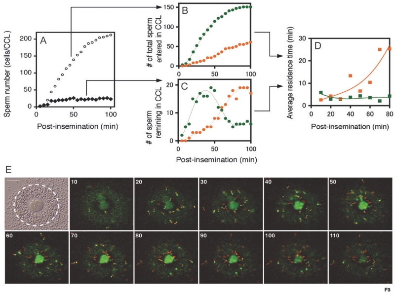
A previous report suggested that most fertilizing sperm undergo AE before binding to the ZP (Jin et al., 2011). In this context, progesterone has remained another favorable candidate for inducing AE. Because the fertilization rate is higher with cumulus-intact oocytes than with cumulus-denuded oocytes (Jin et al., 2011) and progesterone is a major secretory product of the cumulus cells surrounding the ovulated oocytes, we evaluated whether sperm moving through the cumulus matrix in vitro would undergo AE. Capacitated sperm from transgenic male mice were used to inseminate COCs cumulus mass mounted between a glass bottom-culture dish and 3 × 3 mm coverslip and the sperm were monitored continuously using a live imaging system. Videos revealed that both acrosome-intact and acrosome-reacted sperm were capable of entering the cumulus. The total number of sperm that entered the cumulus matrices over the time and the number remaining in the cumulus matrix at given time points were counted (Fig. 1A). Interestingly, the total number of sperm inside the cumulus at any given time point remained fairly constant (black diamonds) while the cumulative number of sperm entering the COCs increased over time (white circles), indicating that large numbers of sperm were able to leave the cumulus. Approximately 90% of sperm entering the cumulus swam out of it by 100-min post-insemination. The rate of entry of acrosome-intact sperm into the cumulus decreased with time, but remained constant for acrosome-reacted sperm (Fig. 1B). Accordingly, a transition from acrosome-intact to acrosome-reacted sperm in the cumulus matrix occurred within ~60 min post-insemination (Fig. 1C). The average residence time in the cumulus, estimated by equations (see SI Materials and Methods), was 7.1 ± 2.2 min for acrosome-intact sperm and 26.4 ± 4.8 min for the acrosome-reacted sperm (Fig. 1D, Table 1). The representative images at different time points after the insemination are shown in figure 1E. After 80 min, we documented the transition from acrosome-intact to an acrosome-reacted population as reflected by the number of sperm containing EGFP in their acrosomes.
Figure 1.
Characterization of sperm migration through the cumulus matrix in vitro according to the acrosomal status. A) The plots represent the cumulative number of sperm entering the COCs (open circles) and the number of sperm remaining in the COCs (closed diamonds) at given times after insemination. The COCs area was drawn as a broken circle as shown in E. B) In the analysis of type of sperm entering the COCs, an apparent logistic increase of acrosome-intact sperm (green), and a relatively low but constant increase of acrosome-reacted sperm (orange), were seen. C) In respect to the sperm number remaining in the COCs, acrosome-intact sperm (green) reached a maximum at ~40 min post-insemination, followed by the rapid accumulation of acrosome-reacted sperm (orange). D) Representative time course of the average residence time of acrosome-intact (green) and acrosome-reacted (orange) sperm in the COCs estimated from equations (Materials & Methods). E) Representative photographs show a transition of the sperm population (from acrosome-intact to acrosome-reacted) as a function of time; scale bar, 100μm.
Table 1.
Acrosomal exocytosis of sperm traveling through the cumulus cells matrix. Data represent 26 experiments (n=26). Average residence time is expressed as the mean ± SEM. AI: acrosome-intact sperm; AR: acrosome-reacted sperm.
| Average residence time of AI sperm in the COCs (min) | 7.1 ± 2.2 |
| Average residence time of AR sperm in the COCs (min) | 26.4 ± 4.8 |
| Total number of sperm that entered the COCs in 90 min | 233 (100 %) |
| Number of AI sperm that entered the COCs | 166 (71.3 %) |
| Number of AR sperm that entered the COCs | 67 (28.7 %) |
| Total number of sperm that underwent AE within the COCs | 8 (4.8 %) |
| Rate of AE in HTF medium (%/ hour) | 10 |
| Rate of AE in cumulus matrix (%/ hour) | 7 |
Next, we quantified the percentage of sperm undergoing AE while travelling through the cumulus. Within the cumulus, 166 sperm with intact acrosomes and 67 sperm without acrosomes reached the ZP (Table 1). However, only 8 out of 166 acrosome intact sperm underwent AE within the cumulus (4.8%). The rate of AE by sperm in the cumulus was only 7%/h during 90 min following insemination. Because the rate of spontaneous AE in HTF-medium was ~10%/h, it is unlikely that induction of AE was promoted by direct interaction with COC under our IVF conditions. Similarly, given that the in vitro fertilization conditions do not completely reflect what occurs in vivo, the question of the site of sperm AE should be addressed within the female reproductive tracts. This led us to examine where AE takes place in the oviduct, before sperm reach the COCs.
Time course of sperm migration and fertilization in vivo
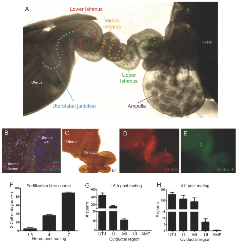
To understand where AE occurs in sperm migrating through the oviduct, the number and acrosomal state of sperm were determined in different regions of the oviduct: utero-tubal junction (UTJ), lower isthmus (LI), middle isthmus (MI), upper isthmus (UI) and ampulla (AMP) (Figure 2A). The ampulla included the ampullary-isthmus junction. Most of the sperm storage reservoir is located in the lower isthmus. Female reproductive tracts were dissected out after copulation of C57BL/6J×BALB/c wild-type females with CAG/su9-DsRed2, Acr3-EGFP double knock-in males. Because of the transparency of the mouse female reproductive tract and the bright green and red fluorescence of the transgenic sperm, it was possible to record precisely the migration of sperm in excised oviducts using live video imaging (Yamaguchi et al., 2009) (Figure 2C-D). After mating, a large number of sperm were deposited in the uterus (Fig.2B) and only a small fraction of uterine sperm entered the oviduct by passing through the UTJ. Sperm were seen within the lumen of the oviduct 30 - 45 min post coitus and populated most of the lower segments of the oviduct after 1.5 h post coitus (Figure 2C-D).
Figure 2.
Evaluation of the timing of in vivo fertilization in mouse. A) Representative diagram showing the different parts of the oviduct that were evaluated in this study; B) Representative image of a cross section of the uterus. Sperm were deposited in the uterus after natural mating. C, D and E) Transgenic sperm in the uterus and the oviduct after mating. The bright field image is shown in C. The DsRed2 (D) and the EGFP (E) fluorescence is visible through the uterus and oviductal walls. Most of the sperm are located in the uterus while very few cross the UTJ and locate in the lower isthmus. F) Time course of in vivo fertilization. The ovulated eggs were removed from the ampulla after 1.5, 4 or 7 h post mating and incubated in culture medium for 24 h. Then, the percentage of two-cell embryos was recorded. G) Number of sperm quantified using z-stacks images of confocal microscopy in each segment of the oviduct after 1.5 h post mating. H) Number of sperm quantified using z-stacks images of confocal microscopy in each segment of the oviduct after 4 h post mating. Data represent the mean ± standard error of the mean (SEM; n = 5 experiments).
In the first set of experiments, we investigated the “fertilization window” with respect to completion of mating. Ovulated unfertilized and fertilized oocytes were retrieved by flushing the ampullae with culture medium at specific time points after mating. The recovered oocytes were incubated for 24 h in 5% CO2 at 37°C and fertilization was evaluated by assessing the percentage of eggs that reached the two-cell stage. We found that at 1.5 h post mating, 0 – 5% of the MII eggs were fertilized. In contrast, at 4 h postmating, ~40% of oocytes recovered were fertilized. By 7 h after mating, more than 90% of the oocytes had had been fertilized (Figure 2E), indicating that fertilization continued for several hours in vivo.
In the next set of experiments, the oviducts were dissected out 1.5 h and 4 h after the mating period and sperm within the oviducts were immediately examined using confocal microscopy to quantify the numbers of sperm in each segment. As expected from the fertilization window experiments, sperm migration through the oviduct was gradual; i.e., most of sperm were found in the lower segments of the oviduct (UTJ to middle isthmus) 1.5 h after mating, and then only small numbers of sperm had reached the upper isthmus and ampulla 4 h after mating (Fig 2F-G).
When oviducts from naturally ovulated and superovulated females were compared and we found no significant differences in the time-course of sperm migration (Supplemental Figure 1). In addition, because under natural conditions, most female mice mate some hours before ovulation, we have also conducted experiments where mating occurred before 4:00 AM. The time course of fertilization as judged by the number of two-cell embryos obtained at 1.5, 4 and 7 h post ovulation as well as the distribution of sperm in the different sections of the oviduct was similar to what occurs when mating occurred at the time of ovulation (Supplementary Figure 2).
Most sperm that migrated through the UTJ and formed the sperm reservoir in the isthmus were acrosome-intact.
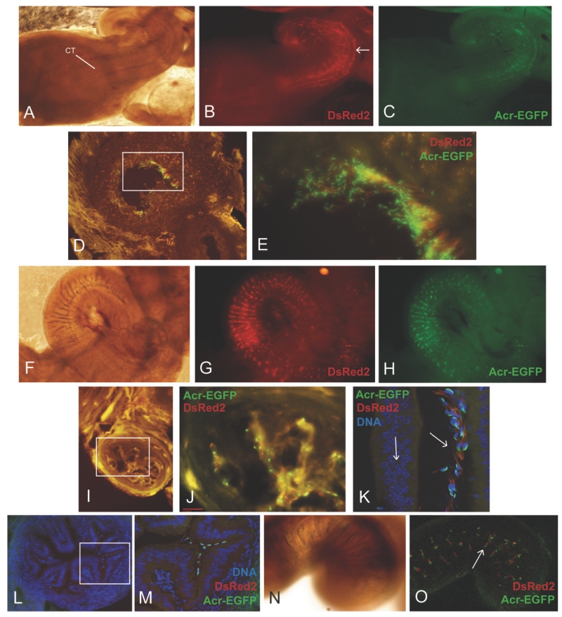
In the UTJ, we observed that sperm migrated through the pockets created by the longitudinal folds (Figure 3 A-C). The heads appeared to adhere to the epithelium but this was not as evident as in the isthmus (Fig. 3D-E). Sperm binding to epithelium was more evident in the upper region of the UTJ. Sperm migration into the UTJ seemed to occur within 1.5 h of the end of the mating period because the number of sperm in this region did not increase during next 2.5 h (Fig. 2F-G). Over 97% of the sperm in the UTJ were acrosome-intact.
Figure 3.
Sperm migration through the UTJ and Isthmus. A) Bright field image of the uterus, UTJ and lower segment of the isthmus. The arrow indicates the colliculus tubarius (CT) protruding into the uterine lumen. B) DsRed2 fluorescence of the image shown in A. Sperm tend to migrate through the UTJ in the pockets between mucosal folds. The arrow in B indicates the mucosal fold were most of the sperm are located. C) EGFP fluorescence of the image shown in A. Most of the sperm in this region have substantially intact acrosomes. D) Cross section of the UTJ after 1.5 h post mating E). Higher magnification of the area depicted in D. Most of the sperm in this region are acrosome intact. F) Bright field representative image the lower isthmus. G and H) DsRed2 and EGFP fluorescence images of 3A respectively. I) Bright field image of a frozen section of the lower isthmus. Sperm were distributed in folds as well as in the central portion of the lumen, in particular, in the region of the lower isthmus that is close to the UTJ. Sperm that locate in the lumen were swimming freely in contrast to the sperm that were located in the folds that were epithelium-bound sperm. J) Higher magnification image of the area depicted in I. K) Representative fluorescent image (merge of DAPI, EGFP and DsRed2) of a cross section of the lower isthmus showing the acrosome intact sperm located in the oviductal folds. The right arrow indicates the oviductal fold. The left arrow indicates the oviductal wall. L) Representative confocal image of a cross section of the middle oviductal isthmus after 4 h post mating. M) Higher magnification image of the area depicted in L. N) Representative bright field image of the middle isthmus. O) Confocal images showing transgenic sperm located in the oviductal folds (arrow).
After crossing the UTJ, sperm moved into the lower isthmus (Figure 3F-G). This region contained the majority of the sperm and is considered to serve as a sperm reservoir (Suarez, 2002). Sperm were distributed in folds as well as in the central portion of the lumen (Supplementary video 1), in particular, proximal to the UTJ (Fig. 3I-K). Sperm in the central portion of the lumen were swimming freely, whereas sperm located in the pockets formed by mucosal folds were bound to the epithelium (Fig. 3H-J).
It was also observed that a number of sperm were not attached to the oviductal epithelium (Supplementary video 2) while others were firmly attached by the convex surface of the falciform head (Supplementary video 3). In other areas of the lower isthmus, we could only observe sperm attached to the epithelium and these were mainly found in the pockets formed by mucosal folds (Supplementary video 4).
In the lower isthmus as in the UTJ, over 95% of the sperm were acrosome-intact regardless mating occurred before or during ovulation (Supplementary Figure 2). The region that is located between the sperm reservoir in the lower isthmus and the upper isthmus was arbitrarily called the middle isthmus. In this region, fewer sperm were observed and they were all located in pockets formed by mucosal folds (Fig. 3L-O and Supplementary video 5). All of the sperm in this region were attached to the epithelium and as observed in the UTJ and lower isthmus, over 95% of them were acrosome-intact.
A significant number of sperm underwent AE in the upper isthmus
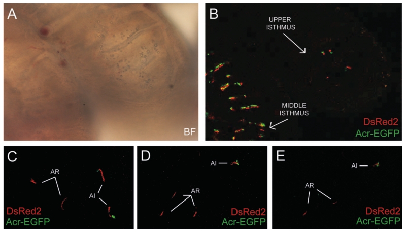
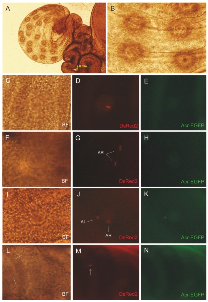
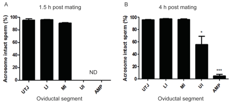
Very few sperm were detected in the upper isthmus at all time points. Most sperm were live and attached to the epithelium, although occasionally we observed some swimming freely (Supplementary video 8 and 9). Interestingly, about 38% of sperm did not show green fluorescence, indicating that they had already undergone AE (Figure 4). Similar results were observed when mating occurred before ovulation (Supplementary Figure 2D-G). In the ampulla, at 1.5 h after the mating period, we did not observe any sperm; however, this did not necessarily mean that the sperm were not arriving at the site of fertilization because about 3% of the eggs had been fertilized by that time. At 4 h, some sperm were detected in each ampulla in a ratio of about 1:1 with the ovulated eggs present. Only 5% of the sperm in the ampulla were acrosome-intact (Fig. 5; Supplementary videos 6 and 7). Occasionally, the red fluorescence of the sperm mitochondria was observed inside the cytoplasm of the oocytes, indicating that fertilization had occurred (Fig.5 C-E). Other times, we observed more than one sperm within the cumulus close to an oocyte (Fig.5 F-K). By analyzing the percentage of acrosome-intact and reacted sperm in the ampulla, we found that the majority of the cells that reached the ZP or its vicinity were motile and acrosome reacted (Supplementary Figure 3). Occasionally, we observed immotile sperm which may represent those who crossed the ZP and started to fuse with the eggs (Satouh et al., 2012). Similarly, all the sperm that were swimming within the cumulus were motile and acrosome-reacted (Supplementary Figure 3). A representative image showing the acrosomal status of sperm in the ampulla and in the upper isthmus where both gametes had lost their acrosomes is shown in Supplementary Figure 4. The percentage of acrosome-intact sperm at different time points in the different regions of the oviduct is shown in Figure 6.
Figure 4.
The number of acrosome reacted sperm increases in the upper isthmus. A (bright field) and B) (fluorescent) confocal images showed very few sperm in the upper isthmus at 4 h post mating. Note that the number of sperm is markedly different compared to what is observed in the middle isthmus (panel A, bottom left corner). C-E) Fluorescent confocal images of sperm in the upper isthmus. In this region, around 40% of the sperm underwent acrosomal exocytosis prior to entering the ampulla. The arrows indicate acrosome reacted (AR) and acrosome intact (AI) sperm.
Figure 5.
Most of the sperm in the ampulla underwent acrosomal exocytosis. A) Representative image of an ampulla after ovulation. B) The eggs were easily observed through the oviductal wall. C-E) Representative images of a fertilized egg where it was possible to observe the fluorescence of the tail. F-H) Representative images of two sperm in the vicinity of an egg. Both sperm lost their acrosomes. I-K) Representative images of two sperm in the vicinity of an egg. In this case, one sperm was acrosome intact (AI) while the other already underwent acrosomal exocytosis (AR). L-N) Representative example of one acrosome reacted sperm swimming through 3 ovulated eggs (indicated by arrows). BF: bright field image,
Figure 6.
Most of the sperm underwent acrosomal exocytosis in the upper segments of the oviductal isthmus. Percentage of sperm that undergo acrosomal exocytosis in the different section of the oviduct after 1.5 h (A) and 4 h (B) post natural mating. UTJ; utero-tubal junction; LI: lower isthmus; MI: middle isthmus; UP: upper isthmus; AMP: ampulla. Results are expressed as the mean ± SEM of 5 independent experiments of the lower segments and 12 of the upper segments. * and *** represents significant difference at P<0.05 and P<0.001 respectively (compared to the middle isthmus).
4. Discussion
Our observations not only provide further challenges to the belief that AE is primarily induced by proteins of the ZP of the oocyte (Arnoult et al., 1996), but surprisingly indicate that a significant proportion of sperm undergo AE before they even reach the cumulus oophorus. It should be reiterated that most of the reports of ZP induction of AE were performed in vitro, outside of the oviduct, and most often in the absence of cumulus (Florman and Storey, 1982; Saling et al., 1979; Storey et al., 1984). In our studies of sperm in the oviduct, the females were inseminated by mating; therefore, the sperm had not only been exposed to oviduct and cumulus, but had also been exposed to seminal plasma and to the lower female tract. This could have primed them differently (and more physiologically) than the sperm collected from the epididymis for use in most studies of ZP-induced AE (McGraw et al., 2015). To our knowledge, this is the first report of assessment of AE in ejaculated sperm swimming within the mouse oviduct.
There are three possible scenarios in which sperm may initiate AE while travelling to the site of fertilization: (i) sperm undergo exocytosis at the ZP surface after stimulation by a cumulus and/or ZP component; (ii) sperm undergo AE within the cumulus by cellular or acellular factors or at the surface of cumulus by cumulus matrix and soluble factors; (iii) sperm undergo AE in the oviduct by factors of the cumulus or oviduct origin (Buffone et al., 2014). In light of recent studies, this first scenario is not the prevalent mechanism in mouse sperm, although this hypothesis remains to be confirmed by in vivo studies. Regarding the second scenario, by using live imaging, we observed that very few sperm underwent AE within the cumulus matrix. If ZP or its surrounding cumulus components are not the primary inducers of AE, what could be the alternative? Progesterone, a major secretory product of the cumulus cells surrounding ovulated oocytes, has been long known to stimulate or prime AE (Osman et al., 1989; Roldan et al., 1994) although this receptor has not yet been identified (Hirohashi et al., 2015). In human sperm, progesterone was demonstrated to modulate the activation of CatSper channels that are essential for the development of hyperactivated motility, but its role in inducing AE is still controversial (Ren et al., 2001). Progesterone is actively produced by cumulus cells after ovulation and its concentration within the cumulus matrix has not been assessed accurately. In mouse, it was recently reported that its concentration within the cumulus cells is in the micromolar range (Abi Nahed et al., 2015). In the oviductal fluid of rabbit and hamster, progesterone was found to be in the range of 2 to 100 nM depending on the phase of the estrous cycle (Libersky and Boatman, 1995; Richardson and Oliphant, 1981). However, the local concentration of this steroid in different segments of the oviduct has not been determined although it is proposed that progesterone secreted by COCs establishes a concentration gradient along the oviduct that is essential for chemotaxis (Guidobaldi et al., 2008). It is plausible that progesterone, given its high concentration in the cumulus, could diffuse to the upper isthmus in sufficient quantities to induce AE although this possibility needs to be explored. It is also possible that exocytosis can be triggered by more than one compound in the middle-to-upper isthmus and lower ampulla. Several molecules were reported to induce acrosome reaction by other groups such as GABA (Shi et al., 1997), Glycine (Bray et al., 2002), atrial natriuretic peptide (Zamir et al., 1995), ATP (Torres-Fuentes et al., 2015), among others. Redundant mechanisms for inducing AE could insure that fertilization is successful. Interestingly, most acrosome-intact sperm were unable to stay in the cumulus permanently in contrast to many acrosome-reacted sperm that remained stacked in it. The molecular mechanisms of how the cumulus cells interact with acrosome-reacted sperm to facilitate their permanence in the matrix remains subject of further investigations. However, this method does not take into consideration that the physiological site for this process is the oviduct where a selected sperm population is capacitated during a close interaction with the epithelium. Thus, we decided to evaluate this process in vivo using natural mating.
We observed that sperm in the UTJ and lower isthmus were acrosome intact. This observation suggests, although it does not prove, that sperm must have intact acrosomes in order to pass into the oviduct. In support of this suggestion, it has been established that mouse sperm must possess certain proteins in the plasma membrane that overlies the acrosome in order to pass through the UTJ (Cho et al., 1998; Ikawa et al., 2011; Marcello et al., 2011; Nishimura et al., 2004; Shamsadin et al., 1999; Tokuhiro et al., 2012). Once sperm reached the upper isthmus, we observed that about 38% of them had undergone AE. Most of the sperm that reached the ampulla had undergone AE, indicating that the physiological AE might take place in the upper segments of the isthmus. Yanagimachi and Mahi (Yanagimachi and Mahi, 1976) demonstrated that the spermatozoa participating in fertilization appeared to undergo the acrosome reaction after they reached the proximal part of the oviduct or when they were very near the eggs. In a previous work from Cummins and Yanagimachi (Cummins and Yanagimachi, 1982), it was reported that in golden hamsters, the majority of sperm in the ampulla had lost their acrosomes or were in the process of undergoing exocytosis. Those sperm that enter the ampulla appeared to be ready to undergo the acrosome reaction and complete it while passing through the cumulus or shortly before. Despite their technical limitations to observe the acrosomal status of the sperm at the time of fertilization, their results are in accordance with our observations. We observed that more than 95% of the sperm that reached the ampulla were already acrosome-reacted when they encountered the cumulus-enclosed eggs. However, It is well established that the sperm population is heterogeneous and, at any given time point, only a fraction of the cells are capacitated while others have completed this process. In the later case, the occurrence of exocytosis might be associated to a biological strategy to prevent them to fertilize the oocyte (Aitken and McLaughlin, 2007; Harper and Publicover, 2005; Uñates et al., 2014), possibly by phagocytosis (Oren-Benaroya et al., 2007). However, because there is currently no way to visually identify the status of sperm capacitation, this hypothesis requires further experimentation.
Jin et al (Jin et al., 2011) showed that most mouse sperm that fertilize cumulus-enclosed oocytes in vitro undergo AE before reaching the ZP. Even though a significant number of the sperm we observed underwent AE in the upper isthmus, we cannot be certain that those sperm go on to fertilize. However, extremely low numbers of sperm are needed in the ampulla in order to ensure successful fertilization. In rodents such as hamster, mouse, rat and Guinea pig, the number of sperm in the ampulla after fertilization is equal to or slightly higher than the number of eggs (Braden and Austin, 1954; Shalgi and Kraicer, 1978; Zamboni, 1972), suggesting that a given sperm that is able to leave the upper isthmus and migrate to the ampulla is highly likely that will end up fertilizing an egg. In addition, we cannot rule out the existence of compensatory or redundant mechanisms where sperm can also undergo AE during their transit through the cumulus or, after binding to the ZP since about 4% of the sperm observed in the ampulla were acrosome intact.
5. Conclusion
We demonstrated for the first time that a significant number of sperm undergo AE in the upper segment of the oviductal isthmus, prior to entering the cumulus mass. This raises new questions about the physiological inducers of the acrosome reaction and whether the acrosomal enzymes serve to do more than simply assist sperm penetration of the ZP.
Supplementary Material
Highlights.
Acrosomal exocytosis rarely occurred within the cumulus cells.
Most sperm that migrated to the lower segments of the oviduct were acrosome-intact.
A significant number of sperm underwent acrosomal exocytosis in the upper isthmus
More than 95% of sperm that reached the ampulla were acrosome reacted.
Acknowledgments
We would like to thank Drs George Gerton, Susan Suarez for their insightful comments and Drs. Shoji Baba, Pablo Pomata, Nicolás Gilio, Juan Cerliani and Trinidad Suarez for their technical assistance. This work was supported by National Institutes of Health (R01TW008662), Agencia Nacional de Promoción Cientifica y Tecnológica (PICT 2013-1175) and Japanese Society for Promotion of Science (JSPS).
Footnotes
Publisher's Disclaimer: This is a PDF file of an unedited manuscript that has been accepted for publication. As a service to our customers we are providing this early version of the manuscript. The manuscript will undergo copyediting, typesetting, and review of the resulting proof before it is published in its final citable form. Please note that during the production process errors may be discovered which could affect the content, and all legal disclaimers that apply to the journal pertain.
Disclosures
No conflict of interest, financial or otherwise, are declared by the authors.
References
- Abi Nahed R, Martinez G, Escoffier J, Yassine S, Karaouzène T, Hograindleur J-P, Turk J, Kokotos G, Ray PF, Bottari S, Lambeau G, Hennebicq S, Arnoult C. Progesterone-induced acrosome exocytosis requires sequential involvement of calcium-independent iPLA2[beta] and group X sPLA2. J. Biol. Chem. 2015 doi: 10.1074/jbc.M115.677799. doi:10.1074/jbc.M115.677799. [DOI] [PMC free article] [PubMed] [Google Scholar]
- Aitken RJ, McLaughlin EA. Molecular mechanisms of sperm capacitation: progesterone-induced secondary calcium oscillations reflect the attainment of a capacitated state. Soc. Reprod. Fertil. Suppl. 2007;63:273–293. [PubMed] [Google Scholar]
- Arnoult C, Zeng Y, Florman HM. ZP3-dependent activation of sperm cation channels regulates acrosomal secretion during mammalian fertilization. J. Cell Biol. 1996;134:637–645. doi: 10.1083/jcb.134.3.637. [DOI] [PMC free article] [PubMed] [Google Scholar]
- Austin CR. Observations on the penetration of the sperm in the mammalian egg. Aust. J. Sci. Res. Ser B Biol. Sci. 1951;4:581–596. doi: 10.1071/bi9510581. [DOI] [PubMed] [Google Scholar]
- Baibakov B, Gauthier L, Talbot P, Rankin TL, Dean J. Sperm binding to the zona pellucida is not sufficient to induce acrosome exocytosis. Dev. Camb. Engl. 2007;134:933–943. doi: 10.1242/dev.02752. doi:10.1242/dev.02752. [DOI] [PubMed] [Google Scholar]
- Braden AW, Austin CR. The number of sperms about the eggs in mammals and its significance for normal fertilization. Aust. J. Biol. Sci. 1954;7:543–551. doi: 10.1071/bi9540543. [DOI] [PubMed] [Google Scholar]
- Bray C, Son J-H, Meizel S. A nicotinic acetylcholine receptor is involved in the arosome reaction of human sperm initiated by recombinant human ZP3. Biol. Reprod. 2002;67:782–788. doi: 10.1095/biolreprod.102.004580. [DOI] [PubMed] [Google Scholar]
- Buffone MG, Hirohashi N, Gerton GL. Unresolved questions concerning mammalian sperm acrosomal exocytosis. Biol. Reprod. 2014;90:112. doi: 10.1095/biolreprod.114.117911. doi:10.1095/biolreprod.114.117911. [DOI] [PMC free article] [PubMed] [Google Scholar]
- Buffone MG, Rodriguez-Miranda E, Storey BT, Gerton GL. Acrosomal exocytosis of mouse sperm progresses in a consistent direction in response to zona pellucida. J. Cell. Physiol. 2009;220:611–620. doi: 10.1002/jcp.21781. doi:10.1002/jcp.21781. [DOI] [PubMed] [Google Scholar]
- Chang MC. Fertilizing capacity of spermatozoa deposited into the fallopian tubes. Nature. 1951;168:697–698. doi: 10.1038/168697b0. [DOI] [PubMed] [Google Scholar]
- Cherr GN, Lambert H, Meizel S, Katz DF. In vitro studies of the golden hamster sperm acrosome reaction: completion on the zona pellucida and induction by homologous soluble zonae pellucidae. Dev. Biol. 1986;114:119–131. doi: 10.1016/0012-1606(86)90388-x. [DOI] [PubMed] [Google Scholar]
- Cho C, Bunch DO, Faure JE, Goulding EH, Eddy EM, Primakoff P, Myles DG. Fertilization defects in sperm from mice lacking fertilin beta. Science. 1998;281:1857–1859. doi: 10.1126/science.281.5384.1857. [DOI] [PubMed] [Google Scholar]
- Cummins JM, Yanagimachi R. Sperm-Egg Ratios and the Site of the Acrosome Reaction During In Vivo Fertilization in the Hamster. Gamete Res. 1982;5:239–256. [Google Scholar]
- Dam AHDM, Feenstra I, Westphal JR, Ramos L, van Golde RJT, Kremer J. a. M. Globozoospermia revisited. Hum. Reprod. Update. 2007;13:63–75. doi: 10.1093/humupd/dml047. doi:10.1093/humupd/dml047. [DOI] [PubMed] [Google Scholar]
- Florman HM, Storey BT. Mouse gamete interactions: the zona pellucida is the site of the acrosome reaction leading to fertilization in vitro. Dev. Biol. 1982;91:121–130. doi: 10.1016/0012-1606(82)90015-x. [DOI] [PubMed] [Google Scholar]
- Guidobaldi HA, Teves ME, Uñates DR, Anastasía A, Giojalas LC. Progesterone from the cumulus cells is the sperm chemoattractant secreted by the rabbit oocyte cumulus complex. PloS One. 2008;3:e3040. doi: 10.1371/journal.pone.0003040. doi:10.1371/journal.pone.0003040. [DOI] [PMC free article] [PubMed] [Google Scholar]
- Harper CV, Publicover SJ. Reassessing the role of progesterone in fertilization--compartmentalized calcium signalling in human spermatozoa? Hum. Reprod. Oxf. Engl. 2005;20:2675–2680. doi: 10.1093/humrep/dei158. doi:10.1093/humrep/dei158. [DOI] [PubMed] [Google Scholar]
- Hasuwa H, Muro Y, Ikawa M, Kato N, Tsujimoto Y, Okabe M. Transgenic mouse sperm that have green acrosome and red mitochondria allow visualization of sperm and their acrosome reaction in vivo. Exp. Anim. Jpn. Assoc. Lab. Anim. Sci. 2010;59:105–107. doi: 10.1538/expanim.59.105. [DOI] [PubMed] [Google Scholar]
- Hirohashi N, Spina FAL, Romarowski A, Buffone MG. Redistribution of the intra-acrosomal EGFP before acrosomal exocytosis in mouse spermatozoa. Reprod. Camb. Engl. 2015;149:657–663. doi: 10.1530/REP-15-0017. doi:10.1530/REP-15-0017. [DOI] [PMC free article] [PubMed] [Google Scholar]
- Ikawa M, Tokuhiro K, Yamaguchi R, Benham AM, Tamura T, Wada I, Satouh Y, Inoue N, Okabe M. Calsperin is a testis-specific chaperone required for sperm fertility. J. Biol. Chem. 2011;286:5639–5646. doi: 10.1074/jbc.M110.140152. doi:10.1074/jbc.M110.140152. [DOI] [PMC free article] [PubMed] [Google Scholar]
- Inoue N, Satouh Y, Ikawa M, Okabe M, Yanagimachi R. Acrosome-reacted mouse spermatozoa recovered from the perivitelline space can fertilize other eggs. Proc. Natl. Acad. Sci. U. S. A. 2011;108:20008–20011. doi: 10.1073/pnas.1116965108. doi:10.1073/pnas.1116965108. [DOI] [PMC free article] [PubMed] [Google Scholar]
- Jin M, Fujiwara E, Kakiuchi Y, Okabe M, Satouh Y, Baba SA, Chiba K, Hirohashi N. Most fertilizing mouse spermatozoa begin their acrosome reaction before contact with the zona pellucida during in vitro fertilization. Proc. Natl. Acad. Sci. U. S. A. 2011;108:4892–4896. doi: 10.1073/pnas.1018202108. doi:10.1073/pnas.1018202108. [DOI] [PMC free article] [PubMed] [Google Scholar]
- Kang-Decker N, Mantchev GT, Juneja SC, McNiven MA, van Deursen JM. Lack of acrosome formation in Hrb-deficient mice. Science. 2001;294:1531–1533. doi: 10.1126/science.1063665. doi:10.1126/science.1063665. [DOI] [PubMed] [Google Scholar]
- Kuzan FB, Fleming AD, Seidel GEJ. Successful fertilization in vitro of fresh intact oocytes by perivitelline (acrosome-reacted) spermatozoa of the rabbit. Fertil. Steril. 1984;41:766–770. doi: 10.1016/s0015-0282(16)47847-7. [DOI] [PubMed] [Google Scholar]
- Libersky EA, Boatman DE. Effects of progesterone on in vitro sperm capacitation and egg penetration in the golden hamster. Biol. Reprod. 1995;53:483–487. doi: 10.1095/biolreprod53.3.483. [DOI] [PubMed] [Google Scholar]
- Lin Y-N, Roy A, Yan W, Burns KH, Matzuk MM. Loss of zona pellucida binding proteins in the acrosomal matrix disrupts acrosome biogenesis and sperm morphogenesis. Mol. Cell. Biol. 2007;27:6794–6805. doi: 10.1128/MCB.01029-07. doi:10.1128/MCB.01029-07. [DOI] [PMC free article] [PubMed] [Google Scholar]
- Marcello MR, Jia W, Leary JA, Moore KL, Evans JP. Lack of tyrosylprotein sulfotransferase-2 activity results in altered sperm-egg interactions and loss of ADAM3 and ADAM6 in epididymal sperm. J. Biol. Chem. 2011;286:13060–13070. doi: 10.1074/jbc.M110.175463. doi:10.1074/jbc.M110.175463. [DOI] [PMC free article] [PubMed] [Google Scholar]
- McGraw LA, Suarez SS, Wolfner MF. On a matter of seminal importance. BioEssays News Rev. Mol. Cell. Dev. Biol. 2015;37:142–147. doi: 10.1002/bies.201400117. doi:10.1002/bies.201400117. [DOI] [PMC free article] [PubMed] [Google Scholar]
- Nishimura H, Kim E, Nakanishi T, Baba T. Possible function of the ADAM1a/ADAM2 Fertilin complex in the appearance of ADAM3 on the sperm surface. J. Biol. Chem. 2004;279:34957–34962. doi: 10.1074/jbc.M314249200. doi:10.1074/jbc.M314249200. [DOI] [PubMed] [Google Scholar]
- Oren-Benaroya R, Kipnis J, Eisenbach M. Phagocytosis of human post-capacitated spermatozoa by macrophages. Hum. Reprod. Oxf. Engl. 2007;22:2947–2955. doi: 10.1093/humrep/dem273. doi:10.1093/humrep/dem273. [DOI] [PubMed] [Google Scholar]
- Osman RA, Andria ML, Jones AD, Meizel S. Steroid induced exocytosis: the human sperm acrosome reaction. Biochem. Biophys. Res. Commun. 1989;160:828–833. doi: 10.1016/0006-291x(89)92508-4. [DOI] [PubMed] [Google Scholar]
- Ren D, Navarro B, Perez G, Jackson AC, Hsu S, Shi Q, Tilly JL, Clapham DE. A sperm ion channel required for sperm motility and male fertility. Nature. 2001;413:603–609. doi: 10.1038/35098027. doi:10.1038/35098027. [DOI] [PMC free article] [PubMed] [Google Scholar]
- Richardson LL, Oliphant G. Steroid concentrations in rabbit oviducal fluid during oestrus and pseudopregnancy. J. Reprod. Fertil. 1981;62:427–431. doi: 10.1530/jrf.0.0620427. [DOI] [PubMed] [Google Scholar]
- Roldan ER, Murase T, Shi QX. Exocytosis in spermatozoa in response to progesterone and zona pellucida. Science. 1994;266:1578–1581. doi: 10.1126/science.7985030. [DOI] [PubMed] [Google Scholar]
- Saling PM, Sowinski J, Storey BT. An ultrastructural study of epididymal mouse spermatozoa binding to zonae pellucidae in vitro: sequential relationship to the acrosome reaction. J. Exp. Zool. 1979;209:229–238. doi: 10.1002/jez.1402090205. doi:10.1002/jez.1402090205. [DOI] [PubMed] [Google Scholar]
- Satouh Y, Inoue N, Ikawa M, Okabe M. Visualization of the moment of mouse sperm-egg fusion and dynamic localization of IZUMO1. J. Cell Sci. 2012;125:4985–4990. doi: 10.1242/jcs.100867. doi:10.1242/jcs.100867. [DOI] [PubMed] [Google Scholar]
- Shalgi R, Kraicer PF. Timing of sperm transport, sperm penetration and cleavage in the rat. J. Exp. Zool. 1978;204:353–360. doi: 10.1002/jez.1402040306. doi:10.1002/jez.1402040306. [DOI] [PubMed] [Google Scholar]
- Shamsadin R, Adham IM, Nayernia K, Heinlein UA, Oberwinkler H, Engel W. Male mice deficient for germ-cell cyritestin are infertile. Biol. Reprod. 1999;61:1445–1451. doi: 10.1095/biolreprod61.6.1445. [DOI] [PubMed] [Google Scholar]
- Shi QX, Yuan YY, Roldan ER. gamma-Aminobutyric acid (GABA) induces the acrosome reaction in human spermatozoa. Mol. Hum. Reprod. 1997;3:677–683. doi: 10.1093/molehr/3.8.677. [DOI] [PubMed] [Google Scholar]
- Sosnik J, Miranda PV, Spiridonov NA, Yoon S-Y, Fissore RA, Johnson GR, Visconti PE. Tssk6 is required for Izumo relocalization and gamete fusion in the mouse. J. Cell Sci. 2009;122:2741–2749. doi: 10.1242/jcs.047225. doi:10.1242/jcs.047225. [DOI] [PMC free article] [PubMed] [Google Scholar]
- Storey BT, Lee MA, Muller C, Ward CR, Wirtshafter DG. Binding of mouse spermatozoa to the zonae pellucidae of mouse eggs in cumulus: evidence that the acrosomes remain substantially intact. Biol. Reprod. 1984;31:1119–1128. doi: 10.1095/biolreprod31.5.1119. [DOI] [PubMed] [Google Scholar]
- Suarez SS. Control of hyperactivation in sperm. Hum. Reprod. Update. 2008;14:647–657. doi: 10.1093/humupd/dmn029. doi:10.1093/humupd/dmn029. [DOI] [PubMed] [Google Scholar]
- Suarez SS. Formation of a reservoir of sperm in the oviduct. Reprod. Domest. Anim. Zuchthyg. 2002;37:140–143. doi: 10.1046/j.1439-0531.2002.00346.x. [DOI] [PubMed] [Google Scholar]
- Tokuhiro K, Ikawa M, Benham AM, Okabe M. Protein disulfide isomerase homolog PDILT is required for quality control of sperm membrane protein ADAM3 and male fertility [corrected] Proc. Natl. Acad. Sci. U. S. A. 2012;109:3850–3855. doi: 10.1073/pnas.1117963109. doi:10.1073/pnas.1117963109. [DOI] [PMC free article] [PubMed] [Google Scholar]
- Torres-Fuentes JL, Rios M, Moreno RD. Involvement of a P2X7 Receptor in the Acrosome Reaction Induced by ATP in Rat Spermatozoa. J. Cell. Physiol. 2015;230:3068–3075. doi: 10.1002/jcp.25044. doi:10.1002/jcp.25044. [DOI] [PubMed] [Google Scholar]
- Uñates DR, Guidobaldi HA, Gatica LV, Cubilla MA, Teves ME, Moreno A, Giojalas LC. Versatile action of picomolar gradients of progesterone on different sperm subpopulations. PloS One. 2014;9:e91181. doi: 10.1371/journal.pone.0091181. doi:10.1371/journal.pone.0091181. [DOI] [PMC free article] [PubMed] [Google Scholar]
- Yamaguchi R, Muro Y, Isotani A, Tokuhiro K, Takumi K, Adham I, Ikawa M, Okabe M. Disruption of ADAM3 impairs the migration of sperm into oviduct in mouse. Biol. Reprod. 2009;81:142–146. doi: 10.1095/biolreprod.108.074021. doi:10.1095/biolreprod.108.074021. [DOI] [PubMed] [Google Scholar]
- Yanagimachi R. Mammalian sperm acrosome reaction: where does it begin before fertilization? Biol. Reprod. 2011;85:4–5. doi: 10.1095/biolreprod.111.092601. doi:10.1095/biolreprod.111.092601. [DOI] [PubMed] [Google Scholar]
- Yanagimachi R, Mahi CA. The sperm acrosome reaction and fertilization in the guinea-pig: a study in vivo. J. Reprod. Fertil. 1976;46:49–54. doi: 10.1530/jrf.0.0460049. [DOI] [PubMed] [Google Scholar]
- Zamboni L. Biology of Mammalian Fertilization and Implantation. Thomas; Springfield, IL. USA: 1972. Fertilization in the mouse; pp. 213–262. [Google Scholar]
- Zamir N, Barkan D, Keynan N, Naor Z, Breitbart H. Atrial natriuretic peptide induces acrosomal exocytosis in bovine spermatozoa. Am. J. Physiol. 1995;269:E216–221. doi: 10.1152/ajpendo.1995.269.2.E216. [DOI] [PubMed] [Google Scholar]
Associated Data
This section collects any data citations, data availability statements, or supplementary materials included in this article.


