Abstract
Purpose:
To provide a detailed analysis of age-related macular degeneration (AMD) with a 5-year follow-up at a Tertiary Eye Care Center in India.
Methods:
In this retrospective institutional study, 408 eyes of 204 subjects (100 males) with a diagnosis of AMD with minimum 5-year follow-up were included. Data collected included demographics, details of the ocular exam, special investigations performed, treatment offered, complications, and systemic diseases, if any.
Results:
The median age was 74.24 ± 8.23 years. Median follow-up was 5.77 years. The visual acuity (VA) at baseline and last visit was 0.74 ± 0.12 (Snellen's equivalent 20/100) and 0.54 ± 0.12 logarithm of the minimum angle of resolution (Snellen's equivalent 20/50; P = 0.032) in patients with choroidal neovascular membrane (CNVM). The most common complaint was decreased vision (94.5%). AMD (any stage) was found to be bilateral in 93% of patients at baseline and 197 patients (96.56%) at 5 years. Seventeen eyes had active CNVM (12 of these were occult) at presentation. At baseline, 43 eyes had a disciform scar. Three hundred twenty-one eyes had dry AMD at baseline (geographic atrophy - 12 [3.7%] eyes). Five-year conversion rate into wet AMD and geographic atrophy was 2.87% and 3.12%. Median number of anti-vascular endothelial growth factor injections administered per patient was 2.8 ± 1.2. CNVM bilaterality was low (7.5%).
Conclusion:
Patients with AMD in India presented later in the course of the disease. Bilateral advanced AMD and geographic atrophy were uncommon. Five-year conversion rate into wet AMD and geographic atrophy was 2.87% and 3.12%.
Keywords: Age-related macular degeneration, anti-vascular endothelial growth factor therapy, hospital-based analysis, India
Age-related macular degeneration (AMD) is an important cause of visual loss in the geriatric population. Several landmark articles describe the patient profile, clinical features, risk factors, and final anatomical and visual outcomes in AMD in the Caucasian and Indian population.[1,2,3,4,5,6] Also, most of these studies are population-based ones, some with a follow-up extending up to two decades. They describe the natural course of the disease, and alterations in the same with intervention. However, there is a relative paucity of literature on the type and stage of AMD in a hospital-based setting, especially in the Indian subcontinent.
The Andhra Pradesh Eye Disease Study was a population-based analysis performed by our institute in the year 1996–2000 that analyzed the incidence and prevalence of AMD in Southern India.[7] The overall prevalence of AMD ranges from 1.8% to 4.7% of the general population in South India.[8] Other studies have also looked at the prevalence of AMD in different geographic regions in India.[4,9] Although population studies are excellent tools for determining the incidence, prevalence and risk factors of AMD, a detailed analysis of these patients (using angiography, optical coherence tomography [OCT], and its interpretation by a retina specialist) is only possible at a Tertiary Eye Care Retina Center. Finally, while trials provide excellent insight into the efficacy and safety of a particular intervention for a particular disease, it is imperative to know the practical points that must be worked out to ensure wider applicability in the population at large.
The current study was performed with the aim of determining the natural history, patient profile, clinical features, stage and type of AMD, and finally a real life scenario of treatment patterns as identified in a Tertiary Eye Care Retina Center when followed up over a period of 5 years. The study also reports the number of patients with early or intermediate AMD, who progress to develop advanced disease and outcomes over a 5-year period in a Tertiary Eye Care Retina Center.
Methods
Patients, who were diagnosed to have AMD in 2008 with a minimum follow-up of 5 years, constitute the study population in this retrospective analysis. Informed consent was obtained from all patients. The study was approved by the Institutional Review Board. The study adhered to the tenets of the Declaration of Helsinki.
For inclusion, patients were required to have any form of AMD, as per age-related eye diseases study (AREDS) protocol[10] with a minimum follow-up of 5 years. A thorough history was obtained from the patients about the onset of symptoms as well as about their past complaints, systemic illnesses, the use of medication, past ocular, and systemic surgery as well as details of habits such as smoking and alcohol consumption. If positive, an assessment was made to determine the extent of use of smoking and alcohol. Details of systemic diseases and therapy were obtained from the patients and examination of their records, and this information was updated at each follow-up. Collected data included demographics, the best-corrected visual acuity (BCVA) at baseline and last visit, details of the ocular and systemic exam, and imaging details including fundus fluorescein angiography (FFA) at baseline, spectral domain (SD)-OCT scans at baseline. Iris color was noted on clinical examination and mentioned in the charts. BCVA assessed using Snellen's chart and was converted to logarithm of the minimum angle of resolution (logMAR) for statistical analysis. Lens grading was performed as per the standard emery and little classification.[11]
The diagnosis was confirmed, and subanalysis performed through fundus photography, including fundus autofluorescence (FAF) images, OCT analysis and, when required, with the help of fluorescein and indocyanine green angiography. The images were obtained and graded independently by one trained retina specialist (AS).
AMD was classified as per AREDS protocol.[10] Patients with multiple small drusen, few intermediate drusen (63–124 microns in diameter), or retinal pigment epithelium abnormalities were considered to have early AMD. Patients with extensive intermediate drusen and at least one large druse (≥125 microns in diameter), or geographic atrophy not involving the center of the fovea were considered to have intermediate AMD. Patients with geographic atrophy involving the fovea and/or any of the features of neovascular AMD were considered to have late AMD.
Imaging procedures
Color fundus photograph
Color fundus photographs were obtained using the standard seven fields as per the Early Treatment Diabetic Retinopathy Study classification, and the image centered on the disk and fovea was used for analysis using a mydriatic camera (Zeiss FF450, Carl Zeiss Meditec, Jena, Germany).
Fundus autofluorescence
FAF imaging was performed with a confocal scanning laser ophthalmoscope (Heidelberg Retina Angiograph 2; Heidelberg Engineering, Dossenheim, Germany) and the same operator acquired all images. For the acquisition of FAF images, a standard procedure was followed, which included focusing of the retinal image in the infrared reflection mode at 820 nm, sensitivity adjustment at 488 nm, and acquisition of 30° FAF images, which encompassed the entire macular area and at least part of the optic disc. Images were digitized and saved on hard discs for further analysis and processing.
Spectral domain optical coherence tomography
SD-OCT was performed for both eyes in all patients with dilated pupils (pupillary dilatation with tropicamide 1% eye drop) using RTVue SD-OCT Machine (Optovue, Inc., Freemont, CA, USA) or Cirrus HD-OCT (Carl Zeiss Meditec, Inc., Dublin, CA, USA). Six mm line scans were performed through the central macula along with a two-dimensional raster scan and a three-dimensional reconstruction of macula centered over the fovea.
Fundus fluorescein angiography
FFA was performed using 20% fluorescein dye and the Zeiss Visucam Lite (Zeiss Meditec, India).
Treatment procedures
Photodynamic therapy
Photodynamic therapy (PDT) with verteporfin (Novartis Pharmaceuticals, East Hanover, New Jersey, USA) (6 mg/m2 intravenously) followed by irradiation (spot size of 1000 microns guided by fluorescein angiography at 600 mW/cm2 with 692 nm of light for a total fluence of 50 J/cm2) for 83 s, 15 min after infusion initiation (similar to the protocol for treatment of neovascular macular degeneration) was performed.
Anti-vascular endothelial growth factor therapy
Treatment consisted of intravitreal injection of 1.25 mg/0.05 mL of bevacizumab (Avastin; Genentech, South San Francisco, CA, USA) or 0.5 mg/0.05 mL of ranibizumab (Lucentis; Genentech, South San Francisco, CA, USA). After obtaining informed consent, injection was given as per standard protocol. Before the injection, topical anesthesia and ocular surface disinfection with 5% povidone-iodine were performed under topical anesthesia followed by a short course of topical antibiotics. The choice of agent was discussed between the patients and the treating physician.
Postoperative period was uneventful in all patients. At each follow-up, SD-OCT scans were repeated. Re-treatment was performed in case of persistence of subretinal haemorrhage on clinical examination and or persistent activity on FFA and/or persistence of subretinal or intra-retinal fluid on SD-OCT scan. In case of poor response, angiography was repeated as per physician's discretion. We defined a complete response as no signs of activity such as intra-retinal or subretinal fluid on SD-OCT scans.
Combination therapy
PDT in combination with anti-vascular endothelial growth factor (anti-VEGF) injections was used for patients with CNVM not responding to anti-VEGF monotherapy as suggested by previous reports.[12]
Statistical analysis
Statistical analysis included descriptive data, the paired t-test and the Chi-square test, wherever appropriate. Generalized estimating equations were used to ensure that data from both eyes could be included for analyses. Based on past data, univariate analysis was performed to determine whether independent variables such as sex, iris color, phakic status, the presence of systemic diseases such as hypercholesterolemia, diabetes mellitus, and hypertension, and a history of smoking and alcoholism influenced the dependent variables such as presence of dry and wet AMD and conversion from the dry form to the wet AMD form. Factors that had significant correlation in univariate analysis were analyzed in a multivariate regression model as well to determine its significance and to eliminate confounding.
Results
The study period was January 2008–December 2013. A total of 239 patients were diagnosed to have AMD as per the AREDS classification.[10] After excluding 8 patients due to poor quality images and 27 patients due to follow-up of <5 years, the final analysis was performed on 204 patients (408 eyes), and this constitutes the study.
Patient profile and complaints
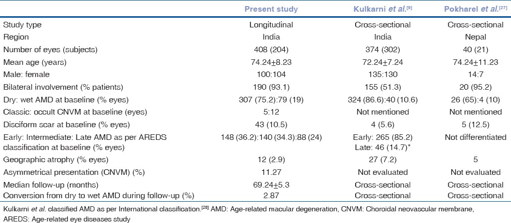
The median age of the patients was 74.24 ± 8.23 years with a range of 63–93 years. Both eyes were included in all analyses. The most common complaint was decreased vision (385 patients; 94.5%), followed by inability to recognize faces (330 patients; 80.42%) and reading disability (78.4%). One-hundred seven patients (26.22%) had metamorphopsia. Only 17 patients (4%) of these were asymptomatic and had been referred here at the behest of an ophthalmologist with a diagnosis of AMD. Table 1 summarizes the demographic, clinical, and investigative characteristics of all patients. Kulkarni et al.[9] classified AMD as per international classification.
Table 1.
Comparative analysis of hospital-based studies from the Indian subcontinent

Systemic disease, smoking, and alcohol status
Twenty-five patients (12.25%, 22 males) had a history of smoking for at least 10 years. Of these 25 patients, only one had evidence of a choroidal neovascular membrane (CNVM); the rest had dry AMD, of which two had geographical atrophy and the rest had intermediate AMD. Forty-nine patients had a history of alcohol intake for the past 10 years at least. Four of these 49 patients had CNVM while two had a disciform scar; seven had geographic atrophy, and the rest had early form of dry AMD.
Twenty-eight patients had Type II diabetes while 31 patients had hypertension. Twenty-nine patients had dyslipidemia. Ten patients had Type II diabetes and hypertension while 13 patients had all three co-morbidities. None of the patients with these systemic co-morbidities had any evidence of retinopathy secondary to these specific conditions. Fourteen patients were on anticoagulants.
Anterior segment examination
Two hundred twenty-six (55.39%) eyes were phakic with 148 (36.27%) of these had some evidence of cataract. One hundred seventy-four (42.67%) eyes were pseudophakic, and 8 (1.96%) eyes were aphakic. The predominant iris color was brown (384 eyes, 94%) followed by green (21 eyes, 5.2%) and grey/blue (3 eyes, 0.8%) eyes. Of the 148 phakic patients, 67 underwent cataract surgery in 5 years follow-up period, and this was included in the final VA analysis. Twenty-eight pseudophakic patients required a neodymium-doped yttrium aluminum garnet laser capsulotomy in the period of 5 years. This was factored in when determining the final VA.
Wet age-related macular degeneration
Seventeen eyes (4.16%) had active choroidal neovascularization of which 12 (70.58%) had a predominantly occult configuration and the rest had a predominantly classic configuration. Of the 17 eyes with active CNVMs, 6 eyes had a subfoveal lesion while 11 eyes had an extrafoveal lesion. Nineteen eyes (4.65%) had scarring CNVM with minimal activity as detected on fluorescein angiography and a further 43 eyes (10.53%) had a disciform scar. These 19 eyes with scarring CNVM were more likely to have an occult configuration (14/19 eyes) and a subfoveal component (13/19 eyes).
Dry age-related macular degeneration
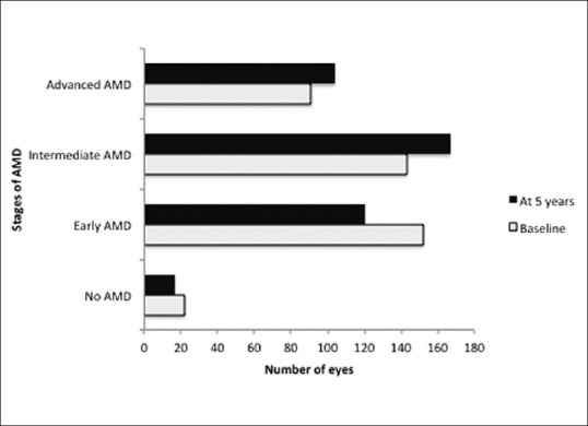
Overall, 307 eyes (75.2%) had dry AMD and 79 eyes (19.36%) had evidence of wet AMD (including the presence of disciform scar). Of these 307 eyes, 12 eyes (3.9%) had geographic atrophy involving the fovea at presentation. At baseline, 152 (37.25%) eyes had early AMD, 143 (35.04%) eyes had intermediate, and 91 (22.3%) eyes had advanced AMD as per the AREDS classification.[10] Twenty-two eyes (5.3%) had no signs of AMD on clinical examination or imaging. Forty-three eyes had a scarred CNVM at baseline.
Patients with early and intermediate AMD (n = 295) had been prescribed oral anti-oxidant therapy. Of these, only 72 patients continued therapy for 5 years. Two-hundred twenty patients had discontinued therapy within 2 years of initiation; however, 103 patients did not receive therapy at all.
Five years follow-up
At 5-year follow-up, 37 (25.0%) eyes with early AMD had progressed to intermediate AMD. At 5-year follow-up, 7 eyes had active CNVM and 85 (20.8%) eyes had a scarred CNVM [Fig. 1]. All seven eyes developed CNVM over the course of 5 years and did not have CNVM at baseline. These seven eyes received anti-VEGF monotherapy. The incidence of bilaterality (some degree of AMD in both eyes) was 93% (190 patients) at baseline and 96.56% (197 patients) at the last follow-up. The most common type of presentation among those with bilateral wet AMD was asymmetry (87.4%) wherein one eye had advanced disease (scarring/scarred CNVM) while the other eye had active choroidal neovascularization. At 5-year follow-up, 37 (18.1%) patients had CNVM in one eye and dry AMD in the other eye. At baseline, bilateral CNVMs (active) were found in 3 (7.5%) patients. At 5-year follow-up, bilateral CNVM (active) was present in 5 (12.5%) patients.
Figure 1.

Distribution of different stages of age-related macular degeneration among study eyes at baseline and at 5 years follow-up
The median follow-up period of these patients was 69.24 ± 5.3 months. The conversion rate of dry AMD to wet AMD was found to be 2.87% (8 eyes) at 5 years. Five eyes (60%) of those who developed CNVM over 5 years had soft drusen and pigmentary abnormalities at baseline. Likewise, the conversion rate for dry, intermediate AMD to geographic atrophy was 3.12% (5 eyes) at 5 years, and 2 eyes of these five eyes had pigmentary abnormalities and soft, indistinct drusen at baseline. None of the patients with early AMD progressed to geographic atrophy or wet AMD over the 5-year follow-up. We failed to find a significant correlation between location of soft, indistinct or reticular drusen and/or pigmentary abnormalities and the development of either geographic atrophy or CNVM (P = 0.69). Finally, two patients who developed CNVM over the course of 2 years were from that sub-group of patients who had received oral anti-oxidant therapy for 5 years.
Visual acuity and investigations
At baseline, the median corrected distance VA (CDVA) among patients with CNVM was and 0.74 ± 0.12 logMAR (Snellen's equivalent 20/100 approximately) with a range of 0.2–2 logMAR. The final CDVA was significantly better, 0.57 ± 0.12 logMAR (Snellen's equivalent 20/60; P = 0.032). The most common SD-OCT findings in dry AMD were drusen (295 eyes; 96.24%) and drusenoid pigment epithelial detachments (234 eyes; 76.4%). The most common SD-OCT findings in eyes with CNVM were Type I CNVM in 19 eyes (60%) and Type II CNVM in 13 eyes (40%).
Treatment
Correspondingly, 32 (54%) eyes had received therapy in the form of intravitreal injections and/or PDT at the time of analysis. Of these, 5 eyes (15%) had received PDT only (until 2008), 25 eyes (78%) had received anti-VEGF monotherapy (2008 onward), and the remaining 2 eyes (7%) had received combination therapy (2008–12). The use of PDT as monotherapy was almost eliminated after 2008 for treatment of CNVM secondary to AMD in our series. About 81.25% of all patients after 2008 who received anti-VEGF monotherapy, received bevacizumab monotherapy, remaining had both, ranibizumab and bevacizumab. The average number of injections (per patient) in the stated period was 2.8 ± 1.2 (range 1–10 injections). Twenty-four patients (75.24%) received incomplete therapy. The most common reason, as stated by the patients for discontinuation of therapy, was the financial burden imposed upon the patient by repeated injections (accounting for 80% of the dropout rate among 75% patients who did not complete treatment). Concurrent health problems, problems with transportation and unsatisfactory outcomes accounted for the remaining 20% of dropouts.
Univariate analyses
Univariate analysis failed to show statistically significant correlation between either dry or wet AMD and the following independent variables: Gender (P = 0.31), profession (P = 0.23), sunlight exposure (P = 0.41), history of smoking/alcohol intake (P = 0.34), iris color (P = 0.41), phakic status (P = 0.44), systemic co-morbidities such as diabetes mellitus (P = 0.42), hypertension (P = 0.51), and dyslipidemia (P = 0.48), and the use of anticoagulants (P = 0.49).
Discussion
The results from our study show that the AMD pattern observed in India closely matches (in most respects) that seen in past reports on AMD in India as well as the Western world.[2,3,7,8,9,10] This study, to the best of our knowledge, is the first from Indian subjects that give a hospital-based longitudinal perspective on the problem of AMD in the specified region. Whereas, most epidemiological studies are based on color fundus photographs; this study uses detailed information obtained from sophisticated imaging technologies to analyze in depth the distribution of various types of AMD, giving us an insight into the presenting features, the course of disease and the outcome of treatment over 5 years. Although the incidence of AMD in India is considered lower than the West, it still adds up to be a significant visual problem in the country, given the large population base.
Our analysis shows that decreased vision and/or vision-related activities were the most common symptoms at also, a fourth of the patients presented later in the course of the disease, making treatment and visual recovery difficult. Poor vision can increase the risk of other health problems, which could be the reason for delayed presentation.[13]
Whereas, in the West, the number of injections per person per year has been reported to be 13 in the “as needed group” versus 23 in the monthly group over 2 years,[19] and the number of injections in our study is significantly lower. This may be attributable to better education, earlier presentation, better insurance cover, and financial capabilities in Western countries.[14,15,16,17,18] This, however, needs to be analyzed further.
The incidence of late AMD as noted in our study (13.81%) matches very closely with another hospital-based study (15%) from India.[9] The study by Kulkarni et al.[9] was cross-sectional in nature and based on clinical photographs and the authors were thus unable to comment on the CNVM type or the conversion rate. Occult CNVM was the most common type of CNVM noted in our analysis, and this is similar to past reports.[22] The incidence of bilaterality of CNVM (any stage) was 11.27% at 5 years. The hospital-based design accounts for the vast difference noted between our study and other population-based analyses.[20,21,23]
The trend of treatment for AMD as observed in our study follows closely that seen in a recent hospital-based study from Singapore and India that reports increasing numbers of injections beginning 2008, with the number of injections doubling over 3 years.[24] This reflects the immense change effected by the introduction of anti-VEGF agents.
Curiously, as opposed to past studies, we could not find any significant correlation between smoking[25] or the presence of systemic disorders such as diabetes or hypertension and AMD. We did not find any relation between the smoking status and the type of AMD (dry or wet). We believe that this is so because the data is from a hospital setting and likely to be biased. Also, smoking exposure was not evaluated through standardized questionnaires. Notwithstanding, there have been other studies from Asia that have failed to demonstrate a strong correlation between smoking and AMD.[9,26]
The retrospective nature of the study, the limited geographic reach and the small numbers of patients are the main limitations of the study. Another limitation is that these data are from a referral center and, hence, the results cannot be applied to the population at large. Finally, with very few patients developing CNVM, it is worthwhile noting that the OCT characteristics we described lack power. In spite of these limitations, we present several features of interest: This study gives us an accurate and real life scenario of the burden of disease and problems with therapy in a Tertiary Eye Care Centre. The hospital-based setting allows in-depth analysis of the various stages of AMD and identifies patients who would benefit with therapy since it incorporates angiography and OCT in the analysis. It makes available data that can form the basis for deciding policy on the treatment of patients with AMD who are amenable to therapy, and to rehabilitate those who have presented too late as well as identify the regions of the country that need the presence of specialists. It can form the basis for grass level education programs that educate people about this problem, encourage them to be more regular with follow-ups.
Conclusion
Our study is a good attempt to explore the patient profile, type and stage of AMD at presentation, conversion to advanced AMD, and treatment pattern at a Tertiary Eye Care Center. Occult CNVM was the most common form of wet AMD at our center, with more asymmetrical presentation at baseline. Geographic atrophy is uncommon in our population, with a conversion rate of around 2.5% from intermediate AMD to advanced form of AMD in 5 years follow-up. Prospective studies that couple a larger sample size with genetic analysis and longer follow-ups are warranted to improve our understanding of AMD in the Indian population.
Financial support and sponsorship
Nil.
Conflicts of interest
There are no conflicts of interest.
References
- 1.Klein R, Klein BE, Knudtson MD, Meuer SM, Swift M, Gangnon RE. Fifteen-year cumulative incidence of age-related macular degeneration: The beaver dam eye study. Ophthalmology. 2007;114:253–62. doi: 10.1016/j.ophtha.2006.10.040. [DOI] [PubMed] [Google Scholar]
- 2.Wang JJ, Rochtchina E, Lee AJ, Chia EM, Smith W, Cumming RG, et al. Ten-year incidence and progression of age-related maculopathy: The blue mountains eye study. Ophthalmology. 2007;114:92–8. doi: 10.1016/j.ophtha.2006.07.017. [DOI] [PubMed] [Google Scholar]
- 3.Mitchell P, Wang JJ, Foran S, Smith W. Five-year incidence of age-related maculopathy lesions: The blue mountains eye study. Ophthalmology. 2002;109:1092–7. doi: 10.1016/s0161-6420(02)01055-2. [DOI] [PubMed] [Google Scholar]
- 4.Nangia V, Jonas JB, Kulkarni M, Matin A. Prevalence of age-related macular degeneration in rural central India: The central india eye and medical study. Retina. 2011;31:1179–85. doi: 10.1097/IAE.0b013e3181f57ff2. [DOI] [PubMed] [Google Scholar]
- 5.Cheung CM, Li X, Cheng CY, Zheng Y, Mitchell P, Wang JJ, et al. Prevalence, racial variations, and risk factors of age-related macular degeneration in Singaporean Chinese, Indians, and Malays. Ophthalmology. 2014;121:1598–603. doi: 10.1016/j.ophtha.2014.02.004. [DOI] [PubMed] [Google Scholar]
- 6.Woo JH, Sanjay S, Au Eong KG. The epidemiology of age-related macular degeneration in the Indian subcontinent. Acta Ophthalmol. 2009;87:262–9. doi: 10.1111/j.1755-3768.2008.01376.x. [DOI] [PubMed] [Google Scholar]
- 7.Krishnaiah S, Das T, Nirmalan PK, Nutheti R, Shamanna BR, Rao GN, et al. Risk factors for age-related macular degeneration: Findings from the Andhra Pradesh eye disease study in South India. Invest Ophthalmol Vis Sci. 2005;46:4442–9. doi: 10.1167/iovs.05-0853. [DOI] [PubMed] [Google Scholar]
- 8.Krishnan T, Ravindran RD, Murthy GV, Vashist P, Fitzpatrick KE, Thulasiraj RD, et al. Prevalence of early and late age-related macular degeneration in India: The INDEYE study. Invest Ophthalmol Vis Sci. 2010;51:701–7. doi: 10.1167/iovs.09-4114. [DOI] [PMC free article] [PubMed] [Google Scholar]
- 9.Kulkarni SR, Aghashe SR, Khandekar RB, Deshpande MD. Prevalence and determinants of age-related macular degeneration in the 50 years and older population: A hospital based study in Maharashtra, India. Indian J Ophthalmol. 2013;61:196–201. doi: 10.4103/0301-4738.99870. [DOI] [PMC free article] [PubMed] [Google Scholar]
- 10.Mitchell P, Foran S. Age-related eye disease study severity scale and simplified severity scale for age-related macular degeneration. Arch Ophthalmol. 2005;123:1598–9. doi: 10.1001/archopht.123.11.1598. [DOI] [PubMed] [Google Scholar]
- 11.Emery J. Kelman phacoemulsification, patient selection. St. Louis (MO): Mosby; 1983. [Google Scholar]
- 12.Englander M, Kaiser PK. Combination therapy for the treatment of neovascular age-related macular degeneration. Curr Opin Ophthalmol. 2013;24:233–8. doi: 10.1097/ICU.0b013e32835f8eaa. [DOI] [PubMed] [Google Scholar]
- 13.Anastasopoulos E, Yu F, Coleman AL. Age-related macular degeneration is associated with an increased risk of hip fractures in the Medicare database. Am J Ophthalmol. 2006;142:1081–3. doi: 10.1016/j.ajo.2006.06.058. [DOI] [PubMed] [Google Scholar]
- 14.Stein JD, Newman-Casey PA, Mrinalini T, Lee PP, Hutton DW. Cost-effectiveness of bevacizumab and ranibizumab for newly diagnosed neovascular macular degeneration (an American Ophthalmological Society thesis) Trans Am Ophthalmol Soc. 2013;111:56–69. [PMC free article] [PubMed] [Google Scholar]
- 15.Levinson D. Medicare payments for drugs used to treat wet age-related macular degeneration. In Edition Office of Inspector General Department of Health and Human Services. [Last accessed on 2014 Jan 24]. Available from: https://www.oig.hhs.gov/oei/reports/oei-03-10-00360.pdf .
- 16.Biswas P, Sengupta S, Choudhary R, Home S, Paul A, Sinha S. Comparative role of intravitreal ranibizumab versus bevacizumab in choroidal neovascular membrane in age-related macular degeneration. Indian J Ophthalmol. 2011;59:191–6. doi: 10.4103/0301-4738.81023. [DOI] [PMC free article] [PubMed] [Google Scholar]
- 17.Azad R, Chandra P, Gupta R. The economic implications of the use of anti-vascular endothelial growth factor drugs in age-related macular degeneration. Indian J Ophthalmol. 2007;55:441–3. doi: 10.4103/0301-4738.36479. [DOI] [PMC free article] [PubMed] [Google Scholar]
- 18.Neubauer AS, Holz FG, Sauer S, Wasmuth T, Hirneiss C, Kampik A, et al. Cost-effectiveness of ranibizumab for the treatment of neovascular age-related macular degeneration in Germany: Model analysis from the perspective of Germany's statutory health insurance system. Clin Ther. 2010;32:1343–56. doi: 10.1016/j.clinthera.2010.07.010. [DOI] [PubMed] [Google Scholar]
- 19.IVAN Study Investigators. Chakravarthy U, Harding SP, Rogers CA, Downes SM, Lotery AJ, et al. Ranibizumab versus bevacizumab to treat neovascular age-related macular degeneration: One-year findings from the IVAN randomized trial. Ophthalmology. 2012;119:1399–411. doi: 10.1016/j.ophtha.2012.04.015. [DOI] [PubMed] [Google Scholar]
- 20.Biarnés M, Monés J, Alonso J, Arias L. Update on geographic atrophy in age-related macular degeneration. Optom Vis Sci. 2011;88:881–9. doi: 10.1097/OPX.0b013e31821988c1. [DOI] [PubMed] [Google Scholar]
- 21.Gregor Z, Joffe L. Senile macular changes in the black African. Br J Ophthalmol. 1978;62:547–50. doi: 10.1136/bjo.62.8.547. [DOI] [PMC free article] [PubMed] [Google Scholar]
- 22.Olsen TW, Feng X, Kasper TJ, Rath PP, Steuer ER. Fluorescein angiographic lesion type frequency in neovascular age-related macular degeneration. Ophthalmology. 2004;111:250–5. doi: 10.1016/j.ophtha.2003.05.030. [DOI] [PubMed] [Google Scholar]
- 23.Wang JJ, Mitchell P, Smith W, Cumming RG. Bilateral involvement by age related maculopathy lesions in a population. Br J Ophthalmol. 1998;82:743–7. doi: 10.1136/bjo.82.7.743. [DOI] [PMC free article] [PubMed] [Google Scholar]
- 24.Shanmugam PM. Changing paradigms of anti-VEGF in the Indian scenario. Indian J Ophthalmol. 2014;62:88–92. doi: 10.4103/0301-4738.126189. [DOI] [PMC free article] [PubMed] [Google Scholar]
- 25.Thornton J, Edwards R, Mitchell P, Harrison RA, Buchan I, Kelly SP. Smoking and age-related macular degeneration: A review of association. Eye (Lond) 2005;19:935–44. doi: 10.1038/sj.eye.6701978. [DOI] [PubMed] [Google Scholar]
- 26.Miyazaki M, Nakamura H, Kubo M, Kiyohara Y, Oshima Y, Ishibashi T, et al. Risk factors for age related maculopathy in a Japanese population: The Hisayama study. Br J Ophthalmol. 2003;87:469–72. doi: 10.1136/bjo.87.4.469. [DOI] [PMC free article] [PubMed] [Google Scholar]
- 27.Pokharel S, Malla OK, Pradhananga CL, Joshi SN. A pattern of age-related macular degeneration. JNMA J Nepal Med Assoc. 2009;48:217–20. [PubMed] [Google Scholar]
- 28.Sallo FB, Peto T, Leung I, Xing W, Bunce C, Bird AC. The International Classification system and the progression of age-related macular degeneration. Curr Eye Res. 2009;34:238–40. doi: 10.1080/02713680802714058. [DOI] [PubMed] [Google Scholar]


