

Abstract
Objective:
This study was conducted to evaluate the relationship between the distance measured from the distal outer of the eye to the parting line of the lips and the occlusal vertical dimension (OVD) measured by two methods.
Methods:
One hundred and fourteen dental students (76 males and 38 females) were recruited for this study with mean age (22.34 ± 1.83) years. The distance from distal canthus of the eye to rima oris (eye-RO) was compared with two different measurements of the OVD (nasal [N] to gnathion [Gn], and subnasal [Sn] to menton [Me]). All distances were measured using modified digital caliper.
Results:
Pearson correlation coefficient test for correlations and paired samples t-test for differences were used with a significant level of (P < 0.05). There was a positive significant correlation between the eye-RO distance and the two measurements of the OVD. However, this correlation was stronger between eye-RO and the distance from the tip of the nose to the tip of the chin than that between eye-RO and the distance from the septum of the nose to the under of the chin (r = 0.313 with P = 0.0007, r = 0.296 with P = 0.0014), respectively.
Conclusion:
The distance from the outer canthus of the eye to the parting of the lips seems to be a reliable method in predicting the OVD and should relate to the distance from the tip of the nose to the tip of the chin.
Keywords: Corner of the mouth, occlusal vertical dimension, outer canthus of the eye, rima oris
INTRODUCTION
Loss of natural dentition has many subsequent complications that can be perceived in function (mastication and swallowing), speech (phonation), esthetics, and a negative psychological as well as social effect.[1,2,3] It can ultimately lead to loss of vertical dimension, shortening of the lower face height that can change one's appearance, cause an inverted smile (corners of the mouth sag), a “toothless” smile, frequent cracking or chapping at the corners of the mouth (angular cheilitis) and problems in chewing due to a decrease in bite force.[4,5] Reestablishing a correct vertical dimension of occlusion for those who have a collapsed vertical dimension is of optimum importance that can help in managing these deficits and, therefore, improving the quality of life.[6,7] Although there are many advances in materials and techniques applied in prosthodontics, in particularly, for the determination of the occlusal vertical dimension (OVD), still there is no scientific, accurate, and accepted method of assessing this component in edentulous patients and the clinical judgment still plays an important role. In the literature, several methods and devices were used to measure the OVD.[8,9,10,11] Measuring the distance between soft tissue landmarks on the face is one of many techniques used for predicting the vertical dimension of occlusion in complete denture construction. This method is based on the harmony of the face and facial proportions that have been advocated to be relatively constant and unchanged with age progress.[12,13] Two different ways of facial measurements can be applied to determine the OVD in edentulous patients: (1) Measuring the resting vertical dimension between two points one on the upper jaw and the other on the lower jaw then subtract 2–4 mm which conform the freeway space. (2) To correlate a proportionate distance between two anatomical landmarks on the face.[11,13,14] The Willis gauge is commonly used to measure the OVD measuring the distance between the septum of the nose (subnasal [Sn]) and the inferior border of the mandible (menton [Me]). Another is pleasure's method that measures the distance between the tip of the nose (nasal [N]) and the tip of the chin (gnathion [Gn]).[9,15,16] The purpose of this study was to evaluate the relation between the distance from outer canthus of the eye to the corner of the mouth (rima oris [RO]) and OVD measured by two methods in a sample of Yemeni dental students.
METHODS
The sample frame included dental students who attended the faculty of dentistry, Thamar University, Dhamar, Yemen. One hundred and fourteen subjects (76 male and 38 female) were randomly recruited for this study, the mean age was (22.34 ± 1.83) and ranged from 19 to 28 years. Ethical approval was obtained from the ethical committee of the college of dentistry and a signed informed consent form was also obtained from each subject prior to conducting this study. Each participant, to be included, had to fulfill the inclusion criteria that are: a full dentition set (except third molars), Class I jaw relation, and a harmonious as well as symmetrical face. The exclusion criteria were missing teeth, malocclusion, Class II or III jaw relation, previous or current orthodontic treatment, previous major stomatognathic or plastic surgery, and disfigurement of the face. Each subject was asked to sit in an erect forward position with an unsupported head. For centric occlusion, the subjects were asked to bring their teeth into contact with their lips relaxed. Two different measurements of the OVD were performed using different landmarks on the face (N, Gn, Sn, and Me). Two dots using indelible pen were placed; one on the tip of the nose (N) and the other on the tip of the chin (Gn), the distance between the dots was recorded [Figure 1]. For the second measurement, the distance between (Sn) and (Me) was also measured and recorded [Figure 2]. The parallelism between the septum of the nose and the under of the chin was achieved using a tongue blade and the subjects were asked to hold it lightly on the inferior border of the chin [Figure 3]. The study variable (eye-RO) was obtained by recording the distance from the outer canthus of the eye to the corner of the mouth [Figure 4]. A digital caliper was used to measure all variables (accurately measure within ± 0.02 mm/0.001 inch). MS. Excel 2013 was used for data entering. Statistical Package for the Social Sciences SPSS version 22.0 (IBM Corp., Released 2013. IBM SPSS Statistics for Windows, Version 22.0. Armonk, NY, USA: IBM Corp.) was used for statistical analysis. Pearson's correlation coefficient test was utilized to study the correlation between the measured parameters at significant level P < 0.05 and 95% confidence level. Independent t-test was used to find out the differences of means between males and females for the parameters of the study, as well as paired t-test, was performed to calculate the differences between the means for the study parameters. All measurements were made by one author.
Figure 1.
Measurement of the distance from tip of the nose to tip of the chin
Figure 2.

Parallelism for measurement
Figure 3.

Measurement of the distance from septum of the nose to under of the chin
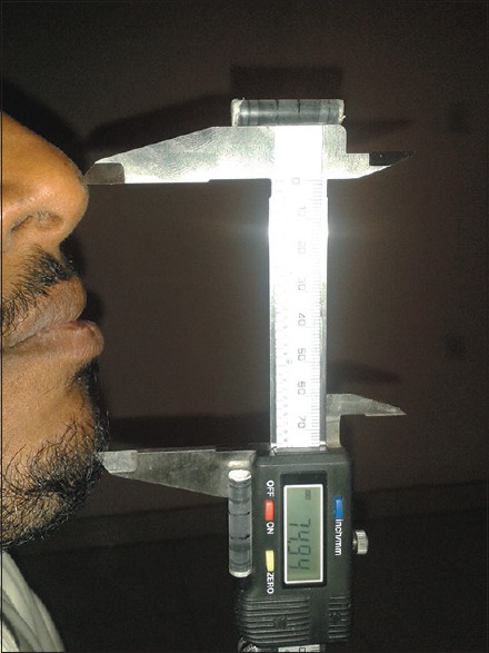
Figure 4.

Measurement of the distance from outer canthus of the eye to rima oris
Reliability test
Sixteen subjects were randomly selected for test-retest method of reliability. The measurements were made twice on the same participants by one author with an interval of 2 weeks in between. Data were entered and analyzed using SPSS program. Intraclass correlation coefficient was performed for intra-examiner reliability. Tables 1 and 2 show the results of the agreement for all variables and between the two readings. Findings suggest that all measurements can be repeated with high significance of reliability and reproducibility ICC = 0.81 with P = 0.0001.
Table 1.
Intraclass correlation coefficient-reliability for all subjects

Table 2.
Intraclass correlation coefficient-reliability for the different measurements

RESULTS
This study consisted of 114 dental students. According to gender, the sample size comprised of 76 males (67%) and 38 females (33%) with an age range of 19–28 years (22.34 ± 1.82). The mean of the distance (eye-RO) was (70.79 ± 4.01) mm with minimum 61.34 mm and maximum 80.47 mm, while (Sn-Me) was (67.24 ± 4.59) mm ranging from 53.97–76.81 mm and (N-Gn) was (69.60 ± 5.32) mm ranging from 57.49–81.77 mm [Table 3]. As shown in [Figure 5], the measurements recorded from the distance (eye-RO) are closer to those recorded from (N-Gn) than those taken from (Sn-Me). To find the correlation between the distances (eye-RO and N-Gn) and (eye-RO and Sn-Me) correlation coefficient of Pearson was initialized [Table 4]. There was a significant correlation (r = 0.313) with P = 0.0007 for the pair (eye-RO and N-Gn), while it was (r = 0.296) for the pair (eye-RO and Sn-Me) with P = 0.0014. The differences between the means were calculated for the two pairs as shown in [Table 5]. The difference between the means for the pair (eye-RO and N-Gn) was lesser than the difference between the means for the pair (eye-RO and Sn-Me). It was (1.19 ± 5.57) with P = 0.025 and (3.55 ± 5.12) with P = 0.0001 respectively. Table 6 shows the differences between males and females for the study parameters, significant differences were found between males and females regarding the recordings measured from the outer canthus of the eye to parting of the lips (2.45 mm, P = 0.002). It was also significant for the readings from the tip of the nose to the tip of the chin (6.23 mm, P = 0.001). While there were no significant differences between the two genders regarding the distance from the septum of the nose to the under of the chin (0.30 mm, P = 0.750).
Table 3.
Descriptive statistics of the facial measurements for total subjects

Figure 5.

Dotplots graph showing all readings
Table 4.
Correlation between (eye-rima oris) and other methods in all subjects

Table 5.
Differences between the means

Table 6.
Group statistics of all measurements comparing the gender

DISCUSSION
Reestablishing the vertical dimension of occlusion not only creates a space between the upper and lower ridges to be occupied by the artificial teeth but also improves the function of the denture prosthesis as well as the esthetic appearance of the patient. Restoring this important component should be in harmony with the facial dimensions with no facial strain or patient discomfort. The absence of any preextraction records for the OVD makes the measurement of the vertical height of the lower third of the face arbitrary. Since there is no superior method to establish the original OVD for all individuals, suggestions have been made to use the facial proportions as alternative methods to predict the missing OVD. This study was set out with the aim of assessing the relation between some distances measured from certain soft tissue landmarks on the face. In this study, the OVD measured from the tip of the nose to the tip of the chin was longer than OVD measured from the septum of the nose to the under of the chin while the distance from the outer canthus of the eye to the corner of the mouth was longer than both of them. However, the difference between the means for (eye-RO) and (Sn-Me) was higher than the one between (eye-RO) and (N-Gn). These results agree with the findings of other studies, in which they found that the distance from the septum of the nose to the under of the chin was smaller than the other distances.[17,18,19,20] Statistically significant correlations were observed between the study variable (eye-RO) and the two readings of the OVD (N-Gn) and (Sn-Me). This correlation was higher, with higher significant level, between the pair (eye-RO) and (N-Gn) than that between (eye-RO) and (Sn-Me). Although these findings seem to be consistent with those found by Nagpal et al.,[21] Delic et al.,[22,23] and Basnet et al.,[24] they differ from a study conducted by Tina-Olaivar et al.[25] in which he found that the difference between (eye-RO) and (N-Gn) was 3 cm, this variation might be because their study was carried out on Mongoloid individuals while this study was conducted on Caucasian subjects. Differences in the distance (between the outer canthus of the eye and parting of the lips) as well as (from tip of the nose to tip of the chin) between males and females were significant. Similar differences between the genders were described by Nagpal et al.,[21] Delic et al.,[22,23] Basent et al.,[24] and Al-Hamdany.[26] It is somewhat surprising that no significant differences were found in the current study between males and females regarding the distance from the septum of the nose to the under of the chin. A possible explanation for this result might be the lack of adequate sample of female subjects (n = 38) compared with the larger sample of males (n = 76). Another possible explanation for this is the variances in soft tissues topography that may reflect such a result. However, several limitations of this study should be noted; the subjects were limited to a single race (Yemenis), collected from convenient sample frame (dental students), and they may have been too young (22.34 years) to be used as considerable data for patients with lost OVD. Thus, further studies using larger sample size from broader age and racial groups are recommended.
CONCLUSION
Within the limitations of the present study, the following conclusion can be drawn:
Eye-RO distance is a reliable method for predicting the OVD for the study group
If this distance is to be used as a method to predict OVD, it should be compared with the distance from the tip of the nose to the tip of the chin rather than the distance for septum of the nose to the under of the chin.
Financial support and sponsorship
Nil.
Conflicts of interest
There are no conflicts of interest.
REFERENCES
- 1.Harper RP. Clinical indications for altering vertical dimension of occlusion. Functional and biologic considerations for reconstruction of the dental occlusion. Quintessence Int. 2000;31:275–80. [PubMed] [Google Scholar]
- 2.Sowmya M, Vinaya B, Krishna P. Psychological impact of edentulousness. JIADS. 2011;2:34–6. [Google Scholar]
- 3.Freeman R. Prosthetics: A study of the emotional reaction to tooth loss. Br Dent J. 2000;188:497. [Google Scholar]
- 4.Khalifa N, Allen PF, Abu-bakr NH, Abdel-Rahman ME. Factors associated with tooth loss and prosthodontic status among Sudanese adults. J Oral Sci. 2012;54:303–12. doi: 10.2334/josnusd.54.303. [DOI] [PubMed] [Google Scholar]
- 5.Allen PF, McMillan AS. A review of the functional and psychosocial outcomes of edentulousness treated with complete replacement dentures. J Can Dent Assoc. 2003;69:662. [PubMed] [Google Scholar]
- 6.Emami E, de Souza RF, Kabawat M, Feine JS. The impact of edentulism on oral and general health. Int J Dent 2013. 2013 doi: 10.1155/2013/498305. 498305. [DOI] [PMC free article] [PubMed] [Google Scholar]
- 7.Alhajj MN. Prosthodontic rehabilitation of severely worn dentition. Int J Dent Clin. 2012;4:35–6. [Google Scholar]
- 8.Fayz F, Eslami A. Determination of occlusal vertical dimension: A literature review. J Prosthet Dent. 1988;59:321–3. doi: 10.1016/0022-3913(88)90182-5. [DOI] [PubMed] [Google Scholar]
- 9.Turrell AJ. Clinical assessment of vertical dimension. J Prosthet Dent. 1972;28:238–46. doi: 10.1016/0022-3913(72)90216-8. [DOI] [PubMed] [Google Scholar]
- 10.Sheppard IM, Sheppard SM. Vertical dimension measurements. J Prosthet Dent. 1975;34:269–77. doi: 10.1016/0022-3913(75)90103-1. [DOI] [PubMed] [Google Scholar]
- 11.Widen HM. Obtaining vertical dimension in edentulous cases. J Am Dent Assoc. 1941;28:240–6. [Google Scholar]
- 12.Misch CE. Clinical indications for altering vertical dimension of occlusion. Objective vs subjective methods for determining vertical dimension of occlusion. Quintessence Int. 2000;31:280–2. [PubMed] [Google Scholar]
- 13.McGee GF. Use of facial measurements in determining vertical dimension. J Am Dent Assoc. 1947;35:342–50. doi: 10.14219/jada.archive.1947.0361. [DOI] [PubMed] [Google Scholar]
- 14.Niswonger ME. Obtaining the vertical relation in edentulous cases that existed prior to extraction. J Am Dent Assoc. 1938;25:1842–7. [Google Scholar]
- 15.Willie RG. Trends in clinical methods of establishing an ideal interarch relationship. J Prosthet Dent. 1958;8:243–51. [Google Scholar]
- 16.Geerts GA, Stuhlinger ME, Nel DG. A comparison of the accuracy of two methods used by pre-doctoral students to measure vertical dimension. J Prosthet Dent. 2004;91:59–66. doi: 10.1016/j.prosdent.2003.10.016. [DOI] [PubMed] [Google Scholar]
- 17.Bhat V, Gopinathan M. Reliability of determining vertical dimension of occlusion in complete dentures: A clinical study. J Indian Prosthodont Soc. 2006;6:38–42. [Google Scholar]
- 18.Miralles R, Dodds C, Palazzi C, Jaramillo C, Quezada V, Ormeño G, et al. Vertical dimension. Part 1: Comparison of clinical freeway space. Cranio. 2001;19:230–6. doi: 10.1080/08869634.2001.11746173. [DOI] [PubMed] [Google Scholar]
- 19.Sakar O, Sülün T, Kurt H, Gençel B. Reliability and comparison of two facial measurements to detect changes of occlusal vertical dimension in complete denture wearers. Gerodontology. 2011;28:205–8. doi: 10.1111/j.1741-2358.2009.00353.x. [DOI] [PubMed] [Google Scholar]
- 20.Unger JW. Comparison of vertical morphologic measurements on dentulous and edentulous patients. J Prosthet Dent. 1990;64:232–4. doi: 10.1016/0022-3913(90)90184-e. [DOI] [PubMed] [Google Scholar]
- 21.Nagpal A, Parkash H, Bhargava A, Chittaranjan B. Reliability of different facial measurements for determination of vertical dimension of occlusion in edentulous using accepted facial dimensions recorded from dentulous subjects. J Indian Prosthodont Soc. 2014;14:233–42. doi: 10.1007/s13191-013-0315-1. [DOI] [PMC free article] [PubMed] [Google Scholar]
- 22.Delic Z, Simunovic-Soskic M, Perinic-Grzic R, Vukovojac S, Rajic Z, Kuna T, et al. Evaluation of craniometric methods for determination of vertical dimension of occlusion. Coll Antropol. 2000;24(Suppl 1):31–5. [PubMed] [Google Scholar]
- 23.Delic Z, Vukovojac S, Grzic R, Maricic D, Kovac Z, Kovacevic D. Evaluation of craniometric methods for determination of vertical dimension of occlusion – Part 2. Coll Antropol. 2003;27(Suppl 1):191–4. [PubMed] [Google Scholar]
- 24.Basnet BB, Singh RK, Parajuli PK, Shrestha P. Correlation between facial measurements and occlusal vertical dimension: An anthropometric study in two ethnic groups of Nepal. Int J Dent Sci Res. 2014;2:171–4. [Google Scholar]
- 25.Tina-Olaivar EO, Olaivar OK. A comparative study of the upper and lower vertical facial measurements of the Filipinos as it is used in the Willis method for determining the vertical dimension of occlusion. J Philipp Dent Assoc. 1998;50:44–8. [PubMed] [Google Scholar]
- 26.Al-Hamdany AK, Kassab NH. Correlation of vertical dimensions of soft tissue facial profiles. Al Rafidain Dent J. 2010;10:243–53. [Google Scholar]