

Case Report
A 6-year-old Chinese girl presented to our institution with fever of 9 days. Her temperature was persistently above 38.5°C, with a maximum temperature of more than 40°C. This was associated with sore throat and cough. There was no recent travel or sick contact, and her vaccinations were up-to-date with the national immunization schedule. She was initially seen by a family physician and given azithromycin on day 4 of illness. She completed the course of antibiotics, despite developing a pruritic and erythematous maculopapular rash 3 hours after the first dose. The rash first started on the limbs, but became generalized over the next 2 days, spreading to the perineum, trunk, neck, and face. On day 6 of illness, the rash started peeling, and she developed left calf pain as well. She was admitted to another hospital and was treated for a drug-induced rash with 5 doses of intravenous (IV) hydrocortisone, but there was minimal improvement. A red tongue with prominent papillae was also noted at the hospital. She then developed left calf swelling on day 8 of illness. She was treated empirically with IV ceftriaxone and amoxicillin/clavulanic acid and was transferred to our institution on day 9 of illness.
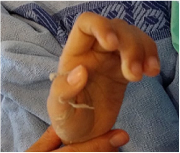
Physical examination revealed a febrile child with a temperature of 39.6°C, heart rate of 148 beats per minute, and blood pressure of 104/95 mm Hg. She was lethargic and irritable, but not in distress. Her neck was supple. An erythematous macular rash was noted over her face, trunk, limbs, and perineum, with peeling over these areas as well. The rash had a sandpaper texture and was blanchable. There was no conjunctival injection, but she had red and swollen lips (Figure 1) with a strawberry tongue. The throat and tonsils were slightly injected, but there was no tonsillar hypertrophy or exudates. Sub-centimeter cervical lymph nodes were palpable bilaterally. On auscultation of the chest, dual heart sounds were present with no murmurs, and lungs were clear. The abdomen was soft but slightly distended, with tender hepatomegaly measuring 2 cm below the costal margin, as well as shifting dullness. Examination of the limbs revealed mild edema over the feet bilaterally, as well as a swollen left calf (Figure 2), which was warm, tender, and erythematous. She was unable to move the left lower limb or ambulate due to pain. Neurological examination was grossly normal.
Figure 1.
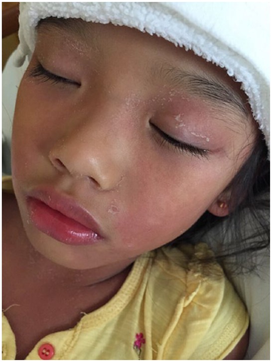
Erythematous rash seen over the face, with swollen red lips.
Figure 2.

Unilateral, left calf swelling seen.
Tables 1 and 2 summarize the blood investigations and microbiological investigations done for the patient respectively. Serum creatine kinase (CK) levels were not raised. Left calf magnetic resonance imaging (MRI) revealed subcutaneous edema with thickening and increased T2 signal to the premuscular and intermuscular fascia in the medial and posterior aspects of the proximal leg, suggestive of possible myositis.
Table 1.
Timeline of Blood Investigations Done for the Patient.
| Day of Illness |
|||||
|---|---|---|---|---|---|
| Day 9 | Day 11 | Day 14 | Day 18 | Day 40 | |
| Hemoglobin (g/dL) | 10.9 | 10.2 | 9.2 | 8.8 | 11.9 |
| White blood cell count (×109/L) | 24.4 | 16.7 | 21.1 | 28.35 | 10.2 |
| Platelet count (×109/L) | 229 | 270 | 683 | 953 | 336 |
| Erythrocyte sedimentation rate (mm/h) | 91 | 116 | >145 | >145 | 77 |
| C-reactive protein (mg/L) | 209.1 | 152.8 | 31.9 | 55.5 | 0.8 |
| Albumin (g/L) | 22 | 17 | 22 | ||
| Alanine transaminase (U/L) | 27 | 31 | 46 | ||
| Aspartate transaminase (U/L) | 30 | 44 | 39 | ||
| Creatine kinase (U/L) | 76 | 25 | 25 | ||
Table 2.
Timeline of Microbiological Investigations Done for the Patient.
| Day of Illness |
|||||
|---|---|---|---|---|---|
| Days 8 to 9 | Day 11 | Day 14 | Day 16 | Day 40 | |
| Measles serology | IgM negative, IgG positive | ||||
| Dengue serology | Negative | ||||
| Mycoplasma pneumoniae serology | IgM nonreactive | ||||
| ASOT | <100 IU/mL | 200 IU/mL | 200 IU/mL | ||
| Cytomegalovirus serology | IgM negative, IgG positive | ||||
| Epstein–Barr virus serology | IgM negative, IgG positive | ||||
| Respiratory viruses multiplex polymerase chain reaction | Not detected for all | ||||
| Widal Weil Felix serology | Not detected for all | ||||
| Rickettsiae serology | Negative | ||||
| Blood cultures (aerobic, anaerobic) | No bacterial growth at 48 hours | ||||
| Urine analysis (/µL) | |||||
| White blood cell | 30 | ||||
| Red blood cell | 0 | ||||
| Epithelial cell | 5 | ||||
| Urine culture | No bacterial growth | ||||
| Fluid from submuscular layer | |||||
| Gram stain smear | No organism seen | ||||
| Acid-fast bacilli (AFB) smear | No AFB seen | ||||
| Bacteria culture | No bacterial growth | ||||
| Fungal culture | No fungal growth | ||||
The patient was treated empirically for an infection with IV clindamycin and penicillin G on day 9 of illness, but her fever persisted with high-temperature spikes. She was treated as for atypical Kawasaki disease on day 11 of illness with intravenous immunoglobulin (IVIG) and aspirin and IV antibiotics were continued. Her fever subsequently improved and subsided by day 13 of illness. Most of her symptoms resolved; however, her left calf swelling deteriorated acutely despite initial improvement, and an incision and drainage (I&D) was done on day 16 of illness, which drained 10 mL of turbid fluid from the submuscular layer. No frank pus was noted intraoperatively, and microbiological investigations revealed no evidence of infection (Table 2). Left calf tissue biopsy revealed mild chronic inflammatory changes.
The patient developed peeling over her fingers (Figure 3) and toes (Figure 4) on day 18 of illness, but was otherwise well. Anti-streptolysin O titer (ASOT) was borderline positive at 200 IU/mL after completing IVIG. However, there was no change in ASOT titer levels on day 40 of illness. This was suggestive of a false-positive ASOT due to presence of anti-streptolysin O antibodies in the donor immunoglobulin product. Her blood results were in keeping with that seen in Kawasaki disease, that is, increasing platelet counts and erythrocyte sedimentation rates (Table 1). Transthoracic 2D-echocardiography showed normal coronary arteries on day 10 and day 16 of illness.
Figure 3.
Peeling over the fingers.
Figure 4.
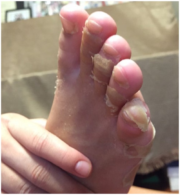
Peeling over the toes.
Discussion
Kawasaki disease was first described in 1967. It is an acute, self-limiting vasculitis of unknown etiology, and it occurs most commonly in infants and young children. It is diagnosed clinically. According to guidelines from the American Academy of Pediatrics1 and the American Heart Association (AHA),2 a case of classical Kawasaki disease can be established if the patient fulfils the following clinical criteria: persistently high fever for 5 or more days and at least 4 of the principle features—(a) changes in extremities, (b) polymorphous exanthem, (c) bilateral conjunctival injection without exudate, (d) changes in the lips and oral cavity, and (e) cervical lymphadenopathy. Other conditions with similar clinical characteristics should be excluded as well. For instance, in a child with fever and rash, other possible causes, such as drug reactions and infections, should be considered and excluded.3 Accurate diagnosis and prompt treatment of Kawasaki disease is important, as it can potentially prevent the feared complication of coronary artery aneurysm or ectasia, which can occur in more than a third of untreated cases.1,2
According to AHA guidelines, incomplete or atypical Kawasaki disease should be considered in patients with an unexplained fever for 5 or more days, and at least 2 or 3 of the 5 clinical features.2 Associated cardiac findings on echocardiography and biochemical results meeting the supplemental laboratory criteria substantiate the diagnosis, but their absences do not rule out this possibility. In our patient’s case, Kawasaki disease was strongly suspected, as her condition failed to improve with IV antibiotic therapy, and initial workup for acute infective causes was unremarkable. In addition, her laboratory results fulfilled all of the AHA supplemental laboratory criteria: she had hypoalbuminemia with serum albumin persistently <30 g/L, anemia, leukocytosis, thrombocytosis, and elevated urine leukocyte count2,4 with sterile pyuria. Other studies have suggested further laboratory tests to substantiate the diagnosis of Kawasaki disease, such as elevated serum erythrocyte sedimentation rate,5,6 which was also evident in our patient. She responded well to IVIG and aspirin, which further supported our diagnosis of Kawasaki disease.
There are several complications, involving various systems, which have been associated with Kawasaki disease. These include various inflammatory conditions, such as myocarditis, coronary artery abnormalities, hydrops gallbladder, and aseptic meningitis.2 However, the relationship between myositis and Kawasaki disease has not been clearly established in existing literature, although there have been a few case reports that report the co-occurrence of these 2 conditions, and suggest a possible association.7-12 In our patient’s case, the left calf myositis was likely a complication of Kawasaki disease rather than a superimposed infection, as microbiological testing of fluid samples obtained during I&D revealed no evidence of infection.
Koutras first reported the occurrence of myositis with Kawasaki disease in a child in 1980.7 A few other cases of myositis with Kawasaki disease in pediatric patients have been published since.8-10 In addition, one case of neuromuscular abnormality has been reported in an adult with Kawasaki disease,11 and one case of orbital myositis in a child with Kawasaki disease has been published.12 Table 3 summarizes all the case reports of Kawasaki disease with myositis in terms of clinical presentations and relevant investigation results.
Table 3.
Summary of Clinical Presentations and Relevant Investigation Findings in Previously Reported Cases of Kawasaki Disease With Myositis.
| Title | Author, Journal, Year | Clinical Presentation | Relevant Investigation Findings | Conclusion |
|---|---|---|---|---|
| Myositis in Kawasaki disease8 | Gama et al, Pediatr Neurol, 1990 | 8-Year-old boy, previously well, presented with clinical features of KD, diffused peripheral weakness, and respiratory failure | • CK: 1509 to 2657 IU | Myositis is one of several neurological complications encountered in KD. The degree of CK elevation may be useful in predicting the severity of myopathy. |
| • EMG: myopathic pattern | ||||
| • Muscle biopsy: atrophy and degeneration | ||||
| A case of polymyositis associated with Kawasaki disease9 | Sugie et al, Brain Dev, 1985 | 3-Year-old boy, previously well, was diagnosed with KD, and developed painful proximal muscle weakness in all extremities | • CK: 152 IU | Polymyositis might be a complication of KD. Proximal muscle weakness suggests inflammatory myopathy, even if serum CK was not significantly elevated. |
| • EMG: myopathic change | ||||
| • Muscle biopsy: mild architecture distortion, fiber atrophy, inflammatory cell infiltrates | ||||
| Myositis with Kawasaki’s disease10 | Koutras, Am J Dis Child, 1982 | 18-Month-old girl, previously well, presented with clinical features of KD and severe proximal muscle weakness and tenderness with dysphonia and dysphagia | • CK: 72 U/L | Clinical presentation suggests a coexistence of KD and myositis. |
| Neuromuscular and immunochemical abnormalities in an adult man with Kawasaki disease11 | Hicks et al, Ann Intern Med, 1982 | A 40-year-old man presented with primary features of KD and distal motor and sensory neuropathy | • Elevated CK | There is a possibility that clinical features and complications of KD are mediated by immune complex deposition in vessels and tissues. |
| • Abnormal EMG | ||||
| • Muscle biopsy: myonecrosis, immunoglobulin deposit, distorted architecture | ||||
| Orbital myositis due to Kawasaki’s disease12 | Lin et al, Pediatr Radiol, 1999 | An 8-month-old boy, previously well, was diagnosed with and treated for KD. He developed unilateral edema and erythema of the upper eye lid with impaired extra-ocular movement 18 days after apparent remission of KD. | • CT orbit: soft tissue swelling of eyelid with thickened orbicularis muscle | Orbital myositis can possibly be a complication of KD |
| • Histology: pan-arteritis and myositis |
Abbreviations: KD, Kawasaki disease; CK, creatine kinase; EMG, electromyography; CT, computed tomography.
The pathophysiology of Kawasaki disease–associated myositis, as suggested by Hicks et al, could be immune-complex depositions in affected vessels and tissues, including muscle tissues.11 In terms of clinical presentation, patients usually present with pain and weakness in the extremities.8-11 In severe cases, there could be generalized weakness involving all 4 limbs and other vital muscles, such as the diaphgramatic8 and oesophageal10 muscles, leading to respiratory failure and dysphagia, respectively. With regard to investigations, serum CK levels may not necessarily be elevated in myositis.8-10 However, there might be a correlation between CK levels and severity of myositis.8 Other possible investigations that can be done include an electromyography and muscle biopsy, whereby the presence of myopathic patterns and inflammatory changes,8,9 respectively, can facilitate diagnosis.
Conclusion
Myositis is a possible complication of Kawasaki disease. Serum CK levels might be elevated in myositis, but it does not necessarily occur in all cases. We believe that this rare case contributes to and value-adds the growing literature that seeks to understand and establish the link between Kawasaki disease and myositis. Future studies can further explore the relationship between these 2 conditions and determine if there is indeed a significant association.
Footnotes
Author Contributions: Lee EYX contributed to the design, analysis, and interpretation of data; literature review; and drafted the manuscript. Oh JY, Chong CY, Choo JTL, and Mahadev A contributed to the analysis and interpretation of data and critically revised the manuscript. Tan NWH contributed to the conception, design, analysis, and interpretation of data and critically revised the manuscript.
Declaration of Conflicting Interests: The author(s) declared no potential conflicts of interest with respect to the research, authorship, and/or publication of this article.
Funding: The author(s) received no financial support for the research, authorship, and/or publication of this article.
Informed Consent: Informed consent was obtained from the patient’s parents for the photographs obtained for publication.
References
- 1. Newburger JW, Takahashi M, Gerber MA, et al. Diagnosis, treatment, and long-term management of Kawasaki disease. Pediatrics. 2004;114:1708-1733. [DOI] [PubMed] [Google Scholar]
- 2. Newburger JW, Takahashi M, Gerber MA, et al. Diagnosis, treatment, and long-term management of Kawasaki disease. Circulation. 2004;110:2747-2771. [DOI] [PubMed] [Google Scholar]
- 3. Saffar MJ, Saffar H, Shahmohammadi S. Fever and rash syndrome: a review of clinical practice guidelines in the differential diagnosis. J Pediatr Rev. 2013;1(2):42-54. [Google Scholar]
- 4. Yu JJ. Diagnosis of incomplete Kawasaki disease. Korean J Pediatr. 2012;55(3):83-87. [DOI] [PMC free article] [PubMed] [Google Scholar]
- 5. Ling XB, Kanegaye JT, Ji J, et al. Point-of-care differentiation of Kawasaki disease from other febrile illnesses. J Pediatr. 2013;162:183-188. [DOI] [PMC free article] [PubMed] [Google Scholar]
- 6. Chaiyarak K, Durongpisitkul K, Atta T, Soongswang J, Laohaprasitiporn D, Nana A. Clinical manifestations of Kawasaki disease: what are the significant parameters? Asian Pac J Allergy Immunol. 2009;27:131-136. [PubMed] [Google Scholar]
- 7. Koutras AK. Myositis with mucocutaneous lymph-node syndrome. N Y State J Med. 1980;80(7 pt 1):1138-1139. [PubMed] [Google Scholar]
- 8. Gama C, Breeden K, Miller R. Myositis in Kawasaki disease. Pediatr Neurol. 1990;6:135-136. [DOI] [PubMed] [Google Scholar]
- 9. Sugie H, Sugie Y, Ichimura M, Mizuno Y, Nishida M, Igarashi Y. A case of polymyositis associated with Kawasaki disease. Brain Dev. 1985;7:513-515. [DOI] [PubMed] [Google Scholar]
- 10. Koutras AK. Myositis with Kawasaki’s disease. Am J Dis Child. 1982;136:78-79. [DOI] [PubMed] [Google Scholar]
- 11. Hicks JT, Korenyi-Both A, Utsinger PD, Baran EM, McLaughlin GE. Neuromuscular and immunochemical abnormalities in an adult man with Kawasaki disease. Ann Intern Med. 1982;96:607-610. [DOI] [PubMed] [Google Scholar]
- 12. Lin H, Burton EM, Felz MW. Orbital myositis due to Kawasaki’s disease. Pediatr Radiol. 1999;29:634-636. [DOI] [PubMed] [Google Scholar]