Abstract
Avian bornaviruses (ABVs) were recently discovered as the causative agents of proventricular dilatation disease (PDD). Although molecular epidemiological studies revealed that ABVs exist in Japan, no Japanese isolate has been reported thus far. In this study, we isolated four strains of Psittaciform 1 bornavirus from psittacine birds affected by PDD using QT6 quail cells. To our knowledge, this is the first report to isolate ABVs in Japan and to show that QT6 cells are available for ABV isolation. These isolates and QT6 cells would be powerful tools for elucidating the fundamental biology and pathogenicity of ABVs.
Keywords: avian bornavirus, isolation, parrot bornavirus, Psittaciform 1 bornavirus, QT6 cells
In 2008, two independent groups discovered novel bornaviruses, named avian bornaviruses (ABVs), from psittacine birds affected by proventricular dilatation disease (PDD) [3, 6], which is often fatal and accompanied with gastrointestinal dysfunction and/or neurologic symptoms. Recent extensive epidemiological studies revealed that 15 types of ABV were detected from many species of birds all over the world [7, 13].
Virus isolation is important for diagnosis of ABV infection as well as fundamental studies of ABVs. For example, Gray et al. used a parrot bornavirus (PaBV) 4 isolate for infection experiments, which fulfilled the Koch’s postulates and demonstrated that ABVs are indeed the causative agent of PDD [2]. In addition, bornaviruses are known to be unique RNA viruses in that they establish non-cytolytic, persistent infections in the host cell nucleus. Therefore, studies of ABVs may also provide interesting insights into interactions between RNA viruses and their hosts as shown by Borna disease virus researches [4, 9]. In Japan, although we and others detected ABV nucleic acids from several birds [5, 12, 16, 17], no one has isolated ABV so far. Ogawa et al. tried to isolate an ABV from 10% cerebrum homogenate of an infected bird using Japanese quail fibroblast cells (QE-1 cells) [12], and we also inoculated a fecal sample, which was positive for PaBV-5 nucleic acid, to QT6 Japanese quail fibroblast-like cells [11], but the attempts failed [5]. Although the above trials failed and there is no report showing that Japanese quail is infected by ABVs, quail-derived cell cultures were reported to be available for isolation of ABVs [14]. Thus, QT6 may be also susceptible to ABV infection.
In the course of molecular epidemiological studies of ABVs, we got four brain samples from parrots affected with PDD (Table 1). We performed RT-PCR using random hexamer and primers MH175 and MH170 which amplify 221 bp segment of ABV N gene [5], and sequencing analyses, revealing that the parrots were infected with Psittaciform 1 bornavirus: two were PaBV-2, and the other two were PaBV-4 (Table 1) (full methods and primer sequences are available in the supplementary information). Therefore, we used these brain samples for isolation of ABVs. Inocula were prepared as described by Rubbenstroth et al. [15] with slight modifications (supplementary information), which were added to semi-confluent QT6 cells. The inoculated cells were passaged as described in the supplementary information.
Table 1. Isolated bornaviruses in this study.
| Inoculum number | Host | Virus species | Virus | Strain | Sampling date |
|---|---|---|---|---|---|
| 1 | Ara militaris | Psittaciform 1 bornavirus | PaBV-2 | IH-1 | August 2011 |
| 2 | Aratinga jandaya | Psittaciform 1 bornavirus | PaBV-2 | KOKO | January 2013 |
| 3 | Diopsittaca nobilis | Psittaciform 1 bornavirus | PaBV-4 | 7I6 | February 2014 |
| 4 | Ara ararauna | Psittaciform 1 bornavirus | PaBV-4 | 7I10 | February 2014 |
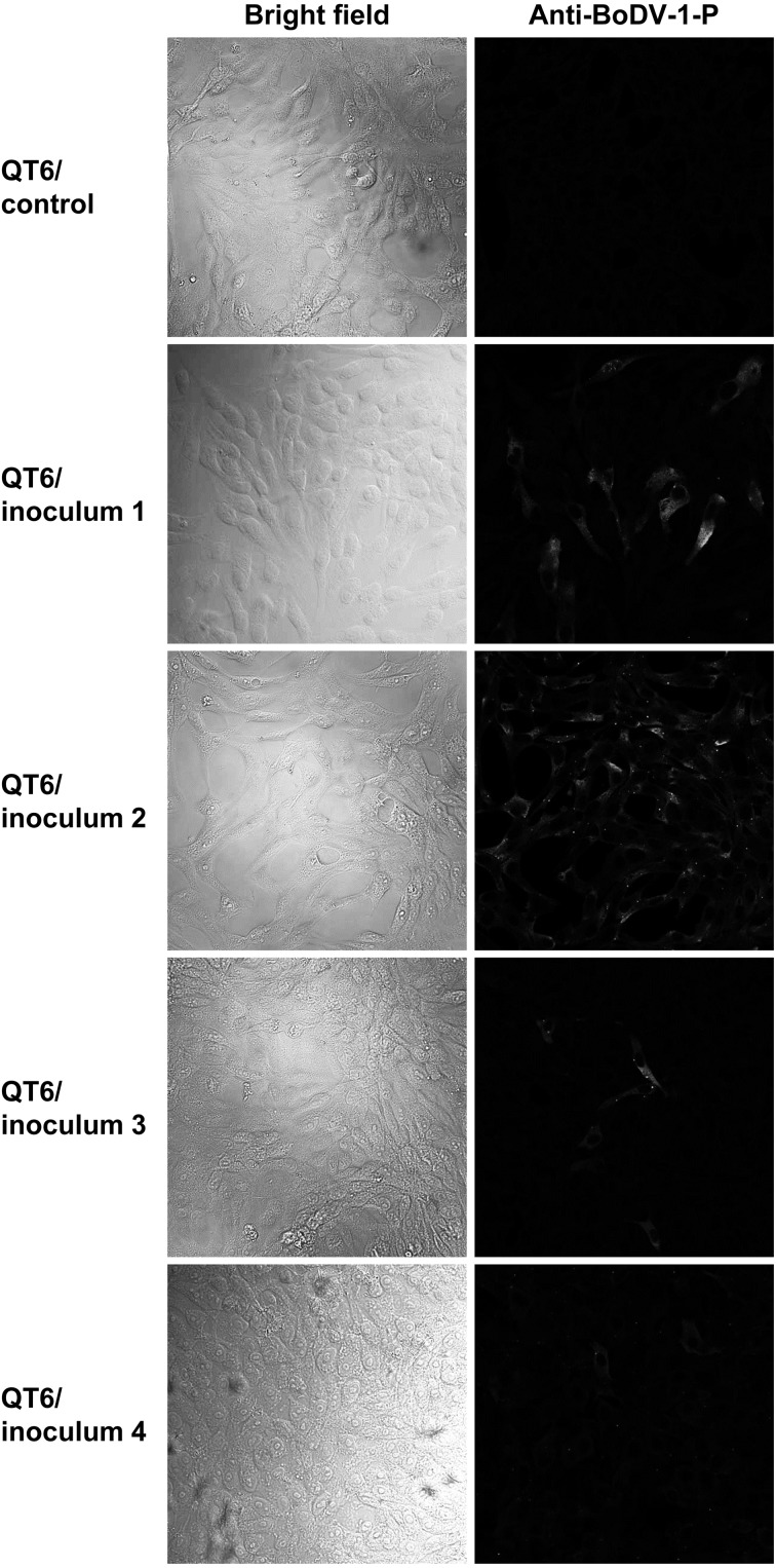
After 3 to 12 weeks post inoculation, we performed indirect immunofluorescent assay (IFA) using anti-Borna disease virus 1 (BoDV-1) P antibodies (supplementary information). Positive signals were found in the inoculated QT6 cells, but not in the control cells, indicating that the QT6 cells were infected with bornaviruses (Fig. 1). We named these isolated strains as IH-1, KOKO, 7I6 and 7I10, respectively (Table 1).
Fig. 1.
Visualization of bornaviral antigens by indirect immunofluorescence assay. Immunofluorescence assays were performed using anti-BoDV-1 P rabbit polyclonal antibodies. Bright fields and fluorescence images are shown. The cells and inocula were indicated on the left side of each panel. Inoculum 1, 12 weeks post inoculation (w. p. i.); inoculum 2, 3 w. p. i.; inoculum 3, 3 w. p. i.; and inoculum 4, 4 w. p. i.
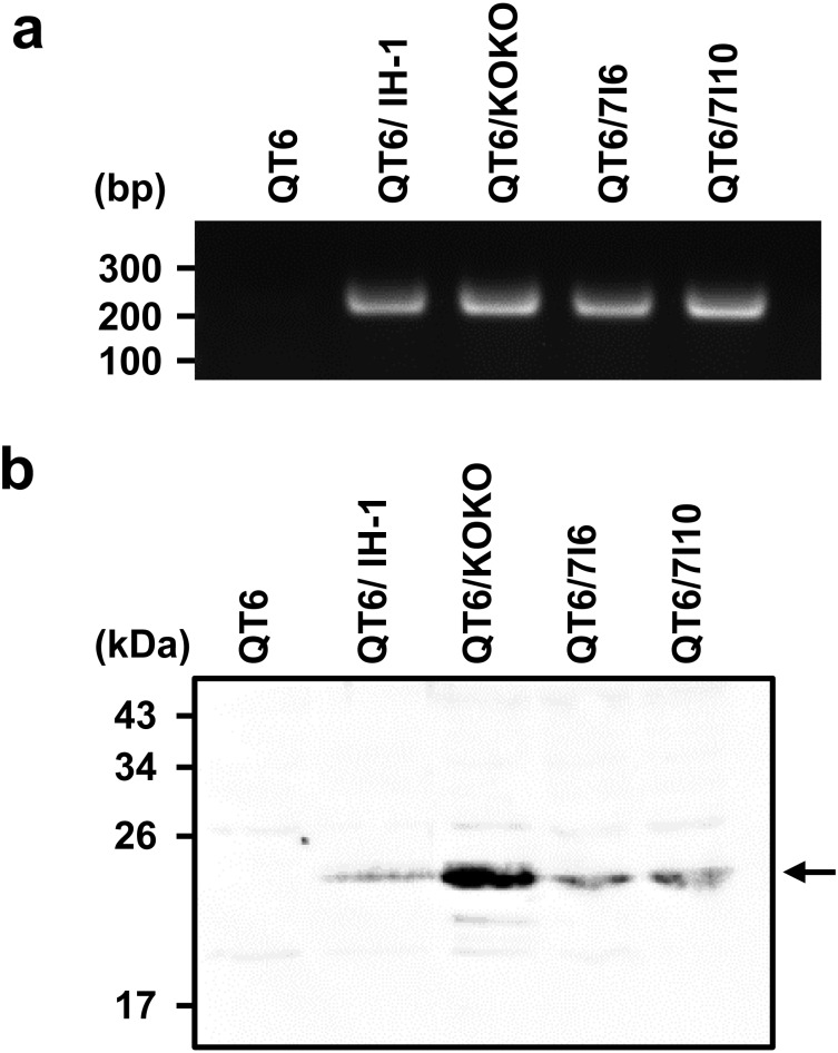
To confirm the above observation, we extracted RNA from the cells and carried out RT-PCR using random hexamer and the primers MH175 and MH170 [5] for reverse-transcription and PCR, respectively (supplementary information). Specific bands were detected in all RNA samples extracted from the inoculated cells (Fig. 2a). We determined the nucleotide sequences of amplicons, showing the sequences were almost identical to those in the brains (data not shown). We further performed western blotting using the rabbit antibodies against BoDV-1 P protein (supplementary information). The western blotting analysis detected specific bands of expected molecular weight in the inoculated cells (Fig. 2b). The difference of band intensities among the samples may be due to different infection rates, protein expression levels and/or affinities to the antibodies. These data demonstrated that we indeed isolated ABVs from the brain samples.
Fig. 2.
Detection of bornaviral nucleic acid and phosphoproteins by RT-PCR and western blotting. (a) RT-PCR with primers MH175 and MH170. Cells and virus strains were indicated above the figure. (b) Western blotting using anti-BoDV-1 P rabbit polyclonal antibodies. Cells and infected virus strains were shown above the figure.
In this study, we isolated four strains of Psittaciform 1 bornavirus using QT6 quail cells. To our knowledge, this is the first report showing that QT6 cells are susceptible to ABVs, at least PaBV-2 and PaBV-4. So far, several avian cell lines were reported to be susceptible to ABVs [14, 15]. However, due to the national regulation for infectious diseases of poultry in Japan, it is time-consuming and sometimes difficult to import chicken and quail cells from abroad. Duck embryonic fibroblast (DEF) was also reported to be available for ABV isolation [2, 15], but it is also laborious to prepare cultures from fertilized eggs. In addition, for propagation of ABVs and preparation of virus stocks, it is usually necessary to passage inoculated cells for long term, since ABVs slowly propagate in cell culture. Because of the limited life span of DEF, low-passage DEF should be regularly provided to propagate ABVs as described in [2]. Therefore, our finding is useful for further isolation of ABVs at least in Japan and maybe also in other countries that have similar regulations. Although it is not sure that QT6 cells are susceptible to other types of ABVs, the cell line is probably available to isolate other types of ABVs because the QT6 cell line was reported to be susceptible to other avian viruses, such as avian influenza viruses [8]. Since it was reported that susceptibilities to ABVs differ among avian cell lines [15], it is interesting to compare the susceptibilities of QT6 cells and the previously reported cell lines. And, more, QT6 cells were also reported to show high transfection efficiency [1]. Therefore, the cells are helpful to study the molecular biology of ABVs as well as to establish reverse genetics system for ABVs.
As described above, we tried to isolate PaBV-5 using QT-6 cells, but it failed. This may be explained by the difference of materials we used to isolate the viruses. When trying to isolate PaBV-5, we inoculated a feces-derived sample to QT6 cells, which showed much higher cytotoxicity than the brain-derived samples. In addition, in infected birds, brains usually contain higher amount of ABVs than feces [14]. Alternatively, QT6 cells might not be susceptible to PaBV-5. Further studies are needed to assess the possibilities.
Our isolates belong to PaBV-2 and PaBV-4, which are dominant types of ABV in the world [2] and were formally shown to cause PDD [2, 10]. Although many PaBV-2 and PaBV-4 have been isolated thus far, there is no report, which systematically investigates differences of viral genome sequences, replication efficiencies and pathogenicity among the strains. Although we have not yet determined the full genome sequences of isolated viruses, it may be interesting to compare their sequences and their properties in vitro and in vivo.
Taken together, we isolated four Japanese strains of ABVs from psittacine birds using QT6 cells. Our isolates and findings are useful for further investigation of the fundamental biology and pathology of ABVs.
Supplementary
Acknowledgments
We are grateful to Kyoko Horie for technical assistance. We would like to thank Dennis Rubbenstroth for helpful comments. This study is partially supported by JSPS KAKENHI grant number 26850208 (MH)and Joint Usage/Research Center program of Institute for Virus Research, Kyoto University (MH).
REFERENCES
- 1.de Wit E., Spronken M. I., Vervaet G., Rimmelzwaan G. F., Osterhaus A. D., Fouchier R. A.2007. A reverse-genetics system for Influenza A virus using T7 RNA polymerase. J. Gen. Virol. 88: 1281–1287. doi: 10.1099/vir.0.82452-0 [DOI] [PubMed] [Google Scholar]
- 2.Gray P., Hoppes S., Suchodolski P., Mirhosseini N., Payne S., Villanueva I., Shivaprasad H. L., Honkavuori K. S., Lipkin W. I., Briese T., Reddy S. M., Tizard I.2010. Use of avian bornavirus isolates to induce proventricular dilatation disease in conures. Emerg. Infect. Dis. 16: 473–479. doi: 10.3201/eid1603.091257 [DOI] [PMC free article] [PubMed] [Google Scholar]
- 3.Honkavuori K. S., Shivaprasad H. L., Williams B. L., Quan P. L., Hornig M., Street C., Palacios G., Hutchison S. K., Franca M., Egholm M., Briese T., Lipkin W. I.2008. Novel borna virus in psittacine birds with proventricular dilatation disease. Emerg. Infect. Dis. 14: 1883–1886. doi: 10.3201/eid1412.080984 [DOI] [PMC free article] [PubMed] [Google Scholar]
- 4.Horie M., Honda T., Suzuki Y., Kobayashi Y., Daito T., Oshida T., Ikuta K., Jern P., Gojobori T., Coffin J. M., Tomonaga K.2010. Endogenous non-retroviral RNA virus elements in mammalian genomes. Nature 463: 84–87. doi: 10.1038/nature08695 [DOI] [PMC free article] [PubMed] [Google Scholar]
- 5.Horie M., Ueda K., Ueda A., Honda T., Tomonaga K.2012. Detection of Avian bornavirus 5 RNA in Eclectus roratus with feather picking disorder. Microbiol. Immunol. 56: 346–349. doi: 10.1111/j.1348-0421.2012.00436.x [DOI] [PubMed] [Google Scholar]
- 6.Kistler A. L., Gancz A., Clubb S., Skewes-Cox P., Fischer K., Sorber K., Chiu C. Y., Lublin A., Mechani S., Farnoushi Y., Greninger A., Wen C. C., Karlene S. B., Ganem D., DeRisi J. L.2008. Recovery of divergent avian bornaviruses from cases of proventricular dilatation disease: identification of a candidate etiologic agent. Virol. J. 5: 88. doi: 10.1186/1743-422X-5-88 [DOI] [PMC free article] [PubMed] [Google Scholar]
- 7.Kuhn J. H., Dürrwald R., Bào Y., Briese T., Carbone K., Clawson A. N., deRisi J. L., Garten W., Jahrling P. B., Kolodziejek J., Rubbenstroth D., Schwemmle M., Stenglein M., Tomonaga K., Weissenböck H., Nowotny N.2015. Taxonomic reorganization of the family Bornaviridae. Arch. Virol. 160: 621–632. doi: 10.1007/s00705-014-2276-z [DOI] [PMC free article] [PubMed] [Google Scholar]
- 8.Lee C. W., Jung K., Jadhao S. J., Suarez D. L.2008. Evaluation of chicken-origin (DF-1) and quail-origin (QT-6) fibroblast cell lines for replication of avian influenza viruses. J. Virol. Methods 153: 22–28. doi: 10.1016/j.jviromet.2008.06.019 [DOI] [PubMed] [Google Scholar]
- 9.Matsumoto Y., Hayashi Y., Omori H., Honda T., Daito T., Horie M., Ikuta K., Fujino K., Nakamura S., Schneider U., Chase G., Yoshimori T., Schwemmle M., Tomonaga K.2012. Bornavirus closely associates and segregates with host chromosomes to ensure persistent intranuclear infection. Cell Host Microbe 11: 492–503. doi: 10.1016/j.chom.2012.04.009 [DOI] [PubMed] [Google Scholar]
- 10.Mirhosseini N., Gray P. L., Hoppes S., Tizard I., Shivaprasad H. L., Payne S.2011. Proventricular dilatation disease in cockatiels (Nymphicus hollandicus) after infection with a genotype 2 avian bornavirus. J. Avian Med. Surg. 25: 199–204. doi: 10.1647/2010-030.1 [DOI] [PubMed] [Google Scholar]
- 11.Moscovici C., Moscovici M. G., Jimenez H., Lai M. M., Hayman M. J., Vogt P. K.1977. Continuous tissue culture cell lines derived from chemically induced tumors of Japanese quail. Cell 11: 95–103. doi: 10.1016/0092-8674(77)90320-8 [DOI] [PubMed] [Google Scholar]
- 12.Ogawa H., Sanada Y., Sanada N., Kudo M., Tuchiya K., Kodama T., Uetsuka K.2011. Proventricular dilatation disease associated with avian bornavirus infection in a Citron-crested Cockatoo that was born and hand-reared in Japan. J. Vet. Med. Sci. 73: 837–840. doi: 10.1292/jvms.10-0465 [DOI] [PubMed] [Google Scholar]
- 13.Philadelpho N. A., Rubbenstroth D., Guimarães M. B., Piantino Ferreira A. J.2014. Survey of bornaviruses in pet psittacines in Brazil reveals a novel parrot bornavirus. Vet. Microbiol. 174: 584–590. doi: 10.1016/j.vetmic.2014.10.020 [DOI] [PubMed] [Google Scholar]
- 14.Rinder M., Ackermann A., Kempf H., Kaspers B., Korbel R., Staeheli P.2009. Broad tissue and cell tropism of avian bornavirus in parrots with proventricular dilatation disease. J. Virol. 83: 5401–5407. doi: 10.1128/JVI.00133-09 [DOI] [PMC free article] [PubMed] [Google Scholar]
- 15.Rubbenstroth D., Rinder M., Kaspers B., Staeheli P.2012. Efficient isolation of avian bornaviruses (ABV) from naturally infected psittacine birds and identification of a new ABV genotype from a salmon-crested cockatoo (Cacatua moluccensis). Vet. Microbiol. 161: 36–42. doi: 10.1016/j.vetmic.2012.07.004 [DOI] [PubMed] [Google Scholar]
- 16.Rubbenstroth D., Rinder M., Stein M., Höper D., Kaspers B., Brosinski K., Horie M., Schmidt V., Legler M., Korbel R., Staeheli P.2013. Avian bornaviruses are widely distributed in canary birds (Serinus canaria f. domestica). Vet. Microbiol. 165: 287–295. doi: 10.1016/j.vetmic.2013.03.024 [DOI] [PubMed] [Google Scholar]
- 17.Sassa Y., Horie M., Fujino K., Nishiura N., Okazaki S., Furuya T., Nagai M., Omatsu T., Kojima A., Mizugami M., Ueda K., Iki H., Ebisawa K., Tomonaga K., Mizutani T.2013. Molecular epidemiology of avian bornavirus from pet birds in Japan. Virus Genes 47: 173–177. doi: 10.1007/s11262-013-0913-3 [DOI] [PubMed] [Google Scholar]
Associated Data
This section collects any data citations, data availability statements, or supplementary materials included in this article.


