Abstract
The TOR (target of rapamycin) signaling pathway first described in the budding yeast Saccharomyces cerevisiae is highly conserved in eukaryotes effector of cell growth, longevity, and stress response. TOR activation by nitrogen sources, in particular amino acids, is well studied; however its interplay with carbohydrates and carbonyl stress is poorly investigated. Fructose is a more potent glycoxidation agent capable of producing greater amounts of reactive carbonyl (RCS) and oxygen species (ROS) than glucose. The increased RCS/ROS production, as a result of glycoxidation in vivo, is supposed to be involved in carbonyl/oxidative stress, metabolic disorders, and lifespan shortening of eukaryotes. In this work we aim to expand our understanding of how TOR is involved in carbonyl/oxidative stress caused by reducing monosaccharides. It was found that in fructose-grown compared with glucose-grown cells the level of carbonyl/oxidative stress markers was higher. The defects in the TOR pathway inhibited metabolic rate and suppressed generation of glycoxidation products in fructose-grown yeast.
1. Introduction
A strong positive correlation between the intake of excessive dietary carbohydrates and metabolic disorders has been observed in many experimental and clinical studies [1–6]. Among the mechanisms thought to be responsible for metabolic disturbances, increased ROS/RCS production, as a result of glycoxidation, is the most well supported one [1, 3]. Glucose is the least reactive reducing monosaccharide, and this characteristic is considered to be responsible for the emergence of glucose as the primary metabolic fuel [7–9]. At the same time, fructose, the intake of which increased considerably during the past several decades [4, 6], is suggested to be more extensively than glucose involved in nonenzymatic processes and generation of reactive species [10–12]. The detrimental effects of long-term application of fructose in different experimental models can be explained by high level of glycoxidation products [4, 13, 14]. For instance, the excessive fructose intake in animals has been demonstrated to cause carbonyl/oxidative stress [10, 12].
It is well documented that signaling through the TOR pathway is activated by various extracellular and intracellular challenges when conditions are favorable for growth [15]. For example, TOR promotes cell growth in response to nutrient availability [16, 17]. Most studies on TOR activation by nutrients were focused on nitrogen sources [15, 18, 19], while carbohydrates have received less attention. In addition, TOR, as a central controller of cell growth, may respond to different types of stress and plays an important role under stressful conditions other than nutrient limitation [18]. However, little is known regarding relationship between TOR and carbonyl stress.
Here, using fructose- and glucose-grown yeast as a model, we have demonstrated that deletion of the TOR genes inhibited metabolic rate and suppressed generation of glycoxidation products in cells cultivated on fructose but not glucose.
2. Material and Methods
2.1. Yeast Strains and Chemicals
The Saccharomyces cerevisiae strains were used as follows: YPH250 (wild type MAT a trp1-Δ1 his3-Δ200 lys2-801 leu2-Δ1 ade2-101 ura3-52), kindly provided by Professor Yoshiharu Inoue (Kyoto University, Japan) [20]; JK9-3da (wild type MATa leu2–3,112 ura3–52 rme1 trp1 his4 GAL+ HMLa) [21] and its derivatives MH349-3d (JK9-3da, tor1::LEU2-4) [22], SH121 (JK9-3da, tor2::ADE2-3/YCplac111::tor2-21ts) [23], and SH221 (JK9-3da, tor1::HIS3-3 tor2::ADE2-3/YCplac111::tor2-21ts) [24], kindly provided by Professor Michael Hall (University of Basel, Switzerland). Chemicals were obtained from Sigma-Aldrich Chemical Co. (USA) and Fluka (Germany). All chemicals were of analytical grade.
2.2. Growth Conditions and Cell Extracts
Yeast cells were grown at 28°С with shaking at 175 r.p.m. in a liquid medium containing 1% yeast extract, 2% peptone, and 1% sucrose (YPS) for 24 h. The obtained culture was split into three groups: one diluted in YPS, another diluted in a medium containing 1% yeast extract, 2% peptone, and 2% glucose (YPD), and the last one diluted in a medium containing 1% yeast extract, 2% peptone, and 2% fructose (YPF). In all diluted cultures (~0.3 × 10⁶ cells/mL) cells were grown under the conditions mentioned above for additional 24 h. Cells from experimental cultures were collected by centrifugation (5 min, 8000 g) and washed with 50 mM potassium phosphate buffer (pH 7.0). The yeast cells from the first group were incubated at 28°С with: (1) 10–60% glucose- or 10–60% fructose-containing buffer solution for 24 h [25]; (2) 10 mM H2O2 for 1 h [26]; and (3) 50 mM glyoxal for 1 h [27].
The yeast pellets from respective experimental cultures were resuspended in lysis buffer (50 mM potassium phosphate buffer, 1 mM phenylmethylsulphonyl chloride, and 0.5 mM EDTA). Cell extracts were prepared by vortexing yeast suspensions with glass beads (0.5 mm) as described earlier [28] and kept on ice for immediate use.
2.3. Evaluation of Yeast Viability and Metabolic Activity
Cell viability was used as a measure of cell survival and was assessed by dilution spots visualization as a qualitative-comparative method [29]. Spot assays were performed by inoculating yeast cells (at the same concentration within each set) and their serial dilutions (1, 10−2, and 10−4) as a single drop of 5 μL on an agar plate. After 3 days of incubation at 28°С, the resulting colonies formed clearly visible culture spots.
To evaluate metabolic activity of yeast 2,3,5-triphenyl tetrazolium chloride was used. Metabolically active cells are capable of reducing the dye to a water-insoluble red formazan that can be extracted from the cells with ethanol/acetone mixture, and the absorbance of this solution was then read at 485 nm [30]. The results are expressed as OD485 units per 108 cells.
2.4. Fluorescent Assay of ROS
The fluorescent, oxidation-sensitive probe 2′,7′-dichlorofluorescein diacetate was used to measure the level of intracellular ROS [26]. The intensity of fluorescence of oxidized dichlorofluorescein diacetate was determined using λ excitation = 500 nm and λ emission = 520 nm with a SpectraMAX Gemini EM 96-well plate spectrofluorometer and Soft Max Pro 4.7 software (both from Molecular Devices, Sunnyvale, CA). The results are expressed in relative fluorescence units per 106 cells.
2.5. Assay of Protein Carbonyls, Glycated Proteins, and α-Dicarbonyl Compounds
The parameters were measured spectrophotometrically with a Spekol 211 spectrophotometer (Carl Zeiss, Germany) and СФ-46 (ЛОМО, USSR). The content of carbonyl groups in proteins was measured by determining the amounts of 2,4-dinitrophenylhydrazone formed under reaction with 2,4-dinitrophenylhydrazine [28]. Carbonyl content was calculated from the absorbance maximum of 2,4-dinitrophenylhydrazone measured at 370 nm using an extinction coefficient of 22 mM−1·cm−1. The results are expressed in nmoles per mg of protein.
The content of glycated proteins was estimated by the fructosamine assay based on the reduction of nitroblue tetrazolium (NBT) at alkaline pH [31]. Cellular homogenates were diluted to a protein concentration of 1 mg/mL and dialyzed against water for 20 h to remove low-molecular-weight compounds. NBT in 100 mM carbonate buffer (pH 10.3) was added to a sample containing 0.2 mg protein to obtain a final NBT concentration of 0.3 mM. The absorbance was measured at 525 nm after 5 h incubation at 37°С. Content of glycated proteins was calculated using an extinction coefficient of 12.6 mM−1·cm−1. The results are expressed in nmoles per mg of protein.
α-Dicarbonyl compounds were measured by the Girard-T reaction [32]. The absorbance of the disubstituted compound, which is formed by binding of two Girard-T reagent molecules to dicarbonyl groups, was measured at a pH of 9.2 and a maximum absorption wavelength of 325 nm using an extinction coefficient of 18.8 mM−1·cm−1 for glyoxal. The results are expressed in nmoles of glyoxal equivalents per mg of protein.
2.6. Protein Concentration Measurement and Statistical Analysis
Protein concentration was determined by the Coomassie brilliant blue G-250 dye-binding method [33] with bovine serum albumin as the standard. Experimental data are expressed as the mean value of 3–7 independent experiments ± the standard error of the mean (SEM), and statistical testing used Student's t-test.
3. Results and Discussion
Fructose is commonly used as a sweetener and its intake has quadrupled since the early 1900s [4, 6]. This parallels the increase in obesity, diabetes mellitus, and other metabolic disorders [3, 5, 34–36]. The enhanced level of glycoxidation products is suggested to be involved in fructose negative effect in different experimental models [4, 13, 14, 36].
To compare glucose and fructose involvement in ROS/RCS generation, recently we used baker's yeast as in vitro and in vivo model and found that in vitro fructose compared to glucose was a more potent initiator of the glycoxidation reactions [25]. However, the vital fructose effects depended very much on the experimental conditions: (i) higher level of carbonyl/oxidative stress markers correlated with a higher aging rate of fructose-grown compared with glucose-grown yeast at the stationary phase (detrimental effect of fructose in long-term model) [11]; (ii) fructose-grown yeast at the exponential phase exposed to H2O2 demonstrated higher survival and lower intracellular concentration of ROS than glucose-grown cells (defensive effect of fructose in short-term model) [26]; and (iii) no difference between the glucose and fructose effects on the yeast survival and intracellular level of glycoxidation products under monosaccharide-induced stress, when intact cells were exposed to high concentrations of the hexoses [25].
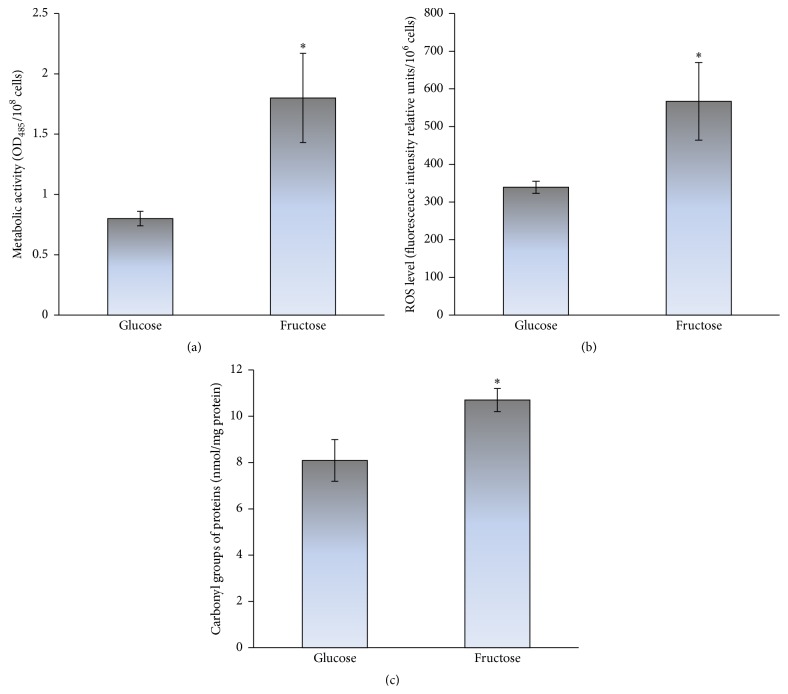
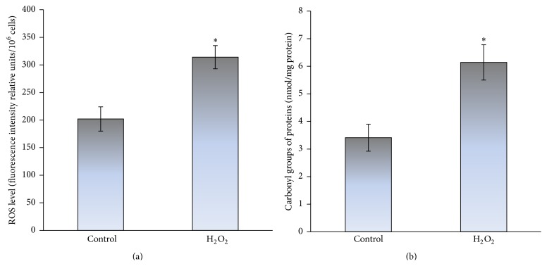
Here, we used yeast cultures grown on glucose or fructose for 24 h, when the exponential phase merged slowly into the stationary phase of growth. Figure 1 shows the influence of glucose and fructose on the intracellular level of the glycoxidation products. Fructose-grown as compared to glucose-grown cells demonstrated 2.5-fold higher metabolic rate (Figure 1(a)), which was consistent with our previous observations [11, 26]. It is well documented that overall metabolic rate, in particular carbohydrate metabolism, largely determines cellular redox balance [37, 38] and lifespan [39]. Significantly higher metabolic activity (Figure 1(a)) correlated with higher level of oxidative stress markers, ROS and protein carbonyls, in fructose-grown than glucose-grown cells (1.7- and 1.3-fold in Figures 1(b) and 1(c), resp.). Figure 2 shows higher level of total ROS (1.6-fold) and carbonyl proteins (1.8-fold) in yeast treated with 10 mM H2O2 relative to the control cells (without H2O2). Therefore, the increased levels of ROS and carbonyl proteins in fructose-grown yeast similar to those in the cells exposed to hydrogen peroxide can be explained by the development of oxidative stress during fructose-supplemented growth.
Figure 1.
The metabolic rate (a) and level of oxidative stress markers: ROS (b) and protein carbonyls (c), in glucose- and fructose-grown S. cerevisiae YPH250. Results are shown as the mean ± SEM (n = 3–6). ∗Significantly different from respective values for glucose with P < 0.05.
Figure 2.
Effect of hydrogen peroxide on the level of oxidative stress markers: ROS (a) and protein carbonyls (b), in S. cerevisiae YPH250. Results are shown as the mean ± SEM (n = 3–7). ∗Significantly different from respective values for control cells (without H2O2) with P < 0.05.
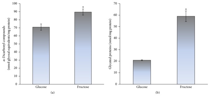
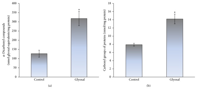
From the previous reports it is well known that the excessive fructose intake in animal [10, 12] and yeast models [11, 36] can also cause carbonyl stress. In addition, the increased protein carbonyls have been shown to accompany a number of physiological and pathological processes and were suggested to be a marker of aging [29, 40–44]. Therefore, next we examined the level of carbonyl stress markers in both the studied types of cells (glucose- and fructose-grown). Figure 3 demonstrates higher level of α-dicarbonyl compounds (a) and glycated proteins (b) in fructose-grown cells than those in cells grown on glucose (1.3- and 2.8-fold, resp.). To compare the ability of fructose and highly reactive carbonyl compound glyoxal to cause carbonyl stress, next we measured the level of carbonyl stress markers in yeast stressed by 50 mM glyoxal. The contents of α-dicarbonyls (Figure 4(a)) and protein carbonyls (Figure 4(b)) were found to be significantly increased after yeast treatment with glyoxal (2.5- and 1.8-fold, resp.). Thus, fructose-supplemented yeast cultivation can cause both oxidative and carbonyl stresses.
Figure 3.
The level of carbonyl stress markers: α-dicarbonyl compounds (a) and glycated proteins (b), in glucose- and fructose-grown S. cerevisiae YPH250. Results are shown as the mean ± SEM (n = 4–6). ∗Significantly different from respective values for glucose with P < 0.05.
Figure 4.
Effect of glyoxal on the level of carbonyl stress markers: α-dicarbonyl compounds (a) and protein carbonyls (b), in S. cerevisiae YPH250. Results are shown as the mean ± SEM (n = 3–7). ∗Significantly different from respective values for control cells (without glyoxal) with P < 0.05.
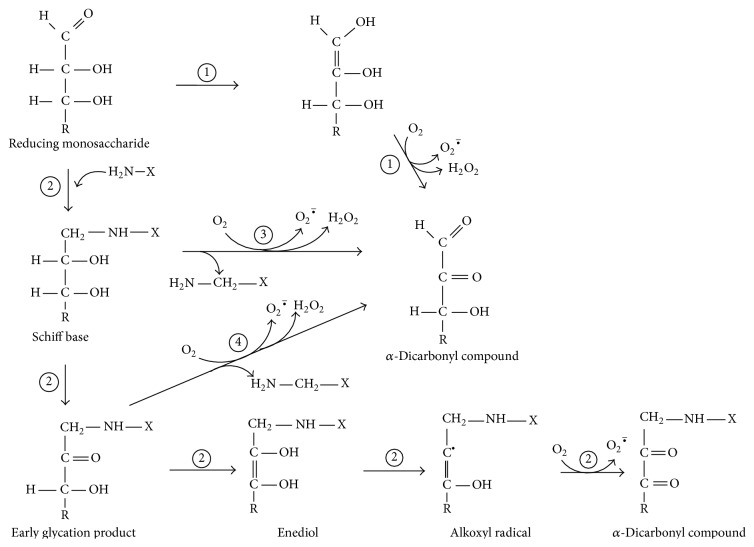
Figure 5 demonstrates how glucose and fructose are involved in ROS/RCS formation and oxidative/carbonyl stress. Glycoxidation can be initiated by both the reducing monosaccharides and yields enediols and alkoxyl radicals. Alkoxyl radical readily reacts with molecular oxygen generating superoxide anion radical, which can be converted to hydrogen peroxide and then to hydroxyl radical. At the same time, glycoxidation results in the formation of α-dicarbonyls such as glyoxal and methylglyoxal. Experiments comparing fructose and glucose reactivity in nonenzymatic processes demonstrate higher reactivity of fructose [4, 11, 13, 14, 26, 36]. At first glance, it disagrees with the concept of organic chemistry that aldoses (e.g., glucose) due to a greater electrophilicity and accessibility of their carbonyl group are more reactive than respective ketoses (e.g., fructose). One explanation is that glucose is less reactive due to the formation of very stable ring structures in aqueous solutions which retards its reactivity. Fructose also forms cyclic structures but exists to a greater extent in the open-chain active form than glucose. Therefore, under conditions used in this study fructose compared to glucose is a more potent initiator of glycoxidation in vivo (Figures 1 and 3).
Figure 5.
Involvement of reducing monosaccharides such as glucose and fructose in the glycoxidation reactions resulting in the production of ROS and RCS: (1) Wolff pathway; (2) glycation; (3) Namiki pathway; and (4) Hodge pathway.
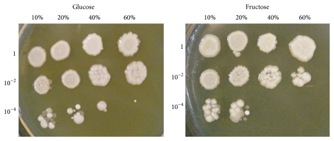
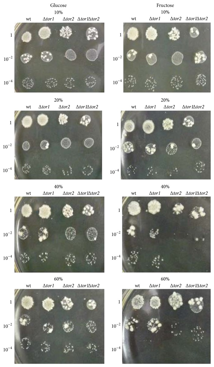
The above mentioned is consistent with somewhat better survival of S. cerevisiae YPH250 after stress induced by 40% glucose than 40% fructose (Figure 6). Similar results were obtained when another wild type strain S. cerevisiae JK9-3da and 60% carbohydrates were used (Figure 7); but, in contrast to S. cerevisiae YPH250, no difference has been observed between glucose- and fructose-stressed S. cerevisiae JK9-3da in the case of 40% monosaccharides. Thus, S. cerevisiae JK9-3da seems to be more resistant to high concentrations of fructose than S. cerevisiae YPH250. It is interesting that derivatives of JK9-3da, single mutant Δtor2 and double mutant Δtor1Δtor2, demonstrated lower survival under stress induced by 40% fructose than 40% glucose (Figure 7). Thus, some involvement of the TOR pathway can be supposed in fructose-induced stress in yeast. The previous work reported the decreased intracellular ROS levels at the inactivation of TOR signaling pathway by caloric restriction or the tor1 gene deletion, while activation of the TOR signaling pathway in S. cerevisiae by high nutrient concentrations increased ROS levels [45]. In addition, recently it has been suggested that TOR inhibition suppressed formation of methylglyoxal and other deleterious RCS [46] produced during carbohydrate metabolism [47, 48]; however any experimental confirmation of these hypotheses had not yet been found.
Figure 6.
Cell viability assay of S. cerevisiae YPH250 cells after exposure to high concentrations of glucose and fructose. In all samples within each plate, cell concentration was equaled and successively diluted by a factor of 100 down to 10−4. Representative results from a set of three experiments are shown.
Figure 7.
Cell viability assay of S. cerevisiae JK9-3da (wild type), Δtor1, Δtor2, and Δtor1Δtor2 cells after exposure to high concentrations of glucose and fructose. In all samples within each plate, cell concentration was equaled and successively diluted by a factor of 100 down to 10−4. Representative results from a set of three experiments are shown.
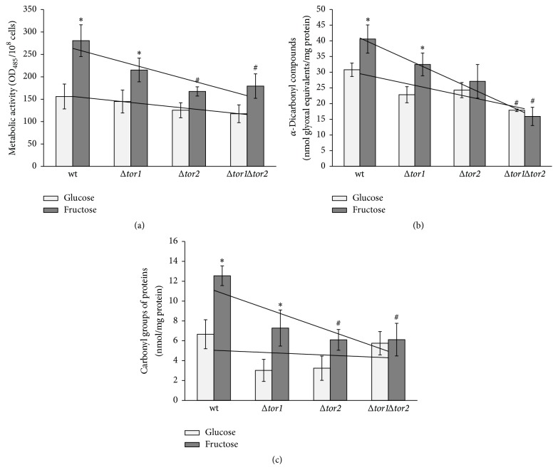
In order to examine the above-mentioned suggestion, the level of oxidative/carbonyl stress markers has been determined in the wild type JK9-3da and mutants defective in the tor1 and tor2 genes grown on glucose and fructose (Figure 8). In accordance with the suggestion that fructose versus glucose is a more potent inducer of oxidative/carbonyl stress and the data obtained on S. cerevisiae YPH250 (Figures 1 and 3), the metabolic rate (Figure 8(a)), level of α-dicarbonyls (Figure 8(b)), and protein carbonyls (Figure 8(c)) were found to be significantly higher in JK9-3da cells grown on fructose than those in the cells grown on glucose (1.8-, 1.3-, and 1.8-fold, resp.). The parameters in the three mutants grown on glucose demonstrated the values similar to those found in glucose-grown wild type cells. In the case of fructose-supplemented cultivation, all of the three parameters determined, metabolic activity, level of α-dicarbonyls, and protein carbonyl groups, were overall lower in the three mutants as compared to wild type (1.7-, 2.6-, and 1.9-fold, resp.). Therefore, the TOR gene deletions decreased the concentration of RCS in fructose-grown yeast. The latter corresponds well to the previous suggestion on suppressed RCS generation by TOR pathway inactivation [46].
Figure 8.
The metabolic rate (a) and level of carbonyl/oxidative stress markers: α-dicarbonyl compounds (b) and protein carbonyls (c), in glucose- and fructose-grown S. cerevisiae JK9-3da and its mutants Δtor1, Δtor2, and Δtor1Δtor2. Results are shown as the mean ± SEM (n = 3–6). ∗Significantly different from respective values for glucose and #wild type with P < 0.05.
4. Conclusion
The TOR signaling pathway first described in S. cerevisiae is highly conserved between yeast, animals, and plants. It received tremendous attention due to its importance in regulating cell growth, metabolism, longevity, and stress response. Our study confirms the previous findings on the higher reactivity of fructose versus glucose, more potent production of RCS/ROS, and development of oxidative/carbonyl stress in different fructose-supplemented models and extends them with the first report of the metabolic rate inhibition and lower generation of glycoxidation products in fructose-grown yeast lacking TOR proteins. Further research is needed to understand how TOR regulation may help prevent metabolic syndrome, diabetes complications, and other disturbances related to chronic consumption of diets high in fructose.
Acknowledgments
The authors are grateful to Professor Yoshiharu Inoue and Professor Michael Hall for providing the yeast strains and Professor Jacek Miedzobrodzki for support and cooperation.
Conflict of Interests
The authors declare that there is no conflict of interests regarding the publication of this paper.
References
- 1.Gaby A. R. Adverse effects of dietary fructose. Alternative Medicine Review. 2005;10(4):294–306. [PubMed] [Google Scholar]
- 2.Tappy L. Q&A: ‘toxic’ effects of sugar: should we be afraid of fructose? BMC Biology. 2012;10, article 42 doi: 10.1186/1741-7007-10-42. [DOI] [PMC free article] [PubMed] [Google Scholar]
- 3.Bray G. A. Energy and fructose from beverages sweetened with sugar or high-fructose corn syrup pose a health risk for some people. Advances in Nutrition. 2013;4(2):220–225. doi: 10.3945/an.112.002816. [DOI] [PMC free article] [PubMed] [Google Scholar]
- 4.Rippe J. M., Angelopoulos T. J. Sucrose, high-fructose corn syrup, and fructose, their metabolism and potential health effects: what do we really know? Advances in Nutrition. 2013;4(2):236–245. doi: 10.3945/an.112.002824. [DOI] [PMC free article] [PubMed] [Google Scholar]
- 5.Stanhope K. L., Schwarz J.-M., Havel P. J. Adverse metabolic effects of dietary fructose: results from the recent epidemiological, clinical, and mechanistic studies. Current Opinion in Lipidology. 2013;24(3):198–206. doi: 10.1097/mol.0b013e3283613bca. [DOI] [PMC free article] [PubMed] [Google Scholar]
- 6.White J. S. Challenging the fructose hypothesis: new perspectives on fructose consumption and metabolism. Advances in Nutrition. 2013;4(2):246–256. doi: 10.3945/an.112.003137. [DOI] [PMC free article] [PubMed] [Google Scholar]
- 7.Bunn H. F., Higgins P. J. Reaction of monosaccharides with proteins: possible evolutionary significance. Science. 1981;213(4504):222–224. doi: 10.1126/science.12192669. [DOI] [PubMed] [Google Scholar]
- 8.Dills W. L., Jr. Protein fructosylation: fructose and the Maillard reaction. The American Journal of Clinical Nutrition. 1993;58(5):779S–787S. doi: 10.1093/ajcn/58.5.779S. [DOI] [PubMed] [Google Scholar]
- 9.Tessier F. J. The Maillard reaction in the human body. The main discoveries and factors that affect glycation. Pathologie Biologie. 2010;58(3):214–219. doi: 10.1016/j.patbio.2009.09.014. [DOI] [PubMed] [Google Scholar]
- 10.Dong Q., Yang K., Wong S. M., O'Brien P. J. Hepatocyte or serum albumin protein carbonylation by oxidized fructose metabolites: glyceraldehyde or glycolaldehyde as endogenous toxins? Chemico-Biological Interactions. 2010;188(1):31–37. doi: 10.1016/j.cbi.2010.06.006. [DOI] [PubMed] [Google Scholar]
- 11.Semchyshyn H. M., Lozinska L. M., Miedzobrodzki J., Lushchak V. I. Fructose and glucose differentially affect aging and carbonyl/oxidative stress parameters in Saccharomyces cerevisiae cells. Carbohydrate Research. 2011;346(7):933–938. doi: 10.1016/j.carres.2011.03.005. [DOI] [PubMed] [Google Scholar]
- 12.Yang K., Feng C., Lip H., Bruce W. R., O'Brien P. J. Cytotoxic molecular mechanisms and cytoprotection by enzymic metabolism or autoxidation for glyceraldehyde, hydroxypyruvate and glycolaldehyde. Chemico-Biological Interactions. 2011;191(1–3):315–321. doi: 10.1016/j.cbi.2011.02.027. [DOI] [PubMed] [Google Scholar]
- 13.Levi B., Werman M. J. Fructose triggers DNA modification and damage in an Escherichia coli plasmid. Journal of Nutritional Biochemistry. 2001;12(4):235–241. doi: 10.1016/s0955-2863(00)00158-3. [DOI] [PubMed] [Google Scholar]
- 14.Lee O., Bruce W. R., Dong Q., Bruce J., Mehta R., O'Brien P. J. Fructose and carbonyl metabolites as endogenous toxins. Chemico-Biological Interactions. 2009;178(1–3):332–339. doi: 10.1016/j.cbi.2008.10.011. [DOI] [PubMed] [Google Scholar]
- 15.Betz C., Hall M. N. Where is mTOR and what is it doing there? Journal of Cell Biology. 2013;203(4):563–574. doi: 10.1083/jcb.201306041. [DOI] [PMC free article] [PubMed] [Google Scholar]
- 16.Schmelzle T., Hall M. N. TOR, a central controller of cell growth. Cell. 2000;103(2):253–262. doi: 10.1016/s0092-8674(00)00117-3. [DOI] [PubMed] [Google Scholar]
- 17.Aung-Htut M. T., Lam Y. T., Lim Y.-L., et al. Maintenance of mitochondrial morphology by autophagy and its role in high glucose effects on chronological lifespan of Saccharomyces cerevisiae . Oxidative Medicine and Cellular Longevity. 2013;2013:13. doi: 10.1155/2013/636287.636287 [DOI] [PMC free article] [PubMed] [Google Scholar]
- 18.Crespo J. L., Hall M. N. Elucidating TOR signaling and rapamycin action: lessons from Saccharomyces cerevisiae . Microbiology and Molecular Biology Reviews. 2002;66(4):579–591. doi: 10.1128/mmbr.66.4.579-591.2002. [DOI] [PMC free article] [PubMed] [Google Scholar]
- 19.Avruch J., Long X., Ortiz-Vega S., Rapley J., Papageorgiou A., Dai N. Amino acid regulation of TOR complex 1. American Journal of Physiology—Endocrinology and Metabolism. 2009;296(4):E592–E602. doi: 10.1152/ajpendo.90645.2008. [DOI] [PMC free article] [PubMed] [Google Scholar]
- 20.Izawa S., Inoue Y., Kimura A. Importance of catalase in the adaptive response to hydrogen peroxide: analysis of acatalasaemic Saccharomyces cerevisiae . Biochemical Journal. 1996;320(1):61–67. doi: 10.1042/bj3200061. [DOI] [PMC free article] [PubMed] [Google Scholar]
- 21.Hietman J., Movva N. R., Hall M. N. Targets for cell cycle arrest by the immunosuppressant rapamycin in yeast. Science. 1991;253(5022):905–909. doi: 10.1126/science.1715094. [DOI] [PubMed] [Google Scholar]
- 22.Helliwell S. B., Wagner P., Kunz J., Deuter-Reinhard M., Henriquez R., Hall M. N. TOR1 and TOR2 are structurally and functionally similar but not identical phosphatidylinositol kinase homologues in yeast. Molecular Biology of the Cell. 1994;5(1):105–118. doi: 10.1091/mbc.5.1.105. [DOI] [PMC free article] [PubMed] [Google Scholar]
- 23.Schmidt A., Kunz J., Hall M. N. TOR2 is required for organization of the actin cytoskeleton in yeast. Proceedings of the National Academy of Sciences of the United States of America. 1996;93(24):13780–13785. doi: 10.1073/pnas.93.24.13780. [DOI] [PMC free article] [PubMed] [Google Scholar]
- 24.Helliwell S. B., Howald I., Barbet N., Hall M. N. TOR2 is part of two related signaling pathways coordinating cell growth in Saccharomyces cerevisiae . Genetics. 1998;148(1):99–112. doi: 10.1093/genetics/148.1.99. [DOI] [PMC free article] [PubMed] [Google Scholar]
- 25.Semchyshyn H. M., Miedzobrodzki J., Bayliak M. M., Lozinska L. M., Homza B. V. Fructose compared with glucose is more a potent glycoxidation agent in vitro, but not under carbohydrate-induced stress in vivo: potential role of antioxidant and antiglycation enzymes. Carbohydrate Research. 2014;384:61–69. doi: 10.1016/j.carres.2013.11.015. [DOI] [PubMed] [Google Scholar]
- 26.Semchyshyn H. M., Lozinska L. M. Fructose protects baker's yeast against peroxide stress: potential role of catalase and superoxide dismutase. FEMS Yeast Research. 2012;12(7):761–773. doi: 10.1111/j.1567-1364.2012.00826.x. [DOI] [PubMed] [Google Scholar]
- 27.Semchyshyn H. M. Defects in antioxidant defence enhance glyoxal toxicity in the yeast Saccharomyces cerevisiae . Ukrainian Biochemical Journal. 2013;85(5):50–60. doi: 10.15407/ubj85.05.050. [DOI] [PubMed] [Google Scholar]
- 28.Lushchak V., Semchyshyn H., Lushchak O., Mandryk S. Diethyldithiocarbamate inhibits in vivo Cu,Zn-superoxide dismutase and perturbs free radical processes in the yeast Saccharomyces cerevisiae cells. Biochemical and Biophysical Research Communications. 2005;338(4):1739–1744. doi: 10.1016/j.bbrc.2005.10.147. [DOI] [PubMed] [Google Scholar]
- 29.Ispolnov K., Gomes R. A., Silva M. S., Freire A. P. Extracellular methylglyoxal toxicity in Saccharomyces cerevisiae: role of glucose and phosphate ions. Journal of Applied Microbiology. 2008;104(4):1092–1102. doi: 10.1111/j.1365-2672.2007.03641.x. [DOI] [PubMed] [Google Scholar]
- 30.Conconi A., Jager-Vottero P., Zhang X., Beard B. C., Smerdon M. J. Mitotic viability and metabolic competence in UV-irradiated yeast cells. Mutation Research. 2000;459(1):55–64. doi: 10.1016/s0921-8777(99)00057-9. [DOI] [PubMed] [Google Scholar]
- 31.Grzelak A., Macierzyńska E., Bartosz G. Accumulation of oxidative damage during replicative aging of the yeast Saccharomyces cerevisiae . Experimental Gerontology. 2006;41(9):813–818. doi: 10.1016/j.exger.2006.06.049. [DOI] [PubMed] [Google Scholar]
- 32.Sakai M., Oimomi M., Kasuga M. Experimental studies on the role of fructose in the development of diabetic complications. Kobe Journal of Medical Sciences. 2002;48(5-6):125–136. [PubMed] [Google Scholar]
- 33.Bradford M. M. A rapid and sensitive method for the quantitation of microgram quantities of protein utilizing the principle of protein-dye binding. Analytical Biochemistry. 1976;72(1-2):248–254. doi: 10.1016/0003-2697(76)90527-3. [DOI] [PubMed] [Google Scholar]
- 34.Tappy L., Lê K.-A. Metabolic effects of fructose and the worldwide increase in obesity. Physiological Reviews. 2010;90(1):23–46. doi: 10.1152/physrev.00019.2009. [DOI] [PubMed] [Google Scholar]
- 35.Tappy L., Lê K. A., Tran C., Paquot N. Fructose and metabolic diseases: new findings, new questions. Nutrition. 2010;26(11-12):1044–1049. doi: 10.1016/j.nut.2010.02.014. [DOI] [PubMed] [Google Scholar]
- 36.Semchyshyn H. M. Fructation in vivo: detrimental and protective effects of fructose. BioMed Research International. 2013;2013:9. doi: 10.1155/2013/343914.343914 [DOI] [PMC free article] [PubMed] [Google Scholar]
- 37.González-Siso M. I., García-Leiro A., Tarrío N., Cerdán M. E. Sugar metabolism, redox balance and oxidative stress response in the respiratory yeast Kluyveromyces lactis . Microbial Cell Factories. 2009;8, article 46 doi: 10.1186/1475-2859-8-46. [DOI] [PMC free article] [PubMed] [Google Scholar]
- 38.Magherini F., Carpentieri A., Amoresano A., et al. Different carbon sources affect lifespan and protein redox state during Saccharomyces cerevisiae chronological ageing. Cellular and Molecular Life Sciences. 2009;66(5):933–947. doi: 10.1007/s00018-009-8574-z. [DOI] [PMC free article] [PubMed] [Google Scholar]
- 39.Van Raamsdonk J. M., Meng Y., Camp D., et al. Decreased energy metabolism extends life span in Caenorhabditis elegans without reducing oxidative damage. Genetics. 2010;185(2):559–571. doi: 10.1534/genetics.110.115378. [DOI] [PMC free article] [PubMed] [Google Scholar]
- 40.Lushchak V. I. Free radical oxidation of proteins and its relationship with functional state of organisms. Biochemistry. 2007;72(8):809–827. doi: 10.1134/s0006297907080020. [DOI] [PubMed] [Google Scholar]
- 41.Tomida H., Fujii T., Furutani N., et al. Antioxidant properties of some different molecular weight chitosans. Carbohydrate Research. 2009;344(13):1690–1696. doi: 10.1016/j.carres.2009.05.006. [DOI] [PubMed] [Google Scholar]
- 42.Madian A. G., Regnier F. E. Proteomic identification of carbonylated proteins and their oxidation sites. Journal of Proteome Research. 2010;9(8):3766–3780. doi: 10.1021/pr1002609. [DOI] [PMC free article] [PubMed] [Google Scholar]
- 43.Lushchak V. I. Oxidative stress in yeast. Biochemistry. 2010;75(3):281–296. doi: 10.1134/s0006297910030041. [DOI] [PubMed] [Google Scholar]
- 44.Trnková L., Dršata J., Boušová I. Oxidation as an important factor of protein damage: implications for Maillard reaction. Journal of Biosciences. 2015;40(2):419–439. doi: 10.1007/s12038-015-9523-7. [DOI] [PubMed] [Google Scholar]
- 45.Wei M., Fabrizio P., Hu J., et al. Life span extension by calorie restriction depends on Rim15 and transcription factors downstream of Ras/PKA, Tor, and Sch9. PLoS Genetics. 2008;4, article e13 doi: 10.1371/journal.pgen.0040013. [DOI] [PMC free article] [PubMed] [Google Scholar]
- 46.Hipkiss A. R. Energy metabolism, proteotoxic stress and age-related dysfunction—protection by carnosine. Molecular Aspects of Medicine. 2011;32(4–6):267–278. doi: 10.1016/j.mam.2011.10.004. [DOI] [PubMed] [Google Scholar]
- 47.Gomes R. A., Oliveira L. M. A., Silva M., et al. Protein glycation in vivo: functional and structural effects on yeast enolase. Biochemical Journal. 2008;416(3):317–325. doi: 10.1042/bj20080632. [DOI] [PubMed] [Google Scholar]
- 48.Inoue Y., Maeta K., Nomura W. Glyoxalase system in yeasts: structure, function, and physiology. Seminars in Cell and Developmental Biology. 2011;22(3):278–284. doi: 10.1016/j.semcdb.2011.02.002. [DOI] [PubMed] [Google Scholar]


