

Abstract
BACKGROUND:
The appropriate sequence of different imagings and indications of thoracic computed tomography (TCT) in evaluating chest trauma have not yet been clarified at present. The current study was undertaken to determine the value of chest X-ray (CXR) in detecting chest injuries in patients with blunt trauma.
METHODS:
A total of 447 patients with blunt thoracic trauma who had been admitted to the emergency department (ED) in the period of 2009–2013 were retrospectively reviewed. The patients met inclusion criteria (age>8 years, blunt injury to the chest, hemodynamically stable, and neurologically intact) and underwent both TCT and upright CXR in the ED. Radiological imagings were re-interpreted after they were collected from the hospital database by two skilled radiologists.
RESULTS:
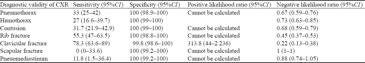
Of the 447 patients, 309 (69.1%) were male. The mean age of the 447 patients was 39.5±19.2 (range 9 and 87 years). 158 (35.3%) patients were injured in motor vehicle accidents (MVA). CXR showed the highest sensitivity in detecting clavicle fractures [95%CI 78.3 (63.6–89)] but the lowest in pneuomediastinum [95%CI 11.8 (1.5–36.4)]. The specificity of CXR was close to 100% in detecting a wide array of entities.
CONCLUSION:
CXR remains to be the first choice in hemodynamically unstable patients with blunt chest trauma. Moreover, stable patients with normal CXR are candidates who should undergo TCT if significant injury has not been ruled out.
KEY WORDS: Chest, Blunt trauma, X-Rays, Computed tomography, Emergency department
INTRODUCTION
Blunt chest trauma accounts for a proportion of trauma mortality and clinicians should rule out chest injury in evaluation of blunt trauma. The evaluation of thoracic injury can lead to appropriate treatment and life-saving. Both conventional radiography (chest X-ray, CXR) and thoracic computed tomography (TCT) are commonly used in the emergency setting.[1,2] CXR has a low rate of sensitivity and specificity in detecting thoracic injuries in hemodynamically stable patients with blunt chest trauma. Thus the rate of TCT orders is high in patients with such a trauma.[3] Generally, history taking and physical examination are helpful in decision-making for imaging. TCT is the preferred method for detecting thoracic injuries with a high sensitivity and specificity.[4,5]
Computed tomography has been improved in quality and availability in the past 15 years. And the use of TCT has raised three problems in practice: increased cancer risk caused by exposure to ionizing radiation; increased costs imposed by TCT; and increased length of ED stay due to unnecessary screening with TCT.[6,7]
Since the appropriate sequence of different imagings and indications of TCT in evaluation of chest trauma are not clear-cut, the present study is undertaken to define the value of CXR in detecting chest injuries in patients with blunt trauma.
METHODS
Sociodemographic and clinical data
This retrospective observational study was conducted in a university-based ED with an annual census of 110 000 in western Turkey. The study was approved by the institutional review board. The patients who had been admitted to the ED because of blunt thoracic trauma during the period of 2009–2013 were retrospectively reviewed.
The inclusion criteria were as follows: patients older than eight years; those who were hemodynamically stable and neurologically intact (GCS=15); those without penetrating thoracic injury; those whose CXR was performed in a recumbent position; and those who had undergone TCT.
Sociodemographic and clinical data of the patients were obtained from hospital charts. The exclusion criteria included the patients who were hemodynamical unstable and had multiple traumas and those whose CXR was taken in an upright position. CXRs were elicited via “Philips Bucky Diagnost TH” whereas TCTs were obtained without contrast media using a 16-detector CT device (Brilliance 16, Philips Medical System, Cleveland, USA).
The radiological data taken from the hospital database were re-interpreted by two experienced radiologists. One radiologist was asked to review CXR and the other interpreted TCT images. The images were screened for the presence of pneumothorax, hemothorax, contusion, rib fracture, clavicular fracture, scapular fracture, and pneuomediastinum.
Statistical analysis
The data of the present study were analyzed with MedCalc software. The numeric variables were presented as mean±standard deviation and frequent data as rates. The diagnostic validity of CXR for chest trauma was defined by sensitivity, specificity, and positive and negative likelihood ratio with 95% confidence intervals.
RESULTS
A total of 2 074 patients with chest trauma referred to the ED were identified in the study period. Eligibility criteria (age>8 years, blunt injury to the chest, hemodynamically stable, neurologically intact/GCS=15) were met by 1 095 patients (Figure 1), of whom, 484 (44.2%) had undergone both CXR and TCT in the ED. Thirty-seven patients were excluded because CXR had been done in the upright position. The remaining 447 patients served as a study group.
Figure 1.
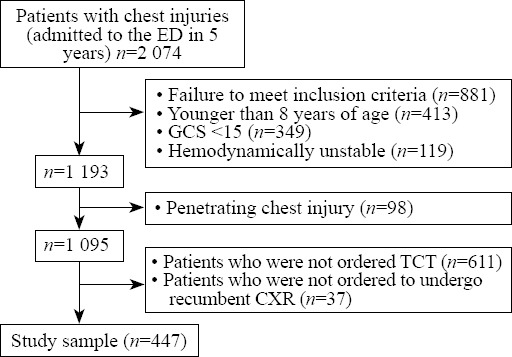
Participant flow through the study.
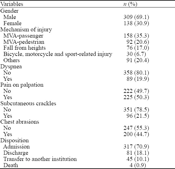
The mean age of these patients was 39.5±19.2 years (range 9–87) in the study group. Most of the patients were male (n=309, 69.1%). The frequent mechanism of injury was motor vehicle accident (MVA) (passengers) (n=158, 35.3%) followed by pedestrians involved in MVA (n=92, 20.6%) and falls from height (n=76, 17.0%) (Table 1).
Table 1.
Sociodemographic characteristics of the participants
Radiological findings
TCT findings in the study group were (in a decreasing order) rib fractures (n=150, 33.6%), pneumothorax (n=118, 26.4%), contusion (n=82, 18.3%), hemothorax (n=63, 14.1%), clavicular fracture (n=46, 10.3%), pneuomediastinum (n=17, 3.8%), and scapular fracture (n=9, 2%).
The sensitivity and specificity of CXR interpretations regarding pneumothorax, hemothorax, contusion, rib fracture, clavicular fracture, scapular fracture and pneuomediastinum are shown in Table 2. CXR showed the highest sensitivity for clavicular fractures [95%CI 78.3 (63.6–89)] and the lowest figure for pneuomediastinum [95%CI 11.8 (1.5–36.4)]. Of note, the specificity of CXR was close to 100% in detecting a wide array of entities (Table 2).
Table 2.
The validity of CXR in detecting injuries caused by chest trauma
DISCUSSION
The present study suggests that CXR has a low sensitivity for nearly all entities in the chest resulting from injury and a specificity of about 100%. The findings indicate that if a pathological change is seen in CXR, it is definitely true. However, the absence of a finding in CXR cannot strongly exclude the presence of an injury.
CXR is the first-line diagnostic tool in the evaluation of the patients with blunt chest trauma in the emergency setting. The diagnostic accuracy of CXR in detecting major injuries is between 6.3% and 12.4%.[1] TCT is viewed as a “gold standard” imaging modality in the ED. The present study investigated the value of CXR in detecting chest injuries in a large number of hemodynamically stable patients with blunt trauma compared to the previous studies.
Studies reported sensitivities of 25.5%[8] and 39.4%[9] for CXR in detecting rib fractures in patients with blunt chest trauma. Others studies reported the sensitivities of 29%[10] and 14.3%[9] in detecting pneumothorax. In the present study, the sensitivity of CXR was 55.3% and 33% in detecting rib fractures and pneumothoraces respectively. The low sensitivity of CXR in detecting rib fractures was consistent with that reported elsewhere.
The sensitivity of CXR in detecting lung contusion was 52% or 53%.[7,8] A study[8] reported that CT identified 11 lung contusions which had been missed by CXR. The sensitivity of CXR in identification of contusions in the present study was 51%. Likewise, the sensitivity of CXR in detecting hemothoraces and clavicular fractures was consistent with that reported in the literature.[8,10] The findings of the present study suggest that CXR is not effective in detecting scapular fractures in patients with blunt chest trauma.
A report[11] advocated that TCT may be helpful in the treatment of hemodynamically stable patients with blunt chest trauma. On the contrary, others[6,12,13] suggested that TCT should be withheld in the initial management of patients. The low sensitivity of CXR suggests that CXR-negative patients should undergo further investigations if they are highly suspected.
Limitations
In the present study, patients with injuries of the diaphragm and vasculature were not included. However, Bhullar and Block[14] found that both CXR and TCT are effective in detecting diafragmatic injuries. The retrospective nature of the present study is also a limitation. Moreover, patients in whom CXR was performed in the upright position were excluded from the study.
CXR remains to be the first choice for hemodynamically unstable patients with blunt chest trauma. But stable patients with CXR should undergo TCT if injury has not been ruled out.
Footnotes
Funding: None.
Ethical approval: The study was approved by the institutional review board.
Conflicts of interest: The authors declare that there are no conflicts of interest related to the publication of this paper.
Contributors: Agladioglu K proposed the study, analyzed the data and wrote the first draft. All authors contributed to the design and interpretation of the study and to further drafts.
REFERENCES
- 1.Calderon G, Perez D, Fortman J, Kea B, Rodriguez RM. Provider perceptions concerning use of chest x-ray studies in adult blunt trauma assessments. J Emerg Med. 2012;43:568–574. doi: 10.1016/j.jemermed.2011.06.045. [DOI] [PubMed] [Google Scholar]
- 2.Pothiawala S, Gogna A. Early diagnosis of bowel obstruction and strangulation by computed tomography in emergency department. World J Emerg Med. 2012;3:227–231. doi: 10.5847/wjem.j.issn.1920-8642.2012.03.012. [DOI] [PMC free article] [PubMed] [Google Scholar]
- 3.Peters S, Nicolas V, Heyer CM. Multidetector computed tomography-spectrum of blunt chest wall and lung injuries in polytraumatized patients. Clin Radiol. 2010;65:333–338. doi: 10.1016/j.crad.2009.12.008. [DOI] [PubMed] [Google Scholar]
- 4.Rodriguez RM, Anglin D, Langdorf MI, Baumann BM, Hendey GW, Bradley RN, et al. NEXUS chest: validation of a decision instrument for selective chest imaging in blunt trauma. JAMA Surg. 2013;148:940–946. doi: 10.1001/jamasurg.2013.2757. [DOI] [PubMed] [Google Scholar]
- 5.Rodriguez RM, Hendey GW, Marek G, Dery RA, Bjoring A. A pilot study to derive clinical variables for selective chest radiography in blunt trauma patients. Ann Emerg Med. 2006;47:415–418. doi: 10.1016/j.annemergmed.2005.10.001. [DOI] [PubMed] [Google Scholar]
- 6.Kea B, Gamarallage R, Vairamuthu H, Fortman J, Lunney K, Hendey GW, et al. What is the clinical significance of chest CT when the chest x-ray result is normal in patients with blunt trauma? Am J Emerg Med. 2013;31:1268–1273. doi: 10.1016/j.ajem.2013.04.021. [DOI] [PMC free article] [PubMed] [Google Scholar]
- 7.Gosk J, Hendrich B, Wiącek R, Sąsiadek M, Rutowski R. Assessment of the usefulness of X-ray myelography and magnetic resonance myelography, performed with an open low-field device, in diagnosing perinatal preganglionic injuries of the brachial plexus. Arch Med Sci. 2012;8:678–683. doi: 10.5114/aoms.2012.28597. [DOI] [PMC free article] [PubMed] [Google Scholar]
- 8.Chardoli M, Hasan-Ghaliaee T, Akbari H, Rahimi-Movaghar V. Accuracy of chest radiography versus chest computed tomography in hemodynamically stable patients with blunt chest trauma. Chin J Traumatol. 2013;16:351–354. [PubMed] [Google Scholar]
- 9.Ziegler K, Feeney JM, Desai C, Sharpio D, Marshall WT, Twohig M. Retrospective review of the use and costs of routine chest x rays in a trauma setting. J Trauma Manag Outcomes. 2013;7:2. doi: 10.1186/1752-2897-7-2. [DOI] [PMC free article] [PubMed] [Google Scholar]
- 10.Traub M, Stevenson M, McEvoy S, Briggs G, Lo SK, Leibman S, et al. The use of chest computed tomography versus chest X-ray in patients with major blunt trauma. Injury. 2007;38:43–47. doi: 10.1016/j.injury.2006.07.006. [DOI] [PubMed] [Google Scholar]
- 11.Trupka A, Waydhas C, Hallfeldt KK, Nast-Kolb D, Pfeifer KJ, Schweiberer L. Value of thoracic computed tomography in the first assessment of severely injured patients with blunt chest trauma: results of a prospective study. J Trauma. 1997;43:405–411. doi: 10.1097/00005373-199709000-00003. discussion 411–412. [DOI] [PubMed] [Google Scholar]
- 12.Marts B, Durham R, Shapiro M, Mazuski JE, Zuckerman D, Sundaram M, et al. Computed-tomography in the diagnosis of blunt thoracic injury. Am J Surg. 1994;168:688–692. doi: 10.1016/s0002-9610(05)80146-1. [DOI] [PubMed] [Google Scholar]
- 13.Daly M, Miller PR, Carr JJ, Gayzik FS, Hoth JJ, Meredith JW, Stitzel JD. Traumatic pulmonary pathology measured with computed tomography and a semiautomated analytic method. Clin Imaging. 2008;32:346–354. doi: 10.1016/j.clinimag.2008.02.026. [DOI] [PubMed] [Google Scholar]
- 14.Bhullar IS, Block EFJ. CT with coronal reconstruction identifies previously missed smaller diaphragmatic injuries after blunt trauma. Am Surg. 2011;77:55–58. [PubMed] [Google Scholar]