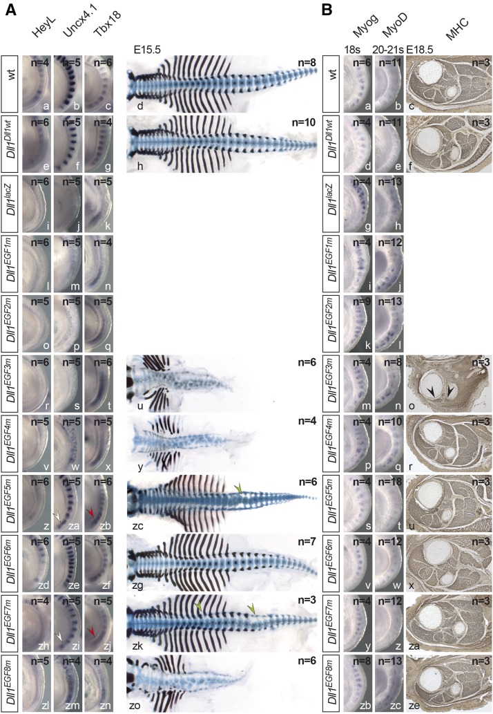
Figure 3.
Somite patterning and skeletal muscle development in mutants homozygous for individual Dll1 EGF alleles. (A) Whole-mount in situ hybridizations (WISH) of E9.5 and skeletal preparations of E15.5 embryos. Alleles are indicated at the left, probes at the top. White and red arrowheads point to irregularities of Uncx4.1 and Tbx18 expression patterns, respectively; green arrowheads point to malformed vertebrae. (B) Muscle differentiation in mutants homozygous for individual EGF alleles. WISH of 18 and 20–21 somite-stage embryos and anti-MHC antibody staining of hind-limb sections of E18.5 embryos. Alleles are indicated at the left, probes/antibodies at the top. Arrowheads in o point to remnants of skeletal muscles. For My32 staining, we analyzed three individual embryos with a minimum of six consecutive sections per genotype.

