Abstract
To investigate whether abdominopelvic hemorrhage shown on computed tomography (CT) images can be diagnosed with the same accuracy on a tablet computer as on a dedicated reading display. One hundred patients with a clinical suspicion of abdominopelvic hemorrhage that underwent biphasic CT imaging were retrospectively read by two readers on a dedicated reading display (reference standard) and on a tablet computer (iPad Air). Reading was performed in a dedicated reading room with ambient light conditions. Image evaluation included signs of an active hemorrhage (extravasation of contrast media) and different signs indicating a condition after abdominopelvic hemorrhage (hematoma, intestinal clots, vessel stump, free abdominopelvic fluid with a mean Hounsfield unit value >20, and asymmetric muscle volume indicating intramuscular hemorrhage). Sensitivity, specificity, and positive and negative predictive values (PPV/NPV) were calculated for the tablet-based reading. Active abdominopelvic hemorrhage (n = 72) was diagnosed with the tablet computer with a sensitivity of 0.96, a specificity of 0.93, a PPV of 0.97, and an NPV of 0.90. The results for the detection of the signs indicating a condition after abdominopelvic hemorrhage range from 0.83 to 1.00 in the case of sensitivity, from 0.95 to 1.00 in the case of specificity, from 0.94 to 1.00 in the case of the PPV, and from 0.96 to 1.00 in the case of the NPV. Abdominopelvic hemorrhage shown on CT images can be diagnosed on a tablet computer with a high diagnostic accuracy allowing mobile on-call diagnoses. This may be helpful because an early and reliable diagnosis at any time is crucial for an adequate treatment strategy.
Keywords: Hemorrhage, Abdominopelvic, Computed tomography, Tablet computer, Display device, Image display, Teleradiology
Introduction
Abdominopelvic hemorrhage is a potentially life-threatening situation. An early and reliable diagnosis is crucial for adequate treatment and patient survival [1–3]. In the case of suspected abdominopelvic hemorrhage, algorithms suggest the prompt performance of computed tomography (CT) angiography after the stabilization of the patient [3]. Depending on the findings, the appropriate treatment option is determined. If active hemorrhage is evident, immediate hemostatic procedures are recommended. Possible procedures are surgery, esophagogastroduodenoscopy (EGD), or angiography. An expert radiologist may not always be present when an active hemorrhage needs to be ruled out or when treatment options need to be discussed. Whenever the radiologist on duty is uncertain about the diagnosis, he/she should be able to discuss the case with an expert. Previous studies have shown that the display quality of tablet computers is sufficient for teleconsultation purposes in various emergency imaging procedures [4–6]. Interestingly, no study has investigated the diagnostic accuracy of detecting abdominopelvic hemorrhage on a tablet computer yet. Reading the studies on a tablet computer would facilitate mobile on-call diagnoses which might be helpful for a quick and reliable diagnosis and for treatment strategy.
Therefore, the aim of this study was to investigate the diagnostic performance of biphasic CT exams displayed on a tablet computer in detecting active abdominopelvic hemorrhage and different signs indicating a condition after hemorrhage compared to using a dedicated reading display.
Materials and Methods
This single-center investigation was approved by the institutional review board of the University Hospital Erlangen, and all procedures were in accordance with the Helsinki Declaration. The need for informed consent was waived.
Patients
We retrospectively collected 100 patients that had been referred to our Department with a suspected abdominopelvic hemorrhage and that were examined with a biphasic CT scan (arterial and portal venous contrast media phases). We searched appropriate patients using our radiological information system (RIS, iSOFT, Mannheim, Germany). Exclusion criterion was an incomplete CT examination. The results obtained using a dedicated workstation/reading display served as reference standard to diagnose active abdominopelvic hemorrhage.
Computed Tomography
CT was performed with 20, 64, 128, or 256 row scanners (Somatom Definition AS20, Somatom Sensation, Somatom Definition AS+, Somatom Force, or Definition Flash, Siemens AG Healthcare, Forchheim, Germany) using power injectors (Accutron CT-D, Medtron, Saarbrücken, Germany) for intravenous contrast media application (Imeron 350, Bracco Imaging, Milan, Italy). The arterial phase was identified by test-bolus measurements; the portal venous phase was set at 70 s after start of contrast media injection.
Image Interpretation
The CT exams were read consensually by two readers in two reading sessions (RS1 and RS2). All CT exams were assigned to both RS. Readers had a work experience of 9 and 5 years. Readers were blinded to the patient’s name, gender, and date of examination. All information provided to the readers was that the referring physicians suspected an abdominopelvic hemorrhage due to clinical circumstances like decreasing concentration of hemoglobin in blood tests or demanding need of catecholamine in intensive care patients.
CT exams were read with a Digital Imaging and Communications in Medicine (DICOM) viewer (syngo.plaza, Siemens AG Healthcare, Erlangen, Germany) running on a workstation with a dedicated reading display (standard setting) as well as with a DICOM viewer (OsiriX HD, Geneva, Switzerland) running on a tablet computer with a 9.7-inch screen with a resolution of 2048 × 1536 pixels at 264 pixels per inch (ppi) (iPad Air, Apple Inc., Cupertino, California, USA). The display connected to the workstation was a 30-inch screen with a resolution of 3280 × 2048 pixels at 127.32 ppi (Coronis Fusion 6MP LED (MDCC-6230), Barco, Kortrijk, Belgium). Detailed specifications of the screen of both devices are shown in Table 1. For standardization, the display brightness of the tablet computer was set to 100 % in the menu of the operating system (iOS). Both DICOM viewers offered the same evaluation tools like magnification of images and windowing or measuring of Hounsfield units (HU). Reading was performed in a dedicated reading room with ambient light conditions.
Table 1.
Technical specifications of the dedicated reading display and of the screen of the tablet computer
| Dedicated reading display | Tablet computer | |
|---|---|---|
| Device | Coronis Fusion 6MP LED (MDCC-6230, Barco, Kortrijk, Belgium) | iPad Air (Apple Inc., Cupertino, CA, USA) |
| Active screen size (diagonal) | 772 mm (30.4 in.) | 246.3 mm (9.7 in.) |
| Aspect ratio (H/V) | 16:10 | 4:3 |
| Resolution | 3280 × 2048 pixels at 127.32 pixels per inch (ppi) | 2048 × 1536 pixels at 264 pixels per inch (ppi) |
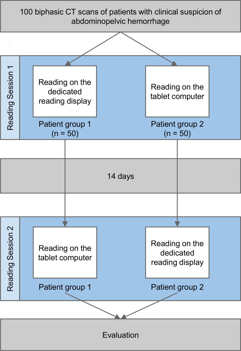
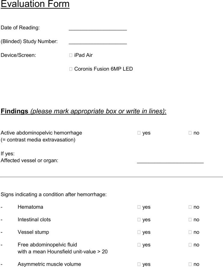
To avoid case recognition and the influence of a training effect, the 100 exams were randomly clustered in two groups and read on the two devices with an interval of 14 days. Patient group 1 (n = 50) was read on the dedicated display and patient group 2 (n = 50) was read on the tablet computer. Fourteen days later, the groups were read on the other device, respectively (study procedure is shown in Fig. 1). To gain a statistical evaluable dataset, a custom-made evaluation form was used (Fig. 2), asking for direct and indirect signs of abdominopelvic hemorrhage. An active abdominopelvic hemorrhage was stated if an extravasation of contrast media extraluminal or intraluminal was observed. Signs of a condition after abdominopelvic hemorrhage were registered with hematoma, free fluid, asymmetric muscle thickness, clots, or vessel stumps. All answers were registered either with present (=1) or absent (=0).
Fig. 1.
Study procedure. One hundred biphasic CT exams of 100 patients were randomly divided in two groups. During the first reading session, 50 CT exams (patient group 1) were read on the dedicated reading display and 50 CT exams (patient group 2) were read on the tablet computer. During the second reading session (14 days later), the same two patient groups were read on the other device
Fig. 2.
Evaluation form. Findings above the horizontal line (contrast media extravasation) indicate active hemorrhage. Findings below the horizontal line are signs indicating a condition after hemorrhage
Statistical Analysis
Statistical analysis was performed using dedicated software (SPSS Statistics v20, IBM, Armonk, USA). Sensitivities, specificities, and positive and negative predictive values with corresponding confidence intervals were calculated for the detection of active abdominal hemorrhage (= contrast media extravasation) and for the detection of different signs indicating a condition after hemorrhage with a tablet computer. The diagnoses done with a dedicated reading display were considered to be the reference standard.
Results
Image quality was satisfactory in all CT exams.
An active abdominopelvic hemorrhage was found in 71 CT exams on the tablet computer and in 72 exams on the dedicated reading display. Active hemorrhage was ruled out on the tablet computer in 29 exams and on the dedicated reading display in 28 exams. Usage of the tablet computer led to two false positive and three false negative diagnoses. This yielded a sensitivity of 0.96 (confidence interval (CI) 0.87–0.99), a specificity of 0.93 (CI 0.75–0.99), a positive predictive value (PPV) of 0.97 (CI 0.89–1.00), and a negative predictive value (NPV) of 0.90 (CI 0.72–0.97).
Detection of different signs indicating a condition after hemorrhage on the tablet computer showed the following results. A hematoma was present in 59 exams and was detected with a sensitivity of 1.00 (CI 0.92–1.00), a specificity of 0.95 (CI 0.82–0.99), a PPV of 0.97 (CI 0.88–0.99), and an NPV of 1.00 (CI 0.89–1.00). Intestinal clots were present in 18 exams and were detected with a sensitivity of 0.83 (CI 0.58–0.96), a specificity of 0.99 (CI 0.92–1.00), a PPV of 0.94 (CI 0.68–1.00), and an NPV of 0.96 (CI 0.89–0.99). A vessel stump was present in three exams and was detected with a sensitivity of 1.00 (CI 0.31–1.00), a specificity of 1.00 (CI 0.95–1.00), a PPV of 1.00 (CI 0.31–1.00), and an NPV of 1.00 (CI 0.95–1.00). Free intra-abdominal fluid with a mean Hounsfield unit value >20 was present in 36 exams and was detected with a sensitivity of 1.00 (CI 0.88–1.00), a specificity of 1.00 (CI 0.93–1.00), a PPV of 1.00 (CI 0.88–1.00), and an NPV of 1.00 (CI 0.93–1.00). Asymmetric muscle volume (indicating intramuscular hemorrhage) was present in 29 exams and was detected with a sensitivity of 1.00 (CI 0.85–1.00), a specificity of 1.00 (CI 0.94–1.00), a PPV of 1.00 (CI 0.85–1.00), and an NPV of 1.00 (CI 0.94–1.00). Results are shown in Table 2.
Table 2.
Diagnostic performance of biphasic CT exams displayed on a tablet computer in detecting active abdominopelvic hemorrhage and signs indicating a condition after hemorrhage
| Diagnostic performance of CT exams displayed on a tablet computer | ||||
|---|---|---|---|---|
| Sensitivity | Specificity | Positive predictive value | Negative predictive value | |
| Active abdominal hemorrhage (=contrast media extravasation) (n = 72) | 0.96 (0.87–0.99) | 0.93 (0.75–0.99 | 0.97 (0.89–1.00) | 0.90 (0.72–0.97) |
| Signs indicating a condition after hemorrhage | ||||
| Hematoma (n = 59) | 1.00 (0.92–1.00) | 0.95 (0.82–0.99) | 0.97 (0.88–0.99) | 1.00 (0.89–1.00) |
| Intestinal clots (n = 18) | 0.83 (0.58–0.96) | 0.99 (0.92–1.00) | 0.94 (0.68–1.00) | 0.96 (0.89–0.99) |
| Vessel stump (n = 3) | 1.00 (0.31–1.00) | 1.00 (0.95–1.00) | 1.00 (0.31–1.00) | 1.00 (0.95–1.00) |
| Free intra-abdominal fluid with a mean Hounsfield unit value > 20) (n = 36) | 1.00 (0.88–1.00) | 1.00 (0.93–1.00) | 1.00 (0.88–1.00) | 1.00 (0.93–1.00) |
| Asymmetric muscle volume (indicating intramuscular hemorrhage) (n = 29) | 1.00 (0.85–1.00) | 1.00 (0.94–1.00) | 1.00 (0.85–1.00) | 1.00 (0.94–1.00) |
Confidence intervals are shown in brackets
Discussion
The diagnostic performance of biphasic CT scans displayed on a tablet computer was convincing for the detection of active abdominopelvic hemorrhage and different signs indicating a condition after hemorrhage. Active abdominopelvic hemorrhage was diagnosed with a sensitivity of 0.96, a specificity of 0.93, a PPV of 0.97, and an NPV of 0.90, and results for detection of the signs indicating a condition after hemorrhage range from 0.83 to 1.00 in the case of sensitivity, from 0.95 to 1.00 in the case of specificity, from 0.94 to 1.00 in the case of the PPV, and from 0.96 to 1.00 in the case of the NPV. Therefore, we assume that reading dedicated CT scans with the question of abdominopelvic hemorrhage can be performed on a tablet computer for teleconsultation purposes.
The use of a tablet computer in emergency radiology has been described previously for different scenarios [4–6]. Intracranial hemorrhage, cerebral infarction, and pulmonary embolism could be detected reliably. Spinal emergency cases have been evaluated as well. All studies show that a tablet computer is a reliable tool in setting up the appropriate diagnoses in emergency cases.
The reasons for abdominopelvic hemorrhage are manifold. Various guidelines are dealing with the issue of the adequate diagnostic procedure [7–10]. Commonly, a prompt and accurate diagnosis is necessary to quickly initiate therapy and to avoid life-threatening complications [11]. CT angiography has proven to play an important role for diagnosis [12, 13].
A contrast media extravasation represents an active hemorrhage out of a vessel or organ and might be treatable through transcatheter arterial embolization [14–17]. Signs of a condition after hemorrhage need to be detected reliably because of the risk of recurrence. In this case, patients will be monitored closely. Especially after hours, mobile on-call diagnoses may be helpful and time-saving. Teleconsultation has already proven to play its role in emergency radiology. In particular, tablet computers seem to be a suitable option as they are mobile and their display quality is comparable to dedicated reading displays [18]. For standardization, reading was performed in a dedicated reading room with ambient light conditions, and the display brightness of the tablet computer was set to 100 %. Previous studies mainly focused on emergency conditions and showed the positive use in brain and spine imaging as well as in pulmonary CT angiography for patients with suspected embolism [8, 9, 19–21]. To our knowledge, this is the first study that investigates abdominopelvic imaging with the question of abdominopelvic hemorrhage. Our results suggest that active hemorrhage as well as a condition after hemorrhage can be diagnosed with a high diagnostic accuracy.
Our study faces some limitations that need to be mentioned.
We are aware that angiography and endoscopic procedures are considered to be the gold standard for diagnosing active abdominopelvic hemorrhage. However, an active hemorrhage found on a CT exam may have terminated before an angiography or an endoscopic procedure is performed. Because of this circumstance, and since the main focus of this study was to investigate whether reading the same CT exam on a dedicated reading display and on a tablet computer leads to the same diagnosis, the reports made with a dedicated reading display were considered to be the reference standard and were compared to the diagnoses made with a tablet computer. Another limitation is that we did not compare different tablet computers regarding diagnostic performance.
Conclusions
As suggested by our data, abdominopelvic hemorrhage shown on CT images can be diagnosed on a tablet computer with a high diagnostic accuracy. This allows mobile on-call diagnoses which might be helpful because abdominopelvic hemorrhage is a potentially life-threatening situation and an early and reliable diagnosis at any time is crucial for an adequate treatment strategy.
Acknowledgments
We thank our patients, the Editors of the Journal of Digital Imaging and those who reviewed this article.
Compliance with Ethical Standards
Conflict of Interest
The authors declare that they have no competing interests.
References
- 1.Bilbao JI, Torres E, Martínez-Cuesta Non-traumatic abdominal emergencies: imaging and intervention in gastrointestinal hemorrhage and ischemia. Eur Radiol. 2002;12:2161–2171. doi: 10.1007/s00330-002-1568-y. [DOI] [PubMed] [Google Scholar]
- 2.Stephen DJ, Kreder HJ, Day AC, McKee MD, Schemitsch EH, ElMaraghy A, Hamilton P, McLellan B. Early detection of arterial hemorrhage in acute pelvic trauma. J Trauma. 1999;47:638–642. doi: 10.1097/00005373-199910000-00006. [DOI] [PubMed] [Google Scholar]
- 3.Artigas JM, Martí M, Soto JA, Esteban H, Pinilla I, Guillén E. Multidetector CT angiography for acute gastrointestinal bleeding: technique and findings. Radiographics. 2013;33:1453–1470. doi: 10.1148/rg.335125072. [DOI] [PubMed] [Google Scholar]
- 4.Park JB, Choi HJ, Lee JH, Kang BS. An assessment of the iPad 2 as a CT teleradiology tool using brain CT with subtle intracranial hemorrhage under conventional illumination. J Digit Imaging. 2013;26:683–690. doi: 10.1007/s10278-013-9580-0. [DOI] [PMC free article] [PubMed] [Google Scholar]
- 5.Tewes S, Rodt T, Marquardt S, Evangelidou E, Wacker FK, von Falck C. Evaluation of the use of a tablet computer with a high-resolution display for interpreting emergency CT scans. Röfo. 2013;185:1063–1069. doi: 10.1055/s-0033-1350155. [DOI] [PubMed] [Google Scholar]
- 6.Johnson PT, Zimmerman SL, Heath D, Eng J, Horton KM, Scott WW, Fishman EK. The iPad as a mobile device for CT display and interpretation: diagnostic accuracy for identification of pulmonary embolism. Emerg Radiol. 2012;19:323–327. doi: 10.1007/s10140-012-1037-0. [DOI] [PubMed] [Google Scholar]
- 7.American Socienty for Gastrointestinal Endoscopy. Available at http://www.asge.org/uploadedFiles/Publications_and_Products/Practice_Guidelines/The%20Role%20of%20Endoscopy%20in%20the%20Management%20of%20obscure%20GI%20bleeding.pdf. Accessed 9 November 2014.
- 8.U.S. Department of Health & Human Services – Agency for Healthcare Research and Quality. Available at http://www.guideline.gov/content.aspx?id=37927. Accessed 9 November 2014.
- 9.American College of Gastroenterology. Available at http://gi.org/guideline/management-of-the-adult-patient-with-acute-lower-gastrointestinal-hemorrhage. Accessed 9 November 2014.
- 10.American College of Gastroenterology. Available at http://gi.org/guideline/management-of-patients-with-ulcer-hemorrhage. Accessed 9 November 2014.
- 11.Cerva DS, Mirvis SE, Shanmuganathan K, Kelly IM, Pais SO. Detection of hemorrhage in patients with major pelvic fracture: value of contrast-enhanced CT. AJR Am J Roentgenol. 1996;166:131–135. doi: 10.2214/ajr.166.1.8571861. [DOI] [PubMed] [Google Scholar]
- 12.Frauenfelder T, Wildermuth S, Marincek B, Boehm T. Nontraumatic emergent abdominal vascular conditions: advantages of multi-detector row CT and three-dimensional imaging. Radiographics. 2004;24:481–496. doi: 10.1148/rg.242025714. [DOI] [PubMed] [Google Scholar]
- 13.Artigas JM, Martí M, Soto JA, Esteban H, Pinilla I, Guillén E. Mulitdetector CT Angiography for acute gastrointestinal hemorrhage: techniques and findings. Radiographics. 2013;33:1453–1470. doi: 10.1148/rg.335125072. [DOI] [PubMed] [Google Scholar]
- 14.Dondelinger RF, Trotteur G, Ghaye B, Szapiro D. Traumatic injuries: radiological hemostatic intervention at admission. Eur Radiol. 2002;12:979–993. doi: 10.1007/s00330-002-1427-x. [DOI] [PubMed] [Google Scholar]
- 15.Shanmuganathan K, Mirvis SE, Sover ER. Value of contrast-enhanced CT in detecting active hemorrhage in patients with blunt abdominal or pelvic trauma. AJR Am J Roentgenol. 1993;161:65–69. doi: 10.2214/ajr.161.1.8517323. [DOI] [PubMed] [Google Scholar]
- 16.Pereira SJ, O'Brien DP, Luchette FA, Choe KA, Lim E, Davis K, Jr, Hurst JM, Johannigman JA, Frame SB. Dynamic helical computed tomography scan accurately detects hemorrhage in patients with pelvic fracture. Surgery. 2000;128:678–685. doi: 10.1067/msy.2000.108219. [DOI] [PubMed] [Google Scholar]
- 17.Aertsen M, Termote B, Souverijns G, Vanrusselt J. The critical role of CT angiography in the detection and therapy of lower gastro-intestinal hemorrhage. JBR-BTR. 2013;96:78–80. doi: 10.5334/jbr-btr.214. [DOI] [PubMed] [Google Scholar]
- 18.Hammon M, Schlechtweg PM, Schulz-Wendtland R, Uder M, Schwab SA. iPads in breast imaging—a phantom study. Geburtshilfe Frauenheilkd. 2014;74:152–156. doi: 10.1055/s-0033-1360184. [DOI] [PMC free article] [PubMed] [Google Scholar]
- 19.Mc Laughlin P, Neill SO, Fanning N, Mc Garrigle AM, Connor OJ, Wyse G, Maher MM. Emergency CT brain: preliminary interpretation with a tablet device: image quality and diagnostic performance of the Apple iPad. Emerg Radiol. 2012;19:127–133. doi: 10.1007/s10140-011-1011-2. [DOI] [PubMed] [Google Scholar]
- 20.McNulty JP, Ryan JT, Evanoff MG, Rainford LA. Flexible image evaluation: iPad versus secondary-class monitors for review of MR spinal emergency cases, a comparative study. Acad Radiol. 2012;19:1023–1028. doi: 10.1016/j.acra.2012.02.021. [DOI] [PubMed] [Google Scholar]
- 21.John S, Poh AC, Lim TC, Chan EH, le Chong R. The iPad tablet computer for mobile on-call radiology diagnosis? Auditing discrepancy in CT and MRI reporting. J Digit Imaging. 2012;25:628–634. doi: 10.1007/s10278-012-9485-3. [DOI] [PMC free article] [PubMed] [Google Scholar]


