Abstract
Hematopoietic stem cells (HSCs) are a unique population of bone marrow cells which are responsible for the generation of various blood cell lineages. One of the significant characteristics of these HSCs is to self-renew, while producing differentiating cells for normal hematopoiesis. Deregulation of self-renewal and differentiation leads to the hematological malignancies. Several pathways are known to be involved in the maintenance of HSC fate among which Wnt signaling is a crucial pathway which controls development and cell fate determination. Wnt signaling also plays a major role in differentiation, self-renewal and maintenance of HSCs. Wnt ligands activate three major pathways including planar cell polarity, Wnt/β-catenin and Wnt/Ca2+. It has been shown that Wnt/β-catenin or canonical pathway regulates cell proliferation, survival and differentiation in HSCs, deregulation of this pathway leads to hematological malignancies. Wnt non-canonical pathway regulates calcium signaling and planar cell polarity. In this review, we discuss various signaling pathways induced by Wnt ligands and their potential role in hematopoiesis.
Keywords: Wnt/β-catenin, Planar cell polarity, Wnt/Ca2+
Introduction
Wnt family of proteins are lipid-modified secreted glycoproteins which can activate cell receptor proteins essential for several developmental processes such as cell differentiation, proliferation, apoptosis, polarity, migration and gene expression [1, 2]. Wnt genes were discovered by Nusse and Varmus in their study on mice mouse mammary tumor virus (MMTV) where they investigated the virus oncogenesis and found a novel mouse proto-oncogene-int1 [3]. Interestingly, it was found that int1 is highly conserved among the species and it is the homologue of the Drosophila wingless (Wg) gene which was characterized as segment polarity gene and based on these two gene names Wg and int1 they named these novel genes as Wnt.
Wnt family genes comprise 19 members which are classified as canonical Wnts and non-canonical Wnts. Canonical Wnt ligands (Wnt1, Wnt2, Wnt3, Wnt8a, Wnt8b, Wnt10a and Wnt10b) activate the β-catenin and translocate it into nucleus to induce its target genes whereas non-canonical Wnt ligands (Wnt 4, Wnt5a and Wnt11) activate Wnt/planar cell polarity (PCP) and Wnt/Ca2+ pathways.
Wnt proteins contain an N-terminal signal peptide required for secretion since they are lipid modified secreted proteins. Two types of lipid modifications (acylation) of Wnts identified include addition of palmitate to cysteine77 of Wnt3a and palmitoleic acid at serine 209 residue of Wnt3a. Disruption of Wnt3a cys77 acylation shows no effect on its secretion, but activation of β-catenin was effected and mutation in Wnt3a ser209 acylation resulted in an accumulation of Wnt3a in the endoplasmic reticulum (ER). It indicates that the cys77 acylation is essential for the distribution of Wnt and ser209 acylation is necessary for the secretion of Wnt [4, 5].
Porcupine (Porc) is believed to be the gene responsible for acylation because it is similar to the O-acyl transferase which is an ER enzyme responsible for the acylation of many proteins. It was reported that Porc mutant cells produced lower Wnt ligands on their membranes when compared to wild type cells [6] clearly showing the significance of porcupine gene for lipid modification in ER.
An interesting question here is how Wnt ligands are secreted from a cell after modification in ER. Two studies have shown the requirement of Evi/Wls for the secretion of Wnt ligands from signaling cells [7, 8]. How hydrophobic Wnt ligands move in aqueous extracellular environment is not known but it is proposed that glycosaminoglycans (GAG) have a role to play. This notion is supported by the study conducted by Reichsman et al. wherein it was found that treatment with glycosaminoglycan lyases significantly reduced the Wnt activity. It was also shown that Wnt activity was inhibited by addition of chlorate, which blocks sulfation [9]. It was further confirmed by gene mutation studies in Drosophila sugarless gene that codes for UDP-glucose dehydrogenase, an enzyme required for the biosynthesis of proteoglycans impaired Wnt signaling [10].
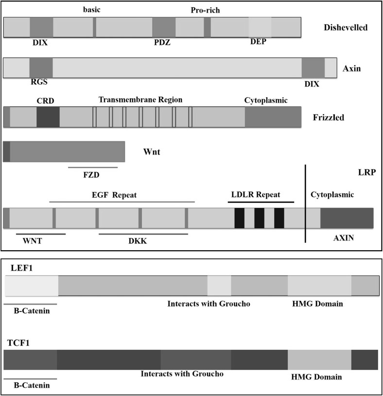
Frizzled, a seven-pass transmembrane protein acts as the receptor for Wnt ligands. [11]. Each Fzd protein contains a cysteine-rich domain (CRD) with approximately 10 cysteine residues. It is known that this CRD domain binds to Wnt proteins with high affinity [12, 13]. Interestingly, Chen et al. identified that CRD domain is dispensable for Wnt signaling in Drosophila, suggesting that other regions of Fzd extracellular domain might be involved in ligand binding [14]. The signaling cascade downstream of Fzd receptor includes several molecules which signal through different branches (Fig. 1; Table 1). These include β-catenin independent (non-canonical) and β-catenin dependent (canonical) pathways [15].
Fig. 1.
Structural features of major components in Wnt signaling pathway. Detailed representation of structural features of molecules involved in canonical Wnt signaling pathway
Table 1.
Major signalling molecules in Wnt-associated pathways
| Gene | Description | Location | Effect on Wnt signaling | Domains | Function | Reference |
|---|---|---|---|---|---|---|
| PORCN | Porcupine (Porc) | Xp11.23 | Activation | Membrane bound O-acyl transferase | Involved in Wnt secretion | [80] |
| Dkk | Dickkopf | 10q21.1 | Inhibition | Binds to LRP | [81] | |
| GSK3β | Glycogen synthase kinase3β | 3q13.33 | Inhibition | Protein kinase-like domain | Phosphorylates β-catenin | [88] |
| Wntless | Evi/Sprinter/Mig-14/Gpr177 | 1p31.3 | Activation | MIG-14 | Secretion of Wnt proteins | [7] |
| Rspo | R-spondin | 1p34.3 | Activation | Thrombospondin domain, furin domains | Enhance Wnt signaling | [83] |
| LGR | Rspo uses Lgr as their receptors | 12q22-q23 | Activation | G protein receptor, rhodopsin like | Acts as receptor for Rspo proteins | [89] |
Molecules involved in the Wnt signaling and their effect on different signaling pathways of Wnt
Wnt/β-Catenin Signaling
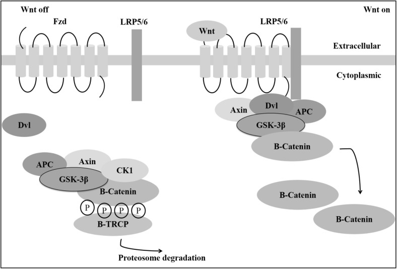
The central molecule involved in Wnt canonical signaling is β-catenin, initially reported to be associated with cell adhesion where it interact with uvomorulin (E-Cadherin) and later on it was shown to be associated with Wnt signaling [16]. In the absence of Wnt ligands, β-catenin is bound by GSK3β (Glycogen synthase kinase 3β), APC (Adenomatous polyposis coli), CK1 (Casein kinase 1) and Axin, collectively called the destruction complex as it destabilizes β-catenin. CK1 and GSK3β phosphorylate β-catenin, CK1 phosphorylates particularly at Ser45 creating a binding site for GSK3β to phosphorylate β-catenins remaining residues Ser33, Ser37 and Thr41. β-Catenin is then recognized by E3-Ubiquitin ligase complex thereby targeting β-catenin for proteasomal degradation [17]. In the presence of Wnt ligands, destruction complex is disassembled leading to the stabilization of β-catenin (Fig. 2) [18, 19].
Fig. 2.
Activation of Wnt/β-catenin signaling. Representation of state of β-catenin in absence and presence of Wnt ligand. In the absence of Wnt ligand, the destruction complex constituting GSK3β (Glycogen synthase kinase 3β), APC (Adenomatous polyposis coli), CK1 (Casein kinase 1) and Axin which results in the proteasomal degradation of β-catenin. Upon Wnt ligation, the destruction complex gets disassembled leading to the stabilization of β-catenin
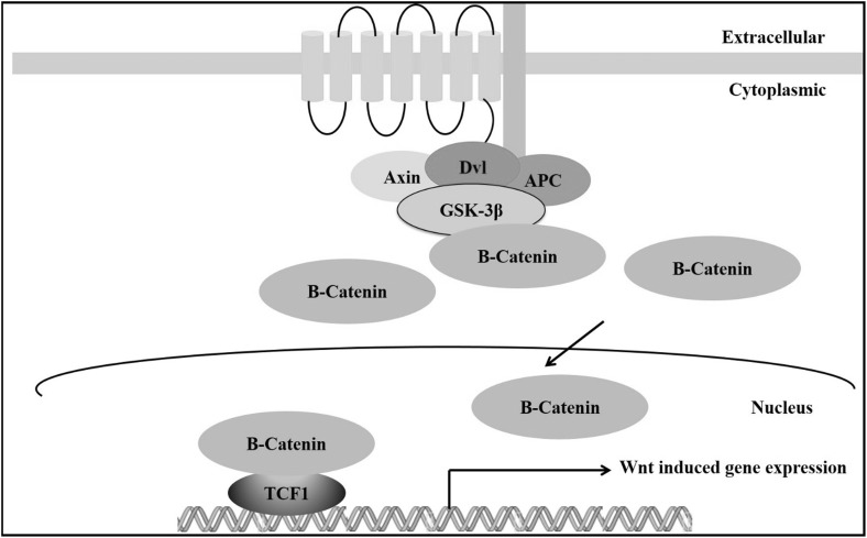
Upon binding of Wnt ligand to the Fzd receptor, the signal is transduced to a protein called Disheveled (Dsh). Interestingly, at the level of Dsh, three different signaling pathways are executed. In all these signaling events, Dsh is the central molecule which execute canonical and non-canonical pathways. The question here is how the same Dsh is involved in different signaling pathways? Molecular mechanisms underlying this are not known, but there are different proposed mechanisms. In mammals, there are three different Dsh proteins called Dsh1, Dsh2 and Dsh3. Dsh contains three conserved domains named DIX, DEP and PDZ. It is assumed that DIX and PDZ domains are responsible for β-catenin signaling, whereas, DEP and PDZ are responsible for non-canonical signaling [20]. There are other findings showing that Wnt binding to Fzd results in clustering of Fzd and LRP along with Dsh [21, 22]. Wnt induced LRP6 phosphorylation is required for initiation of the signaling cascade. LRP5/6 contains PPPSPxS motifs which are the docking sites for Axin which, after Wnt binding recruits Axin to the LRP 5/6. LRP6 is phosphorylated by GSK-3β upon recruitment by Axin and inhibition of GSK-3β at membrane blocks β-catenin signaling indicating that GSK3β is required for LRP6 phosphorylation [23]. Finally β-catenin enters into the nucleus, replaces co-repressor proteins belonging to Groucho which binds to TCF transcription factors, thereby triggering the Wnt targeted genes (Fig. 3) [24].
Fig. 3.
Wnt/β-catenin signaling. Upon ligation with Wnt to frizzled receptor, Wnt/β-catenin signaling pathway gets activated. This results in breakdown of the destruction complex thereby stabilizing β-catenin. Accumulated β-catenin in nucleus acts as a co-activator of TCF family of transcription factors and initiate transcription of Wnt related genes
Non-canonical Pathway
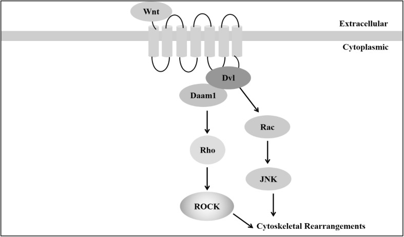
In contrast to Wnt/β-catenin/canonical pathway, there are other mechanisms which are independent of β-catenin known as the non-canonical pathway, comprising Jun N-terminal kinase (JNK) and Wnt/Ca2+ pathways. JNK pathway is essentially similar to the PCP pathway which is described in Drosophila and it involves the activation of Rho, Rac, Rho kinase (ROCK) and JNK etc. [25]. Wnt/JNK signaling like PCP pathway does not use LRP5/6 as co-receptor. Wnt ligand binding to Fzd receptor recruits the disheveled which interacts with Daam1 (Disheveled associated activator of morphogenesis 1) leading to the activation of small GTPases such as Rac, Rho and JNK. JNK activation results in actin polymerization and cytoskeletal modifications (Fig. 4) [26].
Fig. 4.
Wnt/planar cell polarity pathway. Wnt/Planar cell polarity pathway upon binding of Wnt ligand to Fzd receptor recruits disheveled (Dvl), interacts with Daam1 (Disheveled associated activator of morphogenesis 1) leading to the activation of small GTPases such as Rac, Rho and JNK. JNK activation results in actin polymerization and cytoskeletal modifications
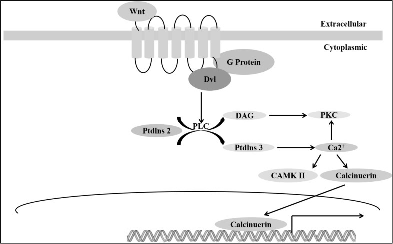
In addition to PCP pathways another significant non-canonical pathway—the Wnt/Ca2+ was identified. In this pathway, the intracellular calcium level is increased as a result of Wnt ligation. Wnt signaling results in an increase in intracellular calcium level. This was established by the studies carried out by Slusarski et al. where they showed that injection of RNA coding for Wnt and Fzd induced calcium release (Fig. 5) [27–29]. This pathway involves the activation of PLC through trimeric G protein which hydrolyses membrane phospholipids to di-acyl glycerol and inositol 1,4,5-triphosphate (IP3). This IP3 induces the release of calcium from the endoplasmic reticulum and this in turn activates PKC [30].
Fig. 5.
Wnt/Ca2+ pathway. Non-canonical Wnt pathway—Wnt/Ca2+ pathway is activated upon Wnt ligation which leads to increase in intracellular calcium level. This pathway involves the activation of PLC through trimeric G protein which hydrolyses membrane phospholipids to di-acyl glycerol (DAG) and inositol 1,4,5-triphosphate (IP3). IP3 induces the release of calcium from endoplasmic reticulum and this in turn activates PKC
Wnt Signaling in Hematopoiesis
Hematopoiesis
Hematopoiesis involves the generation of all types of blood cells from hematopoietic stem cells (HSCs) into different lineages. One in every 10,000 bone marrow cells is a stem cell and in the blood stream this proportion decreases to 1 in 100,000. Daily turnover of HSCs in a human is approximately 1 × 10¹² and there is scarcity of HSCs to replenish the blood cells in a lifetime, which shows the need of maintenance of stable HSC pool. These cells have the capacity to differentiate and self-renew into different lineages. HSC was first identified by Till and McCulloch [31] and now there are many approaches to isolate these primitive cells. The most prominent one among them is selection based on cell surface markers [32].
To maintain their self-renewal property, stem cells can divide by two modes—asymmetric and symmetric divisions. In an asymmetric division, stem cells divide to generate one daughter cell with self renewal capacity and other which differentiates. In a symmetric division, there are two types—proliferation division, in which stem cell divide to generate two stem cells and differentiation division where parent cell gives rise to two differentiated cells [33–35]. Molecular mechanisms which underlie the regulation of self-renewal are yet ripe for research.
It has been shown that many molecules and signaling pathways contribute to HSC self-renewal like p21, Notch, sonic hedgehog etc. [36]. Studies show that Wnt signaling play a significant role in HSC self-renewal as well as differentiation. There are several observations suggesting a clear role of Wnt signaling in hematopoiesis. TCF3, a transcription factor regulated by Wnt signaling is known to control the Nanog, which is required for embryonic stem cell self-renewal [37]. Luis et al. showed that deletion of Wnt3a in mice leads to death at embryonic day 12.5 (E 12.5) and this ligand deficiency results in the reduction of HSCs and progenitor cells in the fetal liver. Reconstitution capacity was also reduced irreversibly as it cannot be restored by transplantation [38]. Constitutive activation of β-catenin induces self-renewal of HSCs and inhibited differentiation [39, 40].
Changes in trabecular bone and HSCs were observed using an osteoblast-specific promoter-driven transgenic expression of DKK1 in Col1α2.3-Dkk1 transgenic (Dkk1 tg) mice as DDK1 is a pan inhibitor of Wnt canonical signaling [41]. Kielman et al. showed that an increase in dosage of β-catenin by mutating APC inhibited the differentiation of mouse embryonic stem cells into three germ layers [42]. In mammals, primitive HSCs first originate in aorta gonad mesonephros (AGM) region in the mesoderm. Likewise, in Xenopus, primitive blood cells originate in ventral blood island (VBI) which is equivalent to the extraembryonic blood islands in the yolk sac. It has been shown that Wnt4 is required for mesoderm development by modulating the BMP4. Inhibition of Wnt/β-catenin signaling results in the reduction of VBI markers indicating that this signaling is required for the primitive hematopoiesis [43]. Multiple Wnt ligands are expressed in bone marrow including Wnt2b, Wnt3a, Wnt5a and Wnt10b [44]. It has been shown that Wnt signaling regulates murine hematopoiesis through bone marrow stromal cells which express Fzd1, Fzd3, Fzd4, Fzd5, Fzd6, Fzd7 and Fzd8 [45]. It was observed that the number of CD34 + cells increased as a result of transfection of Wnt ligands with stromal cells [44]. Wnt5a induces hematopoietic repopulation capacity and it is known to show multi-lineage reconstitution and increase in primitive hematopoietic lineage [46]. Non-canonical Wnt5a signaling plays a critical role in HSC aging as it was observed that reduction of Wnt5a expression results in functionally rejuvenated aged HSCs. Treatment of Young HSCs with Wnt5a resulted in induction of aging like characters and reduction in regenerative capacity [47]. It is reported that Wnt5a acts as a tumor suppressor and inhibits B cell development and it prefers myeloid development [48].
Luis et al. reported a dose-dependent regulation of Wnt signaling in hematopoiesis. Different Apc-mutant mouse models have been used for this study to monitor different levels of Wnt signaling activation. Mild and intermediate Wnt signaling regulates myeloid development, whereas, intermediate level regulates T cell development, but B-cell development is not affected by the different level of Wnt activities [49].
T-Lymphocytes
The role of Wnt signaling was first described in T-cell development in the thymus where T-cells mature. In mice, TCF-1 deficiency reduced thymocyte number and impaired the T-cell development (Table 2) from immature single positive to double positive (DP) stage [50]. Along with this, double negative (DN) stages DN2 and DN4 also drastically reduced in its number (Fig. 6). Interaction between TCF1 and β-catenin is required for thymocyte development at initial stages of T-cell development. Transformation of Wnt genes promotes the accumulation of β-catenin in some cultures of mammalian cells and Wnt-3A mediates TCF dependent transcription. TCF1 −/− leads to defects in early thymocytes [51]. Luis et al. observed that Wnt3a −/− mice impair DP cells and block at the CD8 + ISP stage [38]. TCF1 −/− mice show the reduction in thymic cellularity and block the differentiation at the transition stage of CD8 + ISP stage to DP stage [50]. Xu et al. showed that β-catenin mediated signaling is required for the T-cell development using T cell-specific deletion of β-catenin at the β-selection checkpoint and this lead to a decrease in splenic T cells. This is impaired at β-selection checkpoint with T-cell specific deletion of β-catenin and it is also shown that β-catenin is the target of TCR-CD3 in thymocytes [52]. Further, they have shown that pre-TCR induced signals stabilize β-catenin through the activation of Erk [53]. Disruption of the association between TCF and β-catenin by inhibitor of β-catenin (ICAT) and TCF results in apoptosis of thymocytes and activated T-cells [54]. Recently, Yu et al. identified that TCF1 and β-catenin involvement in T-cells using T cell-specific conditional deletion of β-catenin gene in mice which showed that deletion of β-catenin at the DN3–DN4 stage impaired T-cell development at the β-selection checkpoint. Through pre-T-cell receptor as well as TCR stages, differentiation of CD4 + T-cell into the T-helper lineage takes place [55].
Table 2.
Knock out phenotypes of Wnt signaling molecules in early development
| Gene | Knock out phenotype | Reference |
|---|---|---|
| TCF1 | Differentiation of thymocytes effected | [71] |
| LEF1 | Pro-B cell proliferation and survival effected | [64] |
| Wnt3 | Lack of HSC self renewal Embryos and unable to form primitive streak | [86] |
| Wnt3a | Embryonic lethal | [91] |
| Wnt2 | Placental development is impaired | [92] |
| Fzd9 | Defects in B cell development | [66] |
| Fzd5 | Yolk sac defects | [93] |
| Wnt7b | Abnormal placental development | [94] |
Knock out phenotypes of Wnt signaling molecules and their role in development
Fig. 6.
Wnt signaling in T-cell development. Distinct stages of T cell development indicating various stages influenced by Wnt ligands—Wnt1, Wnt4 and Wnt3a
After allogenic HSC transplantation in adults, recovery of the T-cell population is slow and sometimes inadequate. It impairs thymus, causes a reduction in T-cell regeneration and finally hampers the diversity in T-cell repertoire [56, 57]. Interestingly, Shen et al. has shown that induction of Wnt signaling by inhibiting GSK-3β with 6-bromoindirubin 3′-oxime (BIO) affected the naive T-cell proportion and decreased the differentiation [58].
In a recent study Tiemessen et al. have shown TCF1 −/− mice develops lymphoma. Interestingly, it is due to active Wnt signaling and they identified that a homolog of TCF1, LEF1 is responsible for this. It indicates that TCF1 plays dual roles in T-cell development and acts as a T-cell specific tumor suppressor [59]. TCF1 is needed as a transcriptional activator of Wnt-induced proliferation and helps in early stages of T-cell development in a Wnt-independent way. It also acts as a tumor suppressor and transcriptional repressor to prevent the development of thymic lymphomas and repressing genes of alternate (non-T) cell lineages in turn helps in T-cell development. In T-cell development, TCF1 acts like a molecular switch between proliferation and repression. TCF1 is activated by notch signaling and lack of TCF1 impairs the early T-cell progenitor cells and this role of TCF1 may not be β-catenin dependent because ectopic expression of β-catenin inhibitor ICAT shows no effect on Thy1 + CD25 + cells [60]. The redundancy between TCF1 and LEF1 is supported by LEF1 −/− mice which have normal T-cell development, whereas, LEF1 −/− and TCF1 −/− mice shows a complete block of T-cell differentiation at ISP stage [61].
Along with the positive regulation of Wnt signaling in T-cells, there are also some contradictory reports. Zuklys et al. reported that β-catenin stabilization in thymic epithelial cells commitment to thymic cell fate is inhibited and intrathymic differentiation is also blocked [62]. Liang et al. identified that non-canonical signaling induces apoptosis in fetal thymic cells. Exogenous Wnt5a, a non-canonical Wnt ligand is responsible for this and its deficiency inhibited PKC activation and simultaneously decreased the activity of CamKII [63]. Overexpression of LEF1 by retroviral gene transduction repress Th2 cytokines (IL-4, IL-5, and IL-13) in Th2 cells and they concluded a negative regulatory role of LEF1 in mature T-cells [64]. These studies clearly show the role of Wnt signaling in T-cell development [49].
B-Lymphocytes
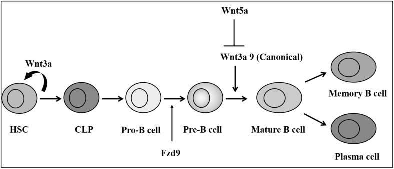
The role of Wnt signaling in B lymphocytes revealed that LEF1 −/− mice have a reduction in B 220 + cells in the fetal liver and perinatal bone marrow and detected increased apoptosis. LEF1 is expressed in B and T-cells. Upon induction of Wnt3a, pro-B cells showed an increase in proliferation, β-catenin stabilization and its nuclear translocation [65]. The role of Fzd receptors in B-cell development is not known, but Ranheim et al. showed that Fzd9 −/− mouse are severely affected in pre and pro B-cell maturation [66]. Conditional inactivation of osteoblasts severely affected the hematopoiesis especially in B-cells, which indicates that osteoblastic niche containing factors are responsible for B-cell development [67].
Wnt signaling in osteoblasts is not a very well understood mechanism, but the osteoblastic niche is required for the HSCs and overexpression of Wnt signaling inhibitor DKK1 reduced the long-term hematopoietic repopulation. Using transgenic mice, induction of DKK1 expression resulted in the reduction of TCF/LEF1 transcription factors which play a crucial role in Wnt signaling pathway. These studies clearly show that the osteoblastic niche is required for stem cell development. Interestingly, it is known that follicular dendritic cells (FDCs) which protect the germinal center B-cells secrete Wnt5a and induce Wnt/Ca2+ pathway [68]. Wnt5a inhibits B cell proliferation by acting as a tumor suppressor by signaling through Wnt/Ca2+ pathway. Wnt5a hemizygous mice develop myeloid leukemia and B-cell lymphoma [48]. Wnt5a −/− mice augment the Wnt/β-catenin signaling indicating Wnt5a is controlling canonical signaling [33]. In another study, Malhotra et al. have shown that Wnt3a inhibited plasmacytoid dendritic cell as well as B-cell and retained HSC markers. Wnt5a thus opposes canonical signaling and induces B-lymphopoiesis indicating that canonical signaling is reduced during differentiation and differential regulation of Wnt signaling in B-cell early development (Fig. 7) [69].
Fig. 7.
Wnt signaling in B-cell development. Various Wnt ligands and Frizzled receptors influencing distinct stages of B cell development
Megakaryocytes
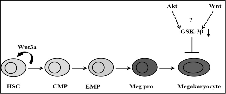
Few studies have been done on Wnt signaling in megakaryocyte development and differentiation. Soda et al. observed that inhibition of GSK3β increased both survival and proliferation of megakaryocytes in a cell line model. In the same study, treatment with Wnt3a in presence and absence of TPO showed no proliferation but there was a subsequent increase in β-catenin, indicating that the increase in β-catenin is independent of Wnt3a (Fig. 8) [70]. In contrary, Macaulay et al. showed that both canonical and non-canonical signaling are active in megakaryocytes in CHRF288-11 cell model and found that increase in β-catenin upon induction of Wnt3a in time and dose dependent manner. A different pattern of gene expression on induction with Wnt3a and Wnt5a is known. In another study, it is shown that GSK-3β negatively regulates megakaryocytes differentiation and platelets production from primary bone marrow cells. Recently it has been shown that negative regulation of Wnt signaling on primitive megakaryocytes derived from embryonic stem cells [71, 72]. K562 differentiation of megakaryocyte with phorbol 12-myristate 13-acetate (PMA) induces three isoforms of Wnt13 (Wnt13a, Wnt13b, Wnt13c) and this up-regulation is correlated to MafB [73].
Fig. 8.
Wnt signaling in megakaryocyte development. Wnt3a and its downstream signaling molecules influence development of megakaryocyte from hematopoietic stem cells
NK Cells
Natural Killer (NK) cells are immune cells involved in the rapid immune response by eradicating the cells lacking MHC-I. They are unique cells which can recognize stressed cells even in the absence of antibody and MHC-1. Initial studies showed that one of the classes of NK cells—Ly49A NK cells depend on TCF1 transcription factor, where TCF1 binds to the Ly49 promoter. In mice lacking TCF1, Ly49A NK cells are reduced and with TCF1 allele the number was slightly increased.This may be due to dose-dependent action of TCF1 in NK cells [74]. To know the effect of LEF1 on the NK cells, Held et al. used LEF −/− mice and has shown that there is no reduction in the cell number indicating that LEF1 has no effect on NK cells [75]. NKT-cell activator glycolipid α-GalCer induces Wnt3a and Wnt5a and inhibited GSK-3β by inducing NKT-cell anergy in liver [76]. Hematopoietic progenitor cells develop into NK cells by cytokines IL-7, IL-15 and SCF, but addition of hydrocortisone as well as stromal cells increased the progenitor cells which could differentiate into NK cells through Wnt signaling [77]. Aoyama et al. identified that both Wnt and notch signaling modulates the NK cell differentiation. They observed that an inhibition in Wnt signaling induced Notch signaling. [78].
Hematological Malignancies
Wnt signaling is of great clinical significance as several studies have shown that deregulation of the same could lead to hematological malignancies. Pellagatti et al. reported the global gene expression in various myelodysplastic syndrome (MDS). Interestingly, it is revealed that cell cycle pathways are deregulated and most significantly, Wnt signaling and thrombopoietin are involved [79]. A commonly deleted region in MDS are 5q region and it contains the APC which is a member of the destructive complex of β-catenin, deletion of this APC in animal model shows HSC defects [80]. Acute myeloid leukemia (AML) cells have more β-catenin compared to the normal cells [81]. Chronic myelogenous leukemia (CML) cells show more Wnt signaling activity and it is indispensable for their growth [82]. This was supported by Tickenbrock et al. as they identified that Fzd4 is overexpressed with the Flt3-ITD mutations in myeloid cell line 32D. Mutations in Flt-3 cause Wnt/β-catenin signaling up regulation and thereby leading to over expression of c-myc [83, 84]. Transfection of AML associated translocation proteins induced the activation of β-catenin, related to the β-catenin. Later transfection studies of γ-catenin showed an increase in the proliferation and survival rate of murine hematopoietic progenitor cells [85]. Elevated levels of nuclear β-catenin were observed in Granulocyte–Macrophage progenitors in CML patients and these cells had the more self-renewal capacity [82]. The regulatory roles of Wnt signaling in myeloid malignancies are not yet clear.
In T-cell leukemias APC is methylated and in a number of T-cell Acute Lymphoblastic Leukemia (T-ALL) cases β-catenin is overexpressed, these may be responsible for the upregulation of β-catenin in T-cell malignancies [86]. It was supported by the findings where inhibition of Wnt signaling by overexpressing the dominant negative form of β-catenin results in reduced proliferation. Recently it has been shown that 85 % of the T-ALL patient samples up-regulated in β-catenin expression and Wnt target genes and they also observed silencing of β-catenin by siRNA induced apoptosis [87].
In pre-B ALL E2A-Pbx1 fusion protein activates the several Wnt related genes. Inhibition of Wnt16 in ALL cells containing t(1:19) translocation induces apoptosis [88]. Abrahamsson et al. identified that GSK-3β mis-splicing results in the generation of leukemia cell because truncated GSK-3β lacks the binding domain for Axin disables it from regulating β-catenin [89]. BCL9 instability is common on chromosome 1 in B cell malignancies and this is overexpressed in pre-B ALL cells with the t(1;14)(q21;q32) translocation. Later, this gene was identified as an orthologous of D. melanogaster legless gene, which was in turn responsible for activation of Wnt signaling [90].
Conclusions
Wnt signaling plays a major role in HSC self-renewal as well as proliferation and differentiation of primitive progenitor cells. Even though Wnt signaling is studied extensively in hematopoiesis, there are controversies regarding the same and several mechanisms are not elucidated till date. Especially different expression profiles and outcomes are shown depending on the dosage of Wnt ligands indicating a dose-dependent regulation of Wnt signaling in hematopoiesis. Non-canonical signaling is also known to play an important role in normal and malignant hematopoiesis but there are several unresolved questions. A thorough understanding of these issues can shed light in the role of Wnt signaling in hematopoiesis, and thereby the mechanisms causing these malignancies.
Acknowledgments
This work was supported by DBT-IYBA, DBT-RGYI, DBT-Developmental Disorders, DST, ICMR and UGC grants of Government of India. The authors appreciate the funding in form of CSIR and UGC fellowships from Government of India.
Compliance with Ethical Standards
Conflict of Interests
The authors declare that there is no conflict of interests regarding the publication of this paper.
References
- 1.Moon RT, Bowerman B, Boutros M, et al. The promise and perils of Wnt signaling through beta-catenin. Science. 2002;296(5573):1644–1646. doi: 10.1126/science.1071549. [DOI] [PubMed] [Google Scholar]
- 2.Reya T, Duncan AW, Ailles L, et al. A role for Wnt signalling in self-renewal of haematopoietic stem cells. Nature. 2003;423(6938):409–414. doi: 10.1038/nature01593. [DOI] [PubMed] [Google Scholar]
- 3.Nusse R, Varmus H. Three decades of Wnts: a personal perspective on how a scientific field developed. EMBO J. 2012;31(12):2670–2684. doi: 10.1038/emboj.2012.146. [DOI] [PMC free article] [PubMed] [Google Scholar]
- 4.Willert K, Brown JD, et al. Wnt proteins are lipid-modified and can act as stem cell growth factors. Nature. 2003;423(6938):448–452. doi: 10.1038/nature01611. [DOI] [PubMed] [Google Scholar]
- 5.Takada R, Satomi Y, Kurata T, et al. Monounsaturated fatty acid modification of Wnt protein: its role in Wnt secretion. Dev Cell. 2006;11(6):791–801. doi: 10.1016/j.devcel.2006.10.003. [DOI] [PubMed] [Google Scholar]
- 6.Zhai L, Chaturvedi D, Cumberledge S. Drosophila wnt-1 undergoes a hydrophobic modification and is targeted to lipid rafts, a process that requires porcupine. J Biol Chem. 2004;279(32):33220–33227. doi: 10.1074/jbc.M403407200. [DOI] [PubMed] [Google Scholar]
- 7.Bänziger C, Soldini D, Schütt C, et al. Wntless, a conserved membrane protein dedicated to the secretion of Wnt proteins from signaling cells. Cell. 2006;125(3):509–522. doi: 10.1016/j.cell.2006.02.049. [DOI] [PubMed] [Google Scholar]
- 8.Bartscherer K, Pelte N, Ingelfinger D, et al. Secretion of Wnt ligands requires Evi, a conserved transmembrane protein. Cell. 2006;125(3):523–533. doi: 10.1016/j.cell.2006.04.009. [DOI] [PubMed] [Google Scholar]
- 9.Reichsman F, Smith L, Cumberledge S. Glycosaminoglycans can modulate extracellular localization of the wingless protein and promote signal transduction. J Cell Biol. 1996;135(3):819–827. doi: 10.1083/jcb.135.3.819. [DOI] [PMC free article] [PubMed] [Google Scholar]
- 10.Häcker U, Lin X, Perrimon N. The Drosophila sugarless gene modulates Wingless signaling and encodes an enzyme involved in polysaccharide biosynthesis. Development. 1997;124(18):3565–3573. doi: 10.1242/dev.124.18.3565. [DOI] [PubMed] [Google Scholar]
- 11.Bhanot P, Brink M, Samos CH, et al. A new member of the frizzled family from Drosophila functions as a Wingless receptor. Nature. 1996;382(6588):225–230. doi: 10.1038/382225a0. [DOI] [PubMed] [Google Scholar]
- 12.Wu CH, Nusse R. Ligand receptor interactions in the Wnt signaling pathway in Drosophila. J Biol Chem. 2002;277(44):41762–41769. doi: 10.1074/jbc.M207850200. [DOI] [PubMed] [Google Scholar]
- 13.Hsieh JC, Rattner A, Smallwood PM, et al. Biochemical characterization of Wnt-frizzled interactions using a soluble, biologically active vertebrate Wnt protein. Proc Natl Acad Sci USA. 1999;96(7):3546–3551. doi: 10.1073/pnas.96.7.3546. [DOI] [PMC free article] [PubMed] [Google Scholar]
- 14.Chen CM, Strapps W, Tomlinson A, et al. Evidence that the cysteine-rich domain of Drosophila Frizzled family receptors is dispensable for transducing Wingless. Proc Natl Acad Sci USA. 2004;101(45):15961–15966. doi: 10.1073/pnas.0407103101. [DOI] [PMC free article] [PubMed] [Google Scholar]
- 15.Logan CY, Nusse R. The Wnt signaling pathway in development and disease. Annu Rev Cell Dev Biol. 2004;20:781–810. doi: 10.1146/annurev.cellbio.20.010403.113126. [DOI] [PubMed] [Google Scholar]
- 16.Ozawa M, Baribault H, Kemler R. The cytoplasmic domain of the cell adhesion molecule uvomorulin associates with three independent proteins structurally related in different species. EMBO J. 1989;8(6):1711–1717. doi: 10.1002/j.1460-2075.1989.tb03563.x. [DOI] [PMC free article] [PubMed] [Google Scholar]
- 17.Liu C, Li Y, Semenov M, et al. Control of beta-catenin phosphorylation/degradation by a dual-kinase mechanism. Cell. 2002;108(6):837–847. doi: 10.1016/S0092-8674(02)00685-2. [DOI] [PubMed] [Google Scholar]
- 18.Mao J, Wang J, Liu B, et al. Low-density lipoprotein receptor-related protein-5 binds to axin and regulates the canonical Wnt signaling pathway. Mol Cell. 2001;7(4):801–809. doi: 10.1016/S1097-2765(01)00224-6. [DOI] [PubMed] [Google Scholar]
- 19.Bilic J, Huang YL, Davidson G, et al. Wnt induces LRP6 signalosomes and promotes dishevelled-dependent LRP6 phosphorylation. Science. 2007;316(5831):1619–1622. doi: 10.1126/science.1137065. [DOI] [PubMed] [Google Scholar]
- 20.Wharton KA., Jr Runnin’ with the Dvl: proteins that associate with Dsh/Dvl and their significance to Wnt signal transduction. Dev Biol. 2003;253(1):1–17. doi: 10.1006/dbio.2002.0869. [DOI] [PubMed] [Google Scholar]
- 21.Bernatík O, Sedová K, Schille C, et al. Functional analysis of dishevelled-3 phosphorylation identifies distinct mechanisms driven by casein kinase 1ϵ and frizzled5. J Biol Chem. 2014;289(34):23520–23533. doi: 10.1074/jbc.M114.590638. [DOI] [PMC free article] [PubMed] [Google Scholar]
- 22.Cong F, Schweizer L, Varmus H. Wnt signals across the plasma membrane to activate the beta-catenin pathway by forming oligomers containing its receptors, Frizzled and LRP. Development. 2004;131(20):5103–5115. doi: 10.1242/dev.01318. [DOI] [PubMed] [Google Scholar]
- 23.Zeng X, Huang H, Tamai K, et al. Initiation of Wnt signaling: control of Wnt coreceptor Lrp6 phosphorylation/activation via frizzled, dishevelled and axin functions. Development. 2008;135(2):367–375. doi: 10.1242/dev.013540. [DOI] [PMC free article] [PubMed] [Google Scholar]
- 24.Nelson WJ, Nusse R. Convergence of Wnt, beta-catenin, and cadherin pathways. Science. 2004;303(5663):1483–1487. doi: 10.1126/science.1094291. [DOI] [PMC free article] [PubMed] [Google Scholar]
- 25.Simons M, Mlodzik M. Planar cell polarity signaling: from fly development to human disease. Annu Rev Genet. 2008;42:517–540. doi: 10.1146/annurev.genet.42.110807.091432. [DOI] [PMC free article] [PubMed] [Google Scholar]
- 26.Gordon MD, Nusse R. Wnt signaling: multiple pathways, multiple receptors, and multiple transcription factors. J Biol Chem. 2006;281(32):22429–22433. doi: 10.1074/jbc.R600015200. [DOI] [PubMed] [Google Scholar]
- 27.Slusarski DC, Corces VG, Moon RT. Interaction of Wnt and a Frizzled homologue triggers G-protein-linked phosphatidylinositol signalling. Nature. 1997;390(6658):410–413. doi: 10.1038/37138. [DOI] [PubMed] [Google Scholar]
- 28.Slusarski DC, Yang-Snyder J, Busa WB, et al. Modulation of embryonic intracellular Ca2+ signaling by Wnt-5A. Dev Biol. 1997;182(1):114–120. doi: 10.1006/dbio.1996.8463. [DOI] [PubMed] [Google Scholar]
- 29.Hikasa H, Shibata M, Hiratani I, et al. The Xenopus receptor tyrosine kinase Xror2 modulates morphogenetic movements of the axial mesoderm and neuroectoderm via Wnt signaling. Development. 2002;129(22):5227–5239. doi: 10.1242/dev.129.22.5227. [DOI] [PubMed] [Google Scholar]
- 30.Kühl M, Sheldahl LC, Malbon CC, et al. Ca(2 +)/calmodulin-dependent protein kinase II is stimulated by Wnt and Frizzled homologs and promotes ventral cell fates in Xenopus. J Biol Chem. 2000;275(17):12701–12711. doi: 10.1074/jbc.275.17.12701. [DOI] [PubMed] [Google Scholar]
- 31.Till JE, McCulloch EA. A direct measurement of the radiation sensitivity of normal mouse bone marrow cells. Radiat Res. 1961;178(2):AV3–AV7. doi: 10.1667/RRAV01.1. [DOI] [PubMed] [Google Scholar]
- 32.Visser JW, Bauman JG, Mulder AH, et al. Isolation of murine pluripotent hemopoietic stem cells. J Exp Med. 1984;159(6):1576–1590. doi: 10.1084/jem.159.6.1576. [DOI] [PMC free article] [PubMed] [Google Scholar]
- 33.Suda J, Suda T, Ogawa M. Analysis of differentiation of mouse hemopoietic stem cells in culture by sequential replating of paired progenitors. Blood. 1984;64(2):393–399. [PubMed] [Google Scholar]
- 34.Leary AG, Ogawa M, Strauss LC, et al. Single cell origin of multilineage colonies in culture. Evidence that differentiation of multipotent progenitors and restriction of proliferative potential of monopotent progenitors are stochastic processes. J Clin Invest. 1984;74(6):2193–2197. doi: 10.1172/JCI111645. [DOI] [PMC free article] [PubMed] [Google Scholar]
- 35.Sánchez-Taltavull D, Alarcón T. Robustness of differentiation cascades with symmetric stem cell division. J R Soc Interface. 2014;11(95):20140264. doi: 10.1098/rsif.2014.0264. [DOI] [PMC free article] [PubMed] [Google Scholar]
- 36.Pazianos G, Uqoezwa M, Reya T. The elements of stem cell self-renewal: a genetic perspective. Biotechniques. 2003;35(6):1240–1247. doi: 10.2144/03356ss03. [DOI] [PubMed] [Google Scholar]
- 37.Pereira L, Yi F, Merrill BJ. Repression of Nanog gene transcription by Tcf3 limits embryonic stem cell self-renewal. Mol Cell Biol. 2006;26(20):7479–7491. doi: 10.1128/MCB.00368-06. [DOI] [PMC free article] [PubMed] [Google Scholar]
- 38.Luis TC, Weerkamp F, Naber BA, et al. Wnt3a deficiency irreversibly impairs hematopoietic stem cell self-renewal and leads to defects in progenitor cell differentiation. Blood. 2009;113(3):546–554. doi: 10.1182/blood-2008-06-163774. [DOI] [PubMed] [Google Scholar]
- 39.Kirstetter P, Anderson K, Porse BT, et al. Activation of the canonical Wnt pathway leads to loss of hematopoietic stem cell repopulation and multilineage differentiation block. Nat Immunol. 2006;7(10):1048–1056. doi: 10.1038/ni1381. [DOI] [PubMed] [Google Scholar]
- 40.Scheller M, Huelsken J, Rosenbauer F, et al. Hematopoietic stem cell and multilineage defects generated by constitutive beta-catenin activation. Nat Immunol. 2006;7(10):1037–1047. doi: 10.1038/ni1387. [DOI] [PubMed] [Google Scholar]
- 41.Fleming HE, Janzen V, Lo Celso C, et al. Wnt signaling in the niche enforces hematopoietic stem cell quiescence and is necessary to preserve self-renewal in vivo. Cell Stem Cell. 2008;2(3):274–283. doi: 10.1016/j.stem.2008.01.003. [DOI] [PMC free article] [PubMed] [Google Scholar]
- 42.Kielman MF, Rindapää M, Gaspar C, et al. Apc modulates embryonic stem-cell differentiation by controlling the dosage of beta-catenin signaling. Nat Genet. 2002;32(4):594–605. doi: 10.1038/ng1045. [DOI] [PubMed] [Google Scholar]
- 43.Tran HT, Sekkali B, Van Imschoot G, et al. Wnt/beta-catenin signaling is involved in the induction and maintenance of primitive hematopoiesis in the vertebrate embryo. Proc Natl Acad Sci USA. 2010;107(37):16160–16165. doi: 10.1073/pnas.1007725107. [DOI] [PMC free article] [PubMed] [Google Scholar]
- 44.Van Den Berg DJ, Sharma AK, Bruno E, et al. Role of members of the Wnt gene family in human hematopoiesis. Blood. 1998;92(9):3189–3202. [PubMed] [Google Scholar]
- 45.Yamane T, Kunisada T, Tsukamoto H, et al. Wnt signaling regulates hemopoiesis through stromal cells. J Immunol. 2001;167(2):765–772. doi: 10.4049/jimmunol.167.2.765. [DOI] [PubMed] [Google Scholar]
- 46.Murdoch B, Chadwick K, Martin M, et al. Wnt-5A augments repopulating capacity and primitive hematopoietic development of human blood stem cells in vivo. Proc Natl Acad Sci USA. 2003;100(6):3422–3427. doi: 10.1073/pnas.0130233100. [DOI] [PMC free article] [PubMed] [Google Scholar]
- 47.Florian MC, Nattamai KJ, Dörr K, et al. A canonical to non-canonical Wnt signalling switch in haematopoietic stem-cell ageing. Nature. 2013;503(7476):392–396. doi: 10.1038/nature12631. [DOI] [PMC free article] [PubMed] [Google Scholar]
- 48.Liang H, Chen Q, Coles AH, et al. Wnt5a inhibits B cell proliferation and functions as a tumor suppressor in hematopoietic tissue. Cancer Cell. 2003;4(5):349–360. doi: 10.1016/S1535-6108(03)00268-X. [DOI] [PubMed] [Google Scholar]
- 49.Luis TC, Naber BA, Roozen PP, et al. Canonical wnt signaling regulates hematopoiesis in a dosage-dependent fashion. Cell Stem Cell. 2011;9(4):345–356. doi: 10.1016/j.stem.2011.07.017. [DOI] [PubMed] [Google Scholar]
- 50.Schilham MW, Wilson A, Moerer P, et al. Critical involvement of Tcf-1 in expansion of thymocytes. J Immunol. 1998;161(8):3984–3991. [PubMed] [Google Scholar]
- 51.Staal FJ, Meeldijk J, Moerer P, et al. Wnt signaling is required for thymocyte development and activates Tcf-1 mediated transcription. Eur J Immunol. 2001;31(1):285–293. doi: 10.1002/1521-4141(200101)31:1<285::AID-IMMU285>3.0.CO;2-D. [DOI] [PubMed] [Google Scholar]
- 52.Xu Y, Banerjee D, Huelsken J, et al. Deletion of β-catenin impairs T cell development. Nat Immunol. 2003;4(12):1177–1182. doi: 10.1038/ni1008. [DOI] [PubMed] [Google Scholar]
- 53.Xu M, Sharma A, Wiest DL, et al. Pre-TCR-induced beta-catenin facilitates traversal through beta-selection. J Immunol. 2009;182(2):751–758. doi: 10.4049/jimmunol.182.2.751. [DOI] [PMC free article] [PubMed] [Google Scholar]
- 54.Hossain MZ, Yu Q, Xu M, et al. ICAT expression disrupts beta-catenin-TCF interactions and impairs survival of thymocytes and activated mature T cells. Int Immunol. 2008;20(7):925–935. doi: 10.1093/intimm/dxn051. [DOI] [PMC free article] [PubMed] [Google Scholar]
- 55.Yu Q, Sharma A, Sen JM. TCF1 and beta-catenin regulate T cell development and function. Immunol Res. 2010;47(1–3):45–55. doi: 10.1007/s12026-009-8137-2. [DOI] [PMC free article] [PubMed] [Google Scholar]
- 56.Seggewiss R, Einsele H. Immune reconstitution after allogeneic transplantation and expanding options for immunomodulation: an update. Blood. 2010;115(19):3861–3868. doi: 10.1182/blood-2009-12-234096. [DOI] [PubMed] [Google Scholar]
- 57.Mir MA, Battiwalla M. Immune deficits in allogeneic hematopoietic stem cell transplant (HSCT) recipients. Mycopathologia. 2009;168(6):271–282. doi: 10.1007/s11046-009-9181-0. [DOI] [PubMed] [Google Scholar]
- 58.Shen S, Klamer G, Xu N, et al. GSK-3β inhibition preserves naive T cell phenotype in bone marrow reconstituted mice. Exp Hematol. 2013;41(12):1016–1027. doi: 10.1016/j.exphem.2013.08.006. [DOI] [PubMed] [Google Scholar]
- 59.Tiemessen MM, Baert MR, Schonewille T, Brugman MH, Famili F, Salvatori DC, Meijerink JP, Ozbek U, Clevers H, van Dongen JJ, Staal FJ. The nuclear effector of Wnt-signaling, Tcf1, functions as a T-cell-specific tumor suppressor for development of lymphomas. PLoS Biol. 2012;10(11):e1001430. doi: 10.1371/journal.pbio.1001430. [DOI] [PMC free article] [PubMed] [Google Scholar]
- 60.Germar K, Dose M, Konstantinou T, Zhang J, et al. T-cell factor 1 is a gatekeeper for T-cell specification in response to Notch signaling. Proc Natl Acad Sci USA. 2011;108(50):20060–20065. doi: 10.1073/pnas.1110230108. [DOI] [PMC free article] [PubMed] [Google Scholar]
- 61.Austin TW, Solar GP, Ziegler FC, Liem L, Matthews W. A role for the Wnt gene family in hematopoiesis: expansion of multilineage progenitor cells. Blood. 1997;89(10):3624–3635. [PubMed] [Google Scholar]
- 62.Zuklys S, Gill J, Keller MP, et al. Stabilized beta-catenin in thymic epithelial cells blocks thymus development and function. J Immunol. 2009;182(5):2997–3007. doi: 10.4049/jimmunol.0713723. [DOI] [PubMed] [Google Scholar]
- 63.Liang H, Coles AH, Zhu Z, et al. Noncanonical Wnt signaling promotes apoptosis in thymocyte development. J Exp Med. 2007;204(13):3077–3084. doi: 10.1084/jem.20062692. [DOI] [PMC free article] [PubMed] [Google Scholar]
- 64.Hebenstreit D, Giaisi M, Treiber MK, et al. LEF-1 negatively controls interleukin-4 expression through a proximal promoter regulatory element. J Biol Chem. 2008;283(33):22490–22497. doi: 10.1074/jbc.M804096200. [DOI] [PMC free article] [PubMed] [Google Scholar]
- 65.Reya T, O’Riordan M, Okamura R, et al. Wnt signaling regulates B lymphocyte proliferation through a LEF-1 dependent mechanism. Immunity. 2000;13(1):15–24. doi: 10.1016/S1074-7613(00)00004-2. [DOI] [PubMed] [Google Scholar]
- 66.Ranheim EA, Kwan HC, Reya T, et al. Frizzled 9 knock-out mice have abnormal B-cell development. Blood. 2005;105(6):2487–2494. doi: 10.1182/blood-2004-06-2334. [DOI] [PubMed] [Google Scholar]
- 67.Visnjic D, Kalajzic Z, Rowe DW, et al. Hematopoiesis is severely altered in mice with an induced osteoblast deficiency. Blood. 2004;103(9):3258–3264. doi: 10.1182/blood-2003-11-4011. [DOI] [PubMed] [Google Scholar]
- 68.Kim J, Kim DW, Chang W, Choe J, Kim J, Park CS, Song K, Lee I. Wnt5a is secreted by follicular dendritic cells to protect germinal center B cells via Wnt/Ca2+/NFAT/NF-κB-B cell lymphoma 6 signaling. J Immunol. 2012;188(1):182–189. doi: 10.4049/jimmunol.1102297. [DOI] [PubMed] [Google Scholar]
- 69.Malhotra S, Baba Y, Garrett KP, et al. Contrasting responses of lymphoid progenitors to canonical and noncanonical Wnt signals. J Immunol. 2008;181(6):3955–3964. doi: 10.4049/jimmunol.181.6.3955. [DOI] [PMC free article] [PubMed] [Google Scholar]
- 70.Soda M, Willert K, Kaushansky K, et al. Inhibition of GSK-3beta promotes survival and proliferation of megakaryocytic cells through a beta-catenin-independent pathway. Cell Signal. 2008;20(12):2317–2323. doi: 10.1016/j.cellsig.2008.09.001. [DOI] [PMC free article] [PubMed] [Google Scholar]
- 71.Paluru P, Hudock KM, Cheng X, Mills JA, Ying L, Galvão AM, Lu L, Tiyaboonchai A, Sim X, Sullivan SK, French DL, Gadue P. The negative impact of Wnt signaling on megakaryocyte and primitive erythroid progenitors derived from human embryonic stem cells. Stem Cell Res. 2014;12(2):441–451. doi: 10.1016/j.scr.2013.12.003. [DOI] [PMC free article] [PubMed] [Google Scholar]
- 72.Macaulay IC, Thon JN, Tijssen MR, et al. Canonical Wnt signaling in megakaryocytes regulates proplatelet formation. Blood. 2013;121(1):188–196. doi: 10.1182/blood-2012-03-416875. [DOI] [PMC free article] [PubMed] [Google Scholar]
- 73.Bunaciu RP, Tang T, Mao CD. Differential expression of Wnt13 isoforms during leukemic cell differentiation. Oncol Rep. 2008;20(1):195–201. [PubMed] [Google Scholar]
- 74.Held W, Kunz B, Lowin-Kropf B, et al. Clonal acquisition of the Ly49A NK cell receptor is dependent on the trans-acting factor TCF-1. Immunity. 1999;11(4):433–442. doi: 10.1016/S1074-7613(00)80118-1. [DOI] [PubMed] [Google Scholar]
- 75.Held W, Clevers H, Grosschedl R. Redundant functions of TCF-1 and LEF-1 during T and NK cell development, but unique role of TCF-1 for Ly49 NK cell receptor acquisition. Eur J Immunol. 2003;33(5):1393–1398. doi: 10.1002/eji.200323840. [DOI] [PubMed] [Google Scholar]
- 76.Deng ZB, Zhuang X, Ju S, et al. Intestinal mucus-derived nanoparticle-mediated activation of Wnt/β-catenin signaling plays a role in induction of liver natural killer T cell anergy in mice. Hepatology. 2013;57(3):1250–1261. doi: 10.1002/hep.26086. [DOI] [PMC free article] [PubMed] [Google Scholar]
- 77.Grzywacz B, Kataria N, Kataria N, et al. Natural killer-cell differentiation by myeloid progenitors. Blood. 2011;117(13):3548–3558. doi: 10.1182/blood-2010-04-281394. [DOI] [PMC free article] [PubMed] [Google Scholar]
- 78.Aoyama K, Delaney C, Varnum-Finney B, et al. The interaction of the Wnt and Notch pathways modulates natural killer versus T cell differentiation. Stem Cells. 2007;25(10):2488–2497. doi: 10.1634/stemcells.2007-0102. [DOI] [PubMed] [Google Scholar]
- 79.Pellagatti A, Cazzola M, Giagounidis A, et al. Deregulated gene expression pathways in myelodysplastic syndrome hematopoietic stem cells. Leukemia. 2010;24(4):756–764. doi: 10.1038/leu.2010.31. [DOI] [PubMed] [Google Scholar]
- 80.Lane SW, Sykes SM, Al-Shahrour F, et al. The Apc(min) mouse has altered hematopoietic stem cell function and provides a model for MPD/MDS. Blood. 2010;115(17):3489–3497. doi: 10.1182/blood-2009-11-251728. [DOI] [PMC free article] [PubMed] [Google Scholar]
- 81.Serinsöz E, Neusch M, Büsche G, et al. Aberrant expression of beta-catenin discriminates acute myeloid leukaemia from acute lymphoblastic leukaemia. Br J Haematol. 2004;126(3):313–319. doi: 10.1111/j.1365-2141.2004.05049.x. [DOI] [PubMed] [Google Scholar]
- 82.Jamieson CH, Ailles LE, Dylla SJ, et al. Granulocyte-macrophage progenitors as candidate leukemic stem cells in blast-crisis CML. N Engl J Med. 2004;351(7):657–667. doi: 10.1056/NEJMoa040258. [DOI] [PubMed] [Google Scholar]
- 83.Tickenbrock L, Schwäble J, Wiedehage M, et al. Flt3 tandem duplication mutations cooperate with Wnt signaling in leukemic signal transduction. Blood. 2005;105(9):3699–3706. doi: 10.1182/blood-2004-07-2924. [DOI] [PubMed] [Google Scholar]
- 84.Kiyoi H, Towatari M, Yokota S, et al. Internal tandem duplication of the FLT3 gene is a novel modality of elongation mutation which causes constitutive activation of the product. Leukemia. 1998;12(9):1333–1337. doi: 10.1038/sj.leu.2401130. [DOI] [PubMed] [Google Scholar]
- 85.Müller-Tidow C, Steffen B, Cauvet T, et al. Translocation products in acute myeloid leukemia activate the Wnt signaling pathway in hematopoietic cells. Mol Cell Biol. 2004;24(7):2890–2904. doi: 10.1128/MCB.24.7.2890-2904.2004. [DOI] [PMC free article] [PubMed] [Google Scholar]
- 86.Yang Y, Takeuchi S, Tsukasaki K, et al. Methylation analysis of the adenomatous polyposis coli (APC) gene in adult T-cell leukemia/lymphoma. Leuk Res. 2005;29(1):47–51. doi: 10.1016/j.leukres.2004.05.004. [DOI] [PubMed] [Google Scholar]
- 87.Ng OH, Erbilgin Y, Firtina S, Celkan T, Karakas Z, Aydogan G, Turkkan E, Yildirmak Y, Timur C, Zengin E, van Dongen JJ, Staal FJ, Ozbek U, Sayitoglu M. Deregulated WNT signaling in childhood T-cell acute lymphoblastic leukemia. Blood Cancer J. 2014;14(4):e192. doi: 10.1038/bcj.2014.12. [DOI] [PMC free article] [PubMed] [Google Scholar]
- 88.Mazieres J, You L, He B, et al. Inhibition of Wnt16 in human acute lymphoblastoid leukemia cells containing the t(1;19) translocation induces apoptosis. Oncogene. 2005;24(34):5396–5400. doi: 10.1038/sj.onc.1208568. [DOI] [PubMed] [Google Scholar]
- 89.Abrahamsson AE, Geron I, Gotlib J, et al. Glycogen synthase kinase 3beta missplicing contributes to leukemia stem cell generation. Proc Natl Acad Sci USA. 2009;106(10):3925–3929. doi: 10.1073/pnas.0900189106. [DOI] [PMC free article] [PubMed] [Google Scholar]
- 90.Kramps T, Peter O, Brunner E, et al. Wnt/wingless signaling requires BCL9/legless-mediated recruitment of pygopus to the nuclear beta-catenin-TCF complex. Cell. 2002;109(1):47–60. doi: 10.1016/S0092-8674(02)00679-7. [DOI] [PubMed] [Google Scholar]
- 91.Verbeek S, Izon D, Hofhuis F, et al. An HMG-box-containing T-cell factor required for thymocyte differentiation. Nature. 1995;374(6517):70–74. doi: 10.1038/374070a0. [DOI] [PubMed] [Google Scholar]
- 92.Monkley SJ, Delaney SJ, Pennisi DJ, et al. Targeted disruption of the Wnt2 results in placentation defects. Development. 1996;122(11):3343–3353. doi: 10.1242/dev.122.11.3343. [DOI] [PubMed] [Google Scholar]
- 93.Ishikawa T, Tamai Y, Zorn AM, et al. Mouse Wnt receptor gene Fzd5 is essential for yolk sac and placental angiogenesis. Development. 2001;128(1):25–33. doi: 10.1242/dev.128.1.25. [DOI] [PubMed] [Google Scholar]
- 94.Parr BA, Cornish VA, Cybulsky MI, et al. Wnt7b regulates placental development in mice. Dev Biol. 2001;237(2):324–332. doi: 10.1006/dbio.2001.0373. [DOI] [PubMed] [Google Scholar]


