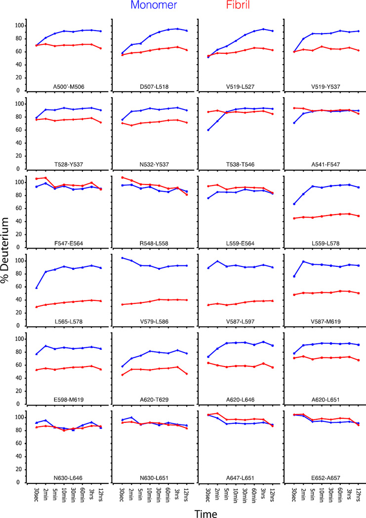
Figure 1.
Fibril formation lowers the hydrogen-deuterium exchange rate of two regions in A546T FAS1-4. HDX-MS analysis measured the hydrogen exchange rate of fibrillated (red) and monomeric A546T FAS1-4 (blue). The samples were allowed to exchange hydrogens from 30 sec up to 12 h, followed by quenching and proteolysis with pepsin. Each diagram shows the degree of deuterium exchange relative to a fully deuterated monomer. Peptides covering the entire sequence of A546T FAS1-4 are shown. The peptides covering the region L565-L597 showed a high degree of protection from the solvent in the fibrils, while the regions A500’-Y537 and E598-T629 displayed less protection.

