Abstract
Objectives
To determine dental maturity (dental age) in cross-sectional sample of Saudi Arabian children by applying the standards established by Demirjian and Golstein and to examine the applicability of these standards in determination of dental maturity among Saudi Arabian children (Qassim region).
Materials & Methods
Dental maturity was assessed from panoramic radiographs of 400 Saudi Arabian children, 222 boys, and 198 girls ranging in age from 4 to 14 years by using these standards. The difference between the dental and chronological age in different age groups in both sexes was statistically compared using ANOVA testat 0.05 level of significance.
Results
The Saudi Arabian children were generally somewhat advanced in dental maturity compared with the French Canadian reference sample with an overall mean difference between the dental and chronological age of 0.279 years in boys and 0.385 years in girls.
Conclusion
The applied standards appear to be adequate for studying dental age in groups of children among Saudi Arabian population.
Keywords: Dental age, chronological age, Demirjian & Goldstein method
Introduction
Dental eruption and dental development are two separate processes. Dental eruption limits itself to a very short period, determined by the time of appearance of a tooth (1) and is influenced by several local factors: ankylosis, early or delayed extraction of deciduous teeth, and impaction or crowding of the permanent teeth. (2, 3, 4, 5) By contrast, the development of the permanent teeth is not affected by the status of the deciduous teeth (2, 6, 7) and can be followed longitudinally and assessed from x-rays for a period of several years, even in periods where no eruption takes place, as between 2.5 – 6 years of age and after the 12th years, if we exclude the third molar. (1) Therefore, dental development is a more reliable indicator of the biological maturity of growing children than dental eruption. (2)
Dental age (DA) estimation has an important field of application in legal and forensic dentistry; it is one of the procedures recommended for forensic age estimation, which carries with it a number of legal implications. This dental age estimation will be useful in those cases where no documentation is available evidencing the age of a person. This would apply both in civil matters (registration of birth out of time or adoptions) as in criminal matters, such as the implementation of specific legislation for minors or modification of sentences based on the victim (for example in case of violations) or the accused(as a mitigating).(8, 9, 10, 11, 12)
Likewise, from a clinical perspective, estimating (DA) in patients of known chronological age(CA) allows us to establish the similarity between (DA) and (CA), an important piece of information when planning certain treatments, determination of dental age is important to orthodontist in planning the treatment of malocclusions relative to maxillofacial growth. It is also important to paediatric dentists who may be concerned about the stage of dental development and possible timing of eruption. DA may be of interest to molecular biologists, because genetic mutation may alter dental morphogenesis. (13) Clinicians also have studied dental development as an index of chronological age, and regarded this as superior to other developing organs or structures in children with unknown or uncertain birth records. (14, 15, 16)
Several methods exist for determining DA using dental radiographs, (16,17,18,19) the best internationally known method is that of Demirjian et al. (20) that later revised by Demirjian and Golstein (21) for estimating overall dental maturity based on a sample comprising 4756 normal French – Canadian children. The method has been widely used in studies of dental maturity and comparisons between various population groups. (3,22,23,24,25,26) Furthermore, this method is the method proposed by Swedish Board for Health and Welfare for age estimation in adopted children of unknown age. (25)
Considerable differences in relation to dental development and eruption of permanent teeth have been reported between populations, so the purpose of the present study was to determine dental maturity (dental age) in cross-sectional sample of Saudi Arabian children by applying the standards established by Demirjian and Golstein (21) and to examine the applicability of these standards for use as reference for dental maturity among Saudi Arabian children.
Subjects and Methods
Panoramic radiographic records of 400 children (222 boys, 189 girls), without any history of systemic diseases, were taken from the records of Faculty of Dentistry, Qassim University, Saudi Arabia after acceptance of the college. They were used for determination of dental maturity (DA). The sample ranged in age from 4.37 to 13.94 years for boys (mean age 9.15 years) and between 4.85 to 13.83 years for girls (average age 9.20 years). The included panoramic radiographs were clear without artefacts in the regions of the roots of the teeth.
Evaluation of dental maturity
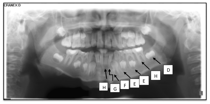
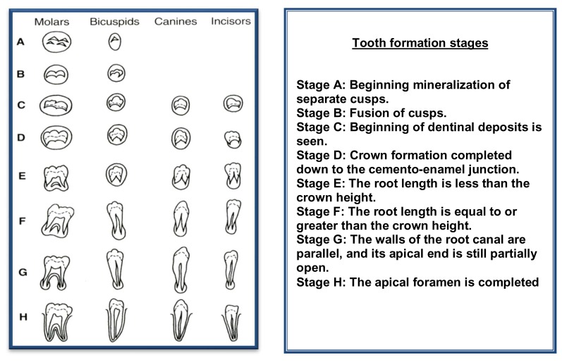
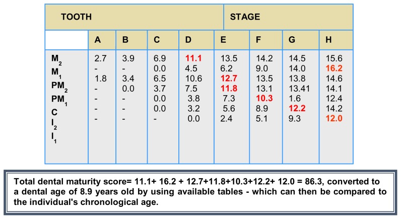
Dental age was assessed twice from the panoramic radiographs using the method of Demirjian and Goldstein. (21) In this method, the individual radiological appearance of the seven permanent teeth (second molar to central incisor) on the left side of the mandible were evaluated according to the developmental criteria to score different stages of a permanent tooth calcification. Eight stages of mineralization (A through H) for each tooth are described by written criteria, which are accompanied by photographic illustrations and schematic drawings. The stage of each tooth was converted to corresponding numeric value, and then all seven values were added to obtain a dental maturity score, which correspond to (DA) by using available tables (Fig: 1A, B & C). The standards are given for boys and girls separately.
Fig 1.
A: Panoramic radiograph of 8.8years (CA) old girl, the corresponding DA is 8.9 years.
B. Determining the developmental stage of the seven left permanent mandibular teeth.
C. Conversion of developmental stage to maturity score
Using the above described method, a dental maturity score was obtained and the (DA) was determined for each subject in both groups and the differences between the chronological age (CA) and DA demonstrated the delay or acceleration in dental maturity.
Reliability was achieved by repeated assessment of all radiographs, the assessments agreed in 94% of the cases and the difference never exceeded one stage.
Statistical Analysis
The difference between DA and CA was tabulated according to sex and age groups, the differences were compared using a one-way ANOVA and post hoc Scheffe’s test at 0.05 level of significance. All data were tabulated using the SPSS ver.19 (IBM Corp., NY, and USA) data processing software.
Results
Dental maturity of the normal permanent teeth assessed by the method of Demirjian and Goldstein (21) resulted in a consistent over estimation of dental age for the Saudi Arabian sample, the overall mean difference of acceleration of dental development was 0 to 0.5 (mean :0.279;sd: 0.128) in boys and 0.1 to 0.6 (mean :0.385;sd: 0.128) in girls. For almost all the 14 age classes in this sample a significant difference was found between the dental ages determined from French – Canadian standers and the chronological age. Both sexes were advanced in dental maturity compared with the reference sample, girls were always more dentally developed than boys in the different age groups and there was a significant statistical difference in the timing of dental development between boys and girls in the studied sample (P<0.05) Table 1.
Table 1.
Comparison of differences between CA and DA in relation to sex
| Males (N1=222) | Females (N2=198) | Test of Sig | 95% Conf. Interval | ||||
|---|---|---|---|---|---|---|---|
| Mean | SD | Mean | SD | t-test | p | Lower | Upper |
| 0.279 | 0.128 | 0.385 | 0.128 | 8.520 | P<0.001* | −0.131 | −0.082 |
statistically significant at p<0.05.
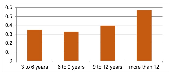
When the sample were categorized into four age groups, in the first group (3–6) years, there was acceleration of dental development amounting (mean: 0.257; sd: 0.047) in boys and (mean: 0.350; sd:51) in girls. In children aged (6 – 9 years) there was an acceleration of dental development amounting (mean: 0.215; sd: 0.095) in boys and (mean: 0.329; sd: 0.113) in girls. In the third age group (9–12 years) the mean differences was (0.296; sd: 0.148) in boys and (mean: 0.396; sd: 0.110) in girls. In the older age group (<12 years), the overestimation was (mean: 0.433; sd: 0.048) in boys and (mean: 0.571; sd: 0.071) in girls (Tables 2&3). The post hoc Schaffer’s test showed that when the boys and girls groups were compared, the Saudi children had a significantly accelerated dental development.
Table 2.
Comparison of differences between CA and DA in different age groups for boys
| 3–6years (N1=42) | 6–9 years (N2=78) | 9–12 years (N3= 72) | More than 12 years (N4=30) | ||||
|---|---|---|---|---|---|---|---|
| Mean | SD | Mean | SD | Mean | SD | Mean | SD |
| 0.257 | 0.074 | 0.215 | 0.095 | 0.296 | 0.148 | 0.433 | 0.048 |
| F= 30.633, p<0.001 | |||||||
F: for one way ANOVA between 4 groups
statistically significant at p<0.05
Table 3.
Post-Hoc test for age groups comparison in boys
| 3–6 years | 6–9 years | 9 – 12 years | More than 12 | |
|---|---|---|---|---|
| 3–6 years | P=0.183 | P=0.255 | p<0.001* | |
| 6–9 years | P<0.01* | p<0.001* | ||
| 9–12 years | p<0.001* |
Post Hoc for multiple comparisons Turkey HSD
statistically significant at p<0.005
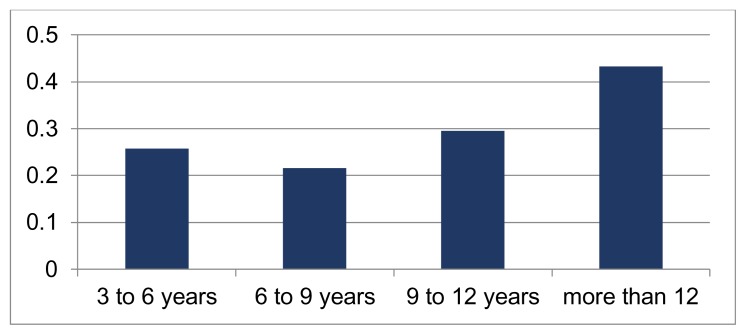
The accelerated dental development may be observed qualitatively from bar graphs of paired dental and chronological age data (Figs: 1 and 2). The most apparent acceleration was found in the older age group (<12 years) than younger age groups in both sexes, and the least was observed in the age groups (6–9 years) in both sexes.
Fig 2.
Comparison of differences between CA and DA at different age groups for boys indicating acceleration of dental development
Discussion
The eight stage system of Demirjian and Goldstein (21) for assessing tooth development was convenient to use and seems to be suitable for making population standards and comparing different population groups with each other.
The present results showed that in general the normal Saudi Arabian children in Qassim region were somewhat advanced in dental maturity compared with the reference sample. This finding is in accordance with the previous studies on different ethnic groups (3,22,23,24,25) in which the Demirjian method has been applied indicating rather good correspondence between dental maturity in the Saudi Arabian children and the French Canadian reference sample.
Although the dental maturity standards used in this study were those of French Canadian children provided in the report of Demirjian and Goldstein, (21) the employment of a race, age and sex matched group in this study should have cancelled sampling error from using the published standards derived from a different population and ethnic groups.
In the present sample, the overall mean differences between the DA determined from the French Canadian standards and the CA were 0.279 years in boys and 0.385 years in girls in accordance with Nykanen et al.(25) and Willam G et al. (26) who reported a mean difference 22 years for boys and 0.3 years for girls in Norwegian children ranging in age from 6 to 12 years, whereas Liversidge et al. (24) reported mean difference of 0.51 years and 0.73 years for boys and girls respectively in British children, however, the mean differences observed in the Somali, (22) Brazilian (3,20) Spanish, (27,28) and Swedish (23) populations were generally greater (up to 2.2 years) than reported in the other studies.
In the present investigation, girls were always more advanced in dental development than boys with statistically significant differences, finding that agree well with other reports on different ethnic groups, (3,22,23,24,25) other authors have reported a close similarity in the early stages of formation of individual teeth in boys and girls but pronounced differences at later stages (5,6,23,29,30) were observed.
It is already an accepted fact that girls are generally more advanced than boys in somatic growth and development up to the pre-adolescent years. This holds for the development and eruption of the permanent teeth occurring between the ages of six and 12 years if we exclude the third molar which is too inconsistent. (1)
Demirjian et al. (17) has observed that the mechanisms controlling dental development are independent of somatic and/or gender maturity and seem to be highly influenced by unknown controlling factors.
One of the greatest advantages of the Demirjian and Goldstein (21) method has been the inter-examiner inter reliability of the method, (1, 24) that confirm and underlines the usefulness of the technique in field where reproducibility rather than accuracy is of prime importance.
Conclusions
Saudi Arabian children (Qassim region) are generally advanced in dental development compared with the French Canadian reference sample by an average of 0.279 years in the boys and 0.385 years in the girls. This is basically due to different rates of dental development in different populations.
Since the average DA in the Saudi Arabian sample did not differ markedly from the reference sample, the applied standards may be adequate as a reference for studying dental maturity in groups of children, particularly in the younger age groups.
Demirjian method, although limited by its accuracy in different populations, offers great scope in fields that require the study of the pattern of growth rather than the accuracy of age estimation.
Fig 3.
Comparison of differences between CA and DA at different age groups for girls indicating acceleration of dental development
Table 4.
Comparison of differences between CA and DA in different age groups for girls
| 3–6years (N1=30) | 6–9 years (N2=85) | 9–12 years (N3= 55) | More than 12 years (N4=28) | ||||
|---|---|---|---|---|---|---|---|
| Mean | SD | Mean | SD | Mean | SD | Mean | SD |
| 0.350 | 0.051 | 0.329 | 0.113 | 0.396 | 0.110 | 0.571 | 0.071 |
| F= | |||||||
F: for one way ANOVA between 4 groups
statistically significant at p<0.05
Table 5.
Post-Hoc test for age groups comparison in girls
| 3–6 years | 6–9 years | 9 – 12 years | More than 12 | |
|---|---|---|---|---|
| 3–6 years | P=0.768 | P=0.178 | p<0.001* | |
| 6–9 years | P=0.01* | p<0.001* | ||
| 9–12 years | p<0.001* |
Post Hoc for multiple comparisons Turkey HSD
statistically significant at p<0.005
Acknowledgment
Great appreciations go to Dr. Ramy Elmoazen, Lecturer of Community Dentistry, Qassim University, KSA, for achieving the statistical analysis for this paper.
References
- 1.Demirjian A, Levesque G. Sexual differences in dental development and prediction of emergence. J Dent Res. 1980;59:1110–1122. doi: 10.1177/00220345800590070301. [DOI] [PubMed] [Google Scholar]
- 2.Demirjian A, Goldstein H, Tanner JM. A new system of dental age assessment. Hum Biol. 1973;45:211–27. [PubMed] [Google Scholar]
- 3.Fanning E. A longitudinal study of tooth formation and root resorption. N Z Dent Res. 1961;51:202–217. [PubMed] [Google Scholar]
- 4.Mc Donald R. Dentistry for child and adolescent. St Louis: CV Mosby Co, Sant Louis; 1969. p. 205. [Google Scholar]
- 5.Posen AL. The effect of premature loss of deciduous molars on premolar eruption. Angle orthod. 1965;35:249–252. doi: 10.1043/0003-3219(1965)035<0249:TEOPLO>2.0.CO;2. [DOI] [PubMed] [Google Scholar]
- 6.Fanning E. Effect of extraction of deciduous molars on the formation and eruption of their successors. Angle Orthod. 1962;32:44–53. [Google Scholar]
- 7.Sapoka AM, Demirjian A. Dental development of the French Canadian children. J Can Dent Assoc. 1971;37:100–104. [PubMed] [Google Scholar]
- 8.Garamendi PM, Landa MI, Ballesteros J, Solono MA. Reliability of the methods applied to assess age minority in living subjects around 18 years old. A survey on a Morocon origin population. Forensic Sci Int. 2005;154:3–12. doi: 10.1016/j.forsciint.2004.08.018. [DOI] [PubMed] [Google Scholar]
- 9.Martin S, Garcia P, Ortega A, Zodocovich S, Valenzuela A. Third molar development according to chronological age in populations from Spanish and Magrebian origin. Forensic Sci Int. 2008;17:47–53. doi: 10.1016/j.forsciint.2007.03.009. [DOI] [PubMed] [Google Scholar]
- 10.Olze A, Reisinger W, Geserick G, Schmeling A. Age estimation of unaccompanied minors. Part II: dental aspects. Forensic Sci Int. 2006;159:3–12. doi: 10.1016/j.forsciint.2006.02.018. [DOI] [PubMed] [Google Scholar]
- 11.Prieto JL, Barberia E, Ortega R, Magana C. Evaluation of chronological age based on third molar development in the Spanish population. Int J Legal Med. 2005;119:349–354. doi: 10.1007/s00414-005-0530-3. [DOI] [PubMed] [Google Scholar]
- 12.Prieto JL, Tortosa C, Bedate A, Segura L, Abenza JM, Mariscal MC, Conejero J, Magana C, Perea B. The importance of the maturity organizationfor identification of victims. Acritical review. Int J Legal Med. 2007;121:517–522. doi: 10.1007/s00414-007-0196-0. [DOI] [PubMed] [Google Scholar]
- 13.Scott K, Kazuhiro I, Karin V. Dental development in Apert syndrome. Cleft palate craniofacial J. 1997;34:117–121. doi: 10.1597/1545-1569_1997_034_0117_ddias_2.3.co_2. [DOI] [PubMed] [Google Scholar]
- 14.Anderson D, Thompson G, Popovich E. Age attainment of mineralized stages of the permanent dentition. Forensic Sci. 1979;12:191–200. [PubMed] [Google Scholar]
- 15.Lewis A, Garn S. The relationship between tooth formation and other nutritional factors. Angle Ortod. 1960;50:70–77. [Google Scholar]
- 16.Moorrees CFA, Fanning EA, Hunt EE. Age variation of formations stages for ten permanent teeth. J Dent Res. 1963;42:1490–1502. doi: 10.1177/00220345630420062701. [DOI] [PubMed] [Google Scholar]
- 17.Demirjian A, Buschhang P, Tanguay R, Kingnorth P. Interrelationship among measures of somatic, skeletal, dental and sexual maturity. Am J Orthod. 1986;88:433–438. doi: 10.1016/0002-9416(85)90070-3. [DOI] [PubMed] [Google Scholar]
- 18.Haavikko K. The formation and the alveolar and clinical eruption of the permanent teeth. Proc Fin Dent Soc. 1970;66:103–107. [PubMed] [Google Scholar]
- 19.Smith BH. Standards of human tooth formation and dental age assessment. In: Kelly MA, Larsen CS, editors. Advances in dental anthropology. New York: Wiley Liss; 1991. pp. 143–168. [Google Scholar]
- 20.Eid R, Simi R, Friggi M, Fisberg M. Assessment of dental maturity of Brazilian children aged 6 to 14 years using Demirjian method. Int J Pediatr Dent. 2002;12:423–428. doi: 10.1046/j.1365-263x.2002.00403.x. [DOI] [PubMed] [Google Scholar]
- 21.Demirjian A, Goldstein H. New systems for dental maturity based onseven and four teeth. Ann Hum Biol. 1976;3:411–21. doi: 10.1080/03014467600001671. [DOI] [PubMed] [Google Scholar]
- 22.Davidson L, Rod H. Interrelationship between dental age and chronological age in Somali children. Community Dent Health. 2001;18:27–30. [PubMed] [Google Scholar]
- 23.Hag U, Matsson Dental maturity as an indicator of chronological age: the accuracy and precision of three methods. Eur J orthod. 1985;7:25–34. doi: 10.1093/ejo/7.1.25. [DOI] [PubMed] [Google Scholar]
- 24.Liversidge H, Seechly T, Hector M. Dental maturation in British children is Demirjian’s standards applicable? Int J Paediatr Dent. 1999;9:263–269. doi: 10.1111/j.1365-263x.1999.00144.x. [DOI] [PubMed] [Google Scholar]
- 25.Nykänen R, Espeland L, Kvaal SI, Krogstad O. Validity of the Demirjian method for dental age estimation when applied to Norwegian children. Acta Odontol Scand. 1998;56:238–44. doi: 10.1080/00016359850142862. [DOI] [PubMed] [Google Scholar]
- 26.Willems G, VanOlmen A, Spiessens B, Carels C. Dental age estimation in Belgian children: Demirjian’s technique revised. J Forensic Sci. 2001;46(4):893–895. [PubMed] [Google Scholar]
- 27.Gonzalo F, Elena B, Joaquin DN, Jose LP. Permanent teeth development in a Spanish sample. Application to dental age estimation. Forensic Sci Int. 2012;214:213–216. doi: 10.1016/j.forsciint.2011.08.024. [DOI] [PubMed] [Google Scholar]
- 28.Gonzalo F, Elena B, Joaquin DN, Jose LP. Dental age estimation in Spanish children. Forensic Sci Int. 2012;223:371–375. doi: 10.1016/j.forsciint.2012.08.021. [DOI] [PubMed] [Google Scholar]
- 29.Garn S, Lewis A, Polacheck D. Variability of tooth formation. J Dent Res. 1959;38:135–148. doi: 10.1177/00220345590380010601. [DOI] [PubMed] [Google Scholar]
- 30.Thompson GW, Anderson DL, Popovich F. Sexual dimorphism in dentition mineralization. Growth. 1975;39:289–301. [PubMed] [Google Scholar]


