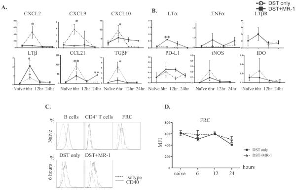
Figure 1. FRC respond to allogeneic stimulation in vivo in a CD40L dependent manner.
(A, B) C57BL/6 mice untreated or treated with DST or DST plus anti-CD40L mAb intravenously, and FRC flow sorted 6, 12 and 24 hours later. DST increased some inflammatory cytokines (A), but did not alter others (B), and anti-CD40L mAb inhibited the inflammatory cytokine response. FRC were sorted as the CD45−gp38+CD31− population, RNA isolated to make cDNA, and qRT-PCR performed for the indicated primers. Results from 3 to 5 samples at each time point, and each sample from 10 mice pooled. * p<0.05, ** p<0.005 vs. naïve. (C.) Surface CD40 stained on CD19+ B220+ naïve B cells, CD4+ T cells and FRC (top). FRC stained 6, 12, and 24 hours after DST or DST plus anti-CD40L mAb administration. Shown here is 6 hours (bottom). (D.) CD40 mean intensity in FRC for each time point after DST (square) or DST plus anti-CD40L (triangle) administration. Results for C and D from 2 to 4 samples per time point.

