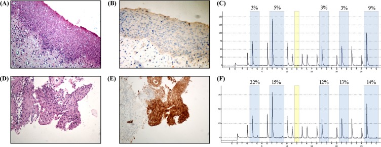
Figure 6. Immunohistochemical staining of P16 in samples from patients with histological diagnosis of CIN2.
(A) Hematoxylin and eosin staining (H/E) for LSIL, and (B) P16, negative in LSIL tissue. (C) Pyrosequencing analysis of LSIL. (D) H/E staining of HSIL, and (E) P16, positive in HSIL tissue. (F) Pyrosequencing analysis of HSIL. Original magnification, A, B, C, D, × 200.

