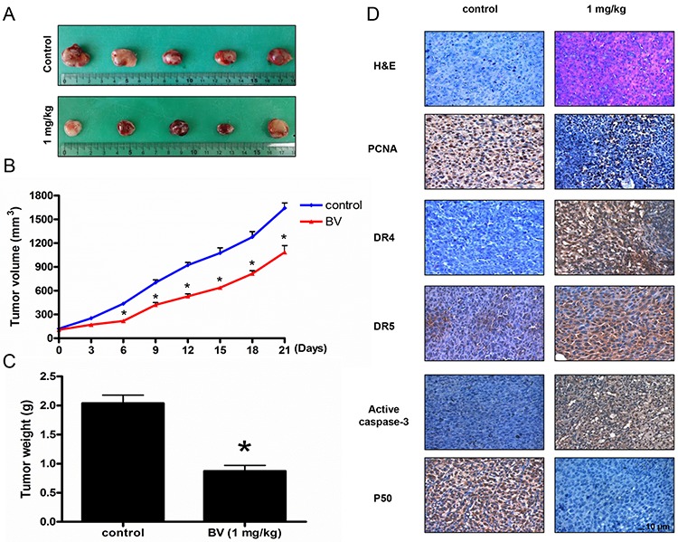
Figure 9. Effect of BV on the tumor growth, on the expression levels of proliferation, apoptosis regulatory proteins and NF-κB p50 subunit in immunohistochemistry.

Growth inhibition (as assessed by tumor volume and weights) of subcutaneously transplanted HCT116 xenografts mice treated with BV (1 mg/kg) twice a week for three weeks. Xenografted mice (n = 6) were administrated intraperitoneally with BV (1 mg/kg). A. Figure represents the tumor photographs at day 21. B. Tumor volumes were measured twice a week for three weeks. C. Tumor weights were measured at study termination on Day 21. Mean ± S.D. estimates of tumor volume and weight from 6 mice in each treatment. D. Immunohistochemistry was used to determine expression levels of H&E, PCNA, DR4, DR5, active caspase-3 and p50 in nude mice xenograft tissues by the different treatments as described in materials and methods. All values represent mean ± S.D. from 5 animal tumor sections. *p < 0.05 indicates significantly different from the control group. Bar indicates 10 μm.
