Abstract
[Purpose] The aim of this study was to identify the most effective method of performing ankle pumping exercises. [Subjects and Methods] The study subjects were 10 men. We measured time-averaged maximum flow velocity and peak systolic velocity in the common femoral vein using a pulse Doppler method with a diagnostic ultrasound system during nine ankle pumping exercises (three different ankle positions and three exercise intervals). Changes of blood flow velocity during ankle pumping exercises with different ankle positions and exercise intervals were compared. [Result] Peak systolic velocity of the leg-up position showed significantly lower values than those of the supine and head-up positions. For all exercise intervals, the increased amount of blood flow velocity in the leg-up position was significantly lower than that in the head-up and supine positions. [Conclusion] Ankle positions and exercise intervals must be considered when performing effective ankle pumping exercises.
Key words: Ankle pumping exercises, Blood flow velocity, Ankle positions
INTRODUCTION
Ankle pumping exercises utilize a calf muscle pump function to pump blood to the heart by muscle contraction. Ankle pumping exercises are often used for the relief of edema3) and the prevention of deep vein thrombosis (DVT)1, 2), which are associated with prolonged bed rest. A 2009 guideline for the Diagnosis, Treatment, and Prevention of Pulmonary Thromboembolism and Deep Vein Thrombosis4) states that ankle pumping exercises are effective for the prevention of DVT. However, there has been much debate about the most effective methods5) and modes of ankle pumping exercises6). Standardized ankle pumping exercises have not been established yet.
Studies have discussed the optimal velocity of muscle contractions7) and the frequency of performing ankle pumping exercises8), but the ankle positions and exercise intervals have not been evaluated in detail9). Therefore, evaluation of the most effective methods and mode of ankle pumping exercises can contribute to standardizing the use of ankle pumping exercises.
The aim of this study was to identify the most effective methods and modes of ankle pumping exercises. Changes of blood flow velocity in the common femoral vein during ankle pumping exercises with different ankle positions and exercise intervals were compared.
SUBJECTS AND METHODS
The study subjects were 10 men without a history of cardio-vascular diseases and no contraindications for exercise testing and training based on a scientific statement from the American Heart Association10).
One experimenter (Y.F.) measured the blood flow velocity in all of these experiments. We measured time-averaged maximum flow velocity (TAMV) and peak systolic velocity (PSV) in the left common femoral vein using a pulse Doppler method with a diagnostic ultrasound system (ACUSON p300, SIEMENS, Germany). TAMV is the averaged blood flow velocity per unit time in the left common femoral vein and PSV is the maximum blood flow velocity in the left common femoral vein during exercise.
The increased amount of blood flow velocity was determined by subtracting TAMV during rest from PSV during exercises. The subject’s heart rate and electrocardiogram were monitored continuously during the muscle pumping exercise. Immediately after each muscle pumping exercise, the subjects rated their local muscle fatigue using a modified Borg scale. Systolic blood pressure (SBP) and diastolic blood pressure (DBP) were also measured before and immediately after the pumping exercises using an automatic blood pressure monitor (SunTech Tango+, SunTech Medical, USA).
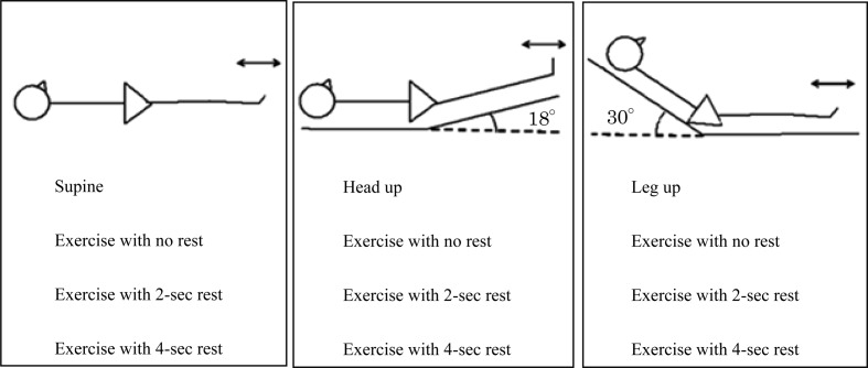
Subjects assumed three different exercise positions: supine (supine position), supine with their legs up after raising the bed to an 18-degree angle (leg-up position), and supine with their head up after raising the bed to a 30-degree angle (head-up position) (Fig. 1). At the beginning of each exercise, the subjects had a 3-min rest period to acclimatize themselves to each position. After the rest, we measured TAMV in the left common femoral vein for 20 sec. The ankle pumping exercises consisted of simple repetitions of dorsiflexion for 1 sec and plantarflexion for 1 sec with three different exercise intervals: repeated dorsiflexion and plantarflexion with no rest (no-rest exercise), repeated dorsiflexion and plantarflexion with a 2-sec rest period (2-sec rest exercise), and repeated dorsiflexion and plantarflexion with a 4-sec rest period (4-sec rest exercise). In total, the subjects performed nine ankle pumping exercises with different positions and exercise intervals. The order of the nine ankle pumping exercises was randomized for each subject. The subjects performed the ankle pumping exercises in rhythm to a metronome. Subjects practiced the ankle pumping exercises before the tests.
Fig. 1.
Experimental protocol
One-way analysis of variance (ANOVA) was used to compare TAMV between the three resting positions. Two-way ANOVA for repeated measures was used to compare PSV and the increased amount of blood flow velocity. Bonferroni adjustments were applied for multiple comparisons. All analyses were performed using SPSS Statistics ver.17.0 (IBM, Tokyo, Japan), and statistical significance was accepted at an alpha level of 0.05.
This study was approved by the Human Ethics Review of Tokyo University of Technology (approval number; E14HS-026). All subjects gave written informed consent prior to data collection. All authors declare that there is no conflict of interest.
RESULTS
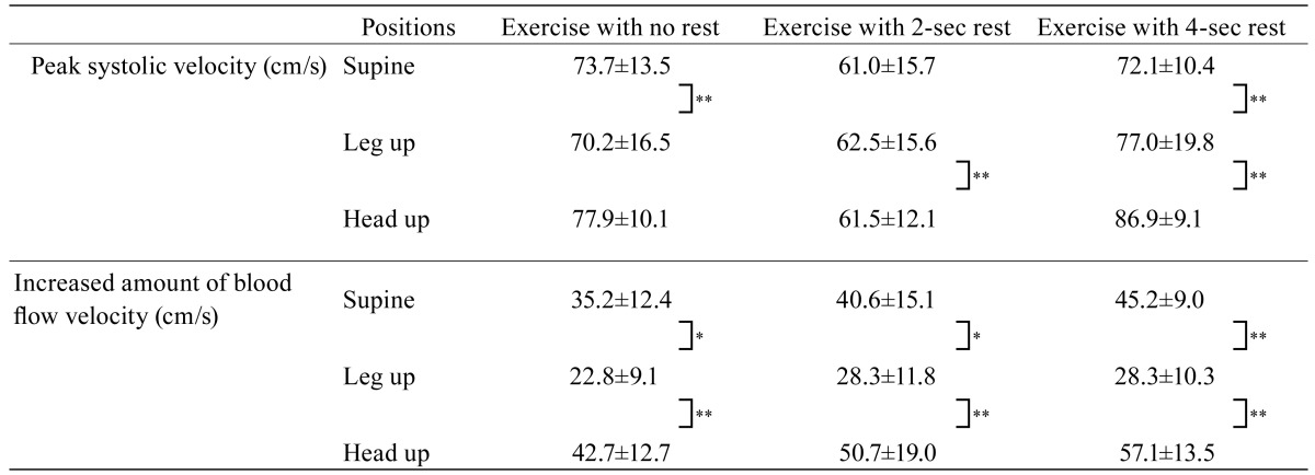
During a rest period, TAMV of the leg-up position (24.4 ± 3.1 cm/sec) was significantly higher than that of the supine (19.9 ± 1.9 cm/sec) and head-up positions (15.0 ± 1.2 cm/sec) (leg up vs. supine, p < 0.05; leg up vs. head up, p < 0.05). TAMV showed no significant difference between the supine position and head-up position (Table 1). For the no-rest exercise, PSV of the leg-up position was significantly lower than that of the supine position (leg up 61.0 ± 15.7 vs. supine 73.7 ± 13.5, p < 0.01). For the 2-sec rest exercise, PSV of the leg-up position was significantly lower than that of the head-up position (leg up 62.5 ± 15.6 vs. head up 77.0 ± 19.8, p < 0.01). For the 4-sec rest exercise, the leg-up position was significantly lower than those of the supine and head-up positions (leg up 61.5 ± 12.1 vs. supine 77.9 ± 10.1, head up 86.9 ± 9.1, p < 0.01). Overall, the PSV of the leg-up position was the lowest in all exercise intervals (Table 2).
Table 1. Time averaged maximum flow velocity in the common femoral vein in three different positions during rest.
Table 2. Peak systolic velocity and increased amount of blood flow velocity in the common femoral vein in three different positions during three different exercise intervals.
For the no-rest exercise, the increased amount of blood flow velocity in the leg-up position was significantly lower than that in the head-up position (leg up 22.8 ± 9.1 vs. head up 42.7 ± 12.7, p < 0.01) and the increased amount of blood flow velocity in the leg-up position was lower than that in the supine position (leg up 22.8 ± 9.1 vs. supine 35.2 ± 12.4, p < 0.05). For the 2-sec rest exercise, the increased amount of blood flow velocity in the leg-up position was lower than that in the head-up position (leg up 28.3 ± 11.8 vs. head up 50.7 ± 19.0, p < 0.01) and the increased amount of blood flow velocity in the leg-up position was lower than that in the supine position (leg up 28.3 ± 11.8 vs. supine 40.6 ± 15.1, p < 0.05). For the 4-sec rest exercise, the increased amount of blood flow velocity in the leg-up position was significantly lower than that in the head-up position (leg up 28.3 ± 10.3 vs. head up 57.1 ± 13.5, p < 0.01) and the increased amount of blood flow velocity in the leg-up position was significantly lower than that in the supine position (leg up 28.3 ± 10.3 vs. supine 45.2 ± 9.0, p < 0.01). Although none of the differences between the exercise intervals reached significance, there was a tendency that the 4-sec rest exercise increased the amount of blood flow velocity the most (Table 2).
DISCUSSION
TAMV of the leg-up position during the resting period was the highest among the three positions. Generally, the venous blood pressure is lower than the arterial pressure, and the venous blood pressure is affected by gravity11), that is, blood flows downhill. The blood flow velocity is calculated by dividing the blood volume (cm3/sec) by the blood vessel dimension (cm2). Therefore, the blood flow velocity positively correlates with the blood volume, and the vessel diameter negatively correlates with the blood flow velocity12).
We thought that the blood flow velocity in the common femoral vein in the leg-up position was the highest because the blood flow volume from the lower limbs to the heart increased by raising the legs. There was no significant difference between TAMV for the supine or head-up position during rest but the TAMV of the head-up position was found to be the lowest of all three positions. Generally, blood pooling in the lower limbs cannot flow back to the heart by only heart pumping. In order to get blood back out of the legs the calf muscle pump is essential13). In the head-up position during a rest, the blood flow volume to lower limbs increased due to gravity, and the muscle pump was not working. These factors resulted in blood pooling in the lower limbs. Therefore, the TAMV of the head-up position during rest was lower than any other position.
Conversely, the PSV of the leg-up position was lower than that of the supine and head-up position. This is because the blood pooling in the lower limbs was reduced by raising the legs. When blood pools in the lower limbs it is essential to send blood out of the lower limbs to the heart using the muscle pump. The blood volume in the lower limb in the leg-up position could have been reduced by gravity even before doing muscle pumping exercises. Therefore, the amount of blood volume output by the muscle pumping exercises was small and the blood flow velocity was not increased very much.
The increased amount of blood flow velocity in the leg-up position was significantly smaller than that of the supine and head-up positions. By raising the legs, venous return increases because of gravity and it is inferred that raising the legs might have prevented pooling of blood in the lower limbs. We thought that the increased amount of blood flow velocity in the leg-up position was the lowest of the three positions due to these reasons.
The short rest time might lead to little pooling of blood in the lower limbs. If the blood volume is related to the increased amount of blood flow velocity, it is assumed that the differences in rest time may cause the changes in the increased amount of blood flow velocity. In this study, there were no significant differences among the three rest exercises, but there was a tendency that the increased amount of blood flow velocity increased with increasing rest time. This suggests that the amount of pooling blood in the lower limb might be important for increasing blood flow. Ankle positions and exercise intervals must be taken into account for performing effective ankle pumping exercises.
The subjects in this study were young health men. Aging causes vascular degeneration and reduced vascular elasticity that can lead to reduced venous return so it takes a longer time for older people to pool blood in the lower limb. This can affect the results and indicates that a study including older people should be done in the future.
REFERENCES
- 1.Ohta S, Yamada N, Tsuji A, et al. : A comparative effect of various mechanical prophylactic methods for the prevention of venous thromboembolism. Jpn J Phlebol, 2004, 15: 89–94. [Google Scholar]
- 2.Nicolaides AN, Kakkar VV, Field ES, et al. : Venous stasis and deep-vein thrombosis. Br J Surg, 1972, 59: 713–717. [DOI] [PubMed] [Google Scholar]
- 3.Ogiwara S: Calf muscle pumping and rest positions during and/or after whirlpool therapy. J Phys Ther Sci, 2001, 13: 99–105. [Google Scholar]
- 4.Editorial Committee on Japanese Guideline for Prevention of Venous Thromboembolism: Guidelines for Diagnosis, Treatment and Prevention of Pulmonary Thromboembolism and Deep Vein Thrombosis (JCS2004). Medical Front International Limited, 2004, pp 11–12. [Google Scholar]
- 5.Uchida M, Katoh M: Verification of the effect of techniques used to prevent deep-vein thrombosis. J Phys Ther Sci, 2011, 23: 243–245. [Google Scholar]
- 6.Eom JH, Chung SH, Shim JH: The effects of squat exercises in postures for toilet use on blood flow velocity of the leg vein. J Phys Ther Sci, 2014, 26: 1485–1487. [DOI] [PMC free article] [PubMed] [Google Scholar]
- 7.Nishiyasu T, Goto S, Nabekura Y, et al. : A Study of muscle pump—relationship between contraction force and blood volume and pumping action—. Jpn J Phys Fit Sports Med, 1987, 36: 195–201. [Google Scholar]
- 8.Kawana T, Egami T, Harada S, et al. : Examination of the optimal movement speed of ankle dorsiflexion and plantar flexion movements in elderly people: examination of femoral-vein flow velocity. J Clin Welf, 2010, 7: 23–26. [Google Scholar]
- 9.Ishii M, Kawaji H, Hamasaki M, et al. : Examination of maximum velocity of femoral vein with various methods for prevention of deep vein thrombosis. Hip joint, 2001, 27: 557–559. [Google Scholar]
- 10.Fletcher GF, Ades PA, Kligfield P, et al. American Heart Association Exercise, Cardiac Rehabilitation, and Prevention Committee of the Council on Clinical Cardiology, Council on Nutrition, Physical Activity and Metabolism, Council on Cardiovascular and Stroke Nursing, and Council on Epidemiology and Prevention: Exercise standards for testing and training: a scientific statement from the American Heart Association. Circulation, 2013, 128: 873–934. [DOI] [PubMed] [Google Scholar]
- 11.Epley D: Pulmonary emboli risk reduction. J Vasc Nurs, 2000, 18: 61–68, quiz 69–70. [DOI] [PubMed] [Google Scholar]
- 12.Guyton AC, Hall JE: Textbook Of Medical Physiology, 11th ed. Philadelphia: Saunders Company, 2006, p 162. [Google Scholar]
- 13.Sakai T, Kawahara K: Structure, function and materials of the human body. Tokyo: Japan Medical Journal, 2012, p 155. [Google Scholar]


