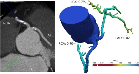
Fractional flow reserve from computed tomography (FFRCT) provides a non-invasive functional assessment of the coronary tree with computed FFR. This imaging modality is particularly useful when invasive FFR measurements are difficult or contraindicated. An otherwise asymptomatic 70-year-old male patient presented with ventricular extrasystoles which were confirmed during exercise stress test. The patient underwent a coronary computed tomography angiography (CCTA). CCTA revealed an abnormal left main origin in the right anterior cusp close to the origin of the right coronary artery with moderate atherosclerosis including calcifications. Invasive coronary angiography showed this abnormality equally well, but wire-based functional evaluation was not performed due to safety considerations. From the CCTA data, virtual FFR evaluation (FFRCT) was computed. The FFRCT revealed a borderline abnormal virtual FFR value of 0.79 in the distal circumflex. However, since coronary anomalies have not been systematically evaluated by FFR, we performed an additional myocardial perfusion imaging test showing no significant myocardial ischaemia. As a result, the patient was discharged with optimal medical therapy. FFRCT is a novel non-invasive functional test providing both anatomical and functional evaluation of the coronary tree. These unique features allow to tackle difficult issues in coronary artery pathology.
On the left side, axial image of coronary CT showing the inter-arterial course of the left main. On the right side, FFRCT analysis showing the FFR value in the distal circumflex (0.79), the distal left anterior descending artery (0.82), and the right coronary artery (0.90).
Funding
Funding to pay the Open Access publication charges for this article was provided by Heartflow, Inc.


