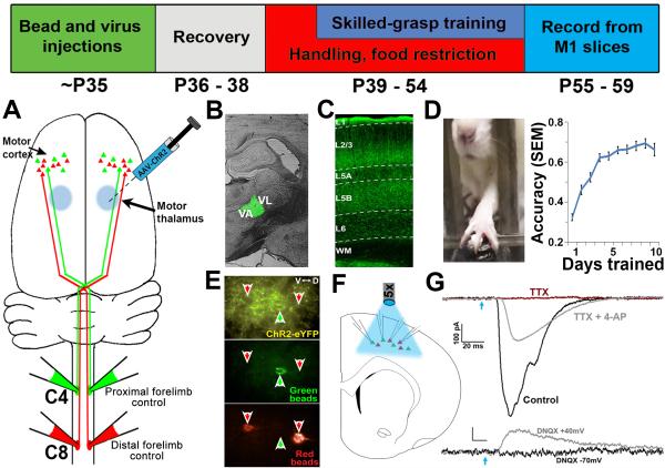
Figure 1. Experimental overview.
Top: Timeline of experiments. (A) Retrograde tracer injections at levels C4 and C8 of the spinal cord label distinct corticospinal projection populations originating in layer 5 of M1. (B) To selectively express ChR2 in motor thalamic nuclei, AAV-ChR2 was targeted to the VA/VL complex of the thalamus. (C) TC axonal expression of ChR2-EYFP was robust in layer 5 (the location of corticospinal cell bodies) of the caudal forelimb region of M1. (D) Skilled grasp training was conducted over 10 days, leading to a significant increase in pellet retrieval accuracy (repeated-measures ANOVA, p < 0.001). (E) Following the completion of training, slices of M1 were prepared and C4- and C8-projecting corticospinal neurons were targeted for simultaneous whole-cell recording. Disambiguation of eYFP signal from green bead fluorescence was achieved via a narrow band GFP filter. (F) Photostimulation at ~470nm was applied across the cortical slice via 5x objective to selectively stimulate thalamocortical axons. (G) Corticospinal neurons receive direct monosynaptic input from TC axons; upper traces: photo-induced EPSCs were abolished in the presence of 1μM TTX (red trace), but rescued following addition of 100 μM 4-AP (grey trace); lower traces: the presence of the AMPAR antagonist DNQX abolished EPSCs when neurons were held at −70mV (black trace). Releasing the Mg2+ block from NMDA channels via depolarization to +40mV unmasked monosynaptic NMDA-mediated currents (grey trace). Blue arrow = light onset.

