Full text
PDF
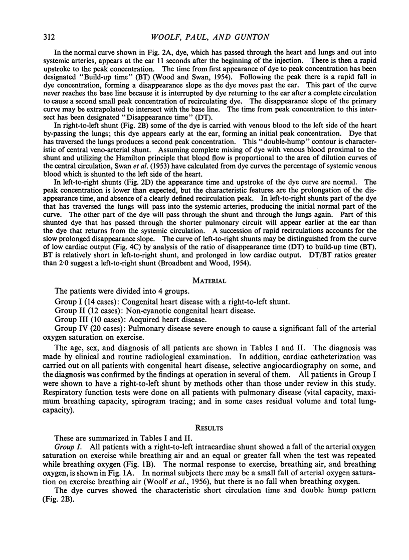
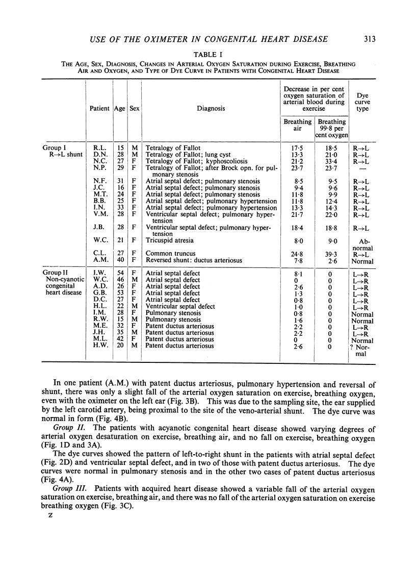
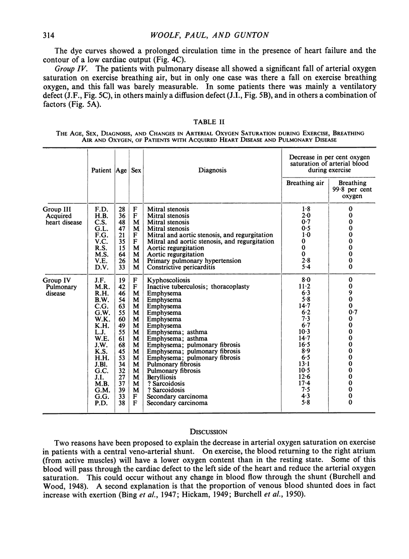
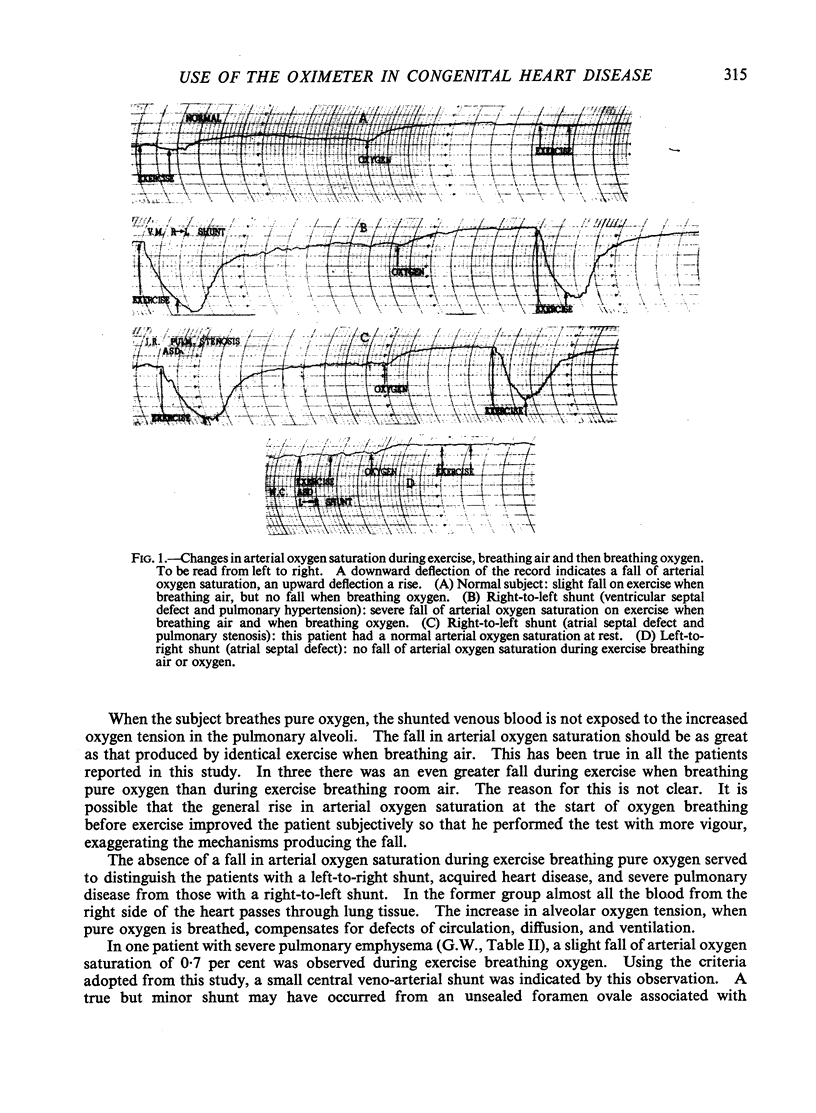
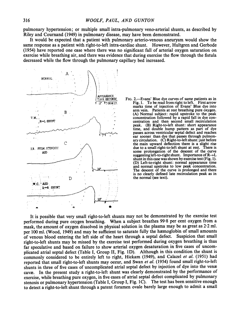
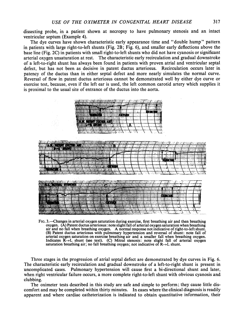
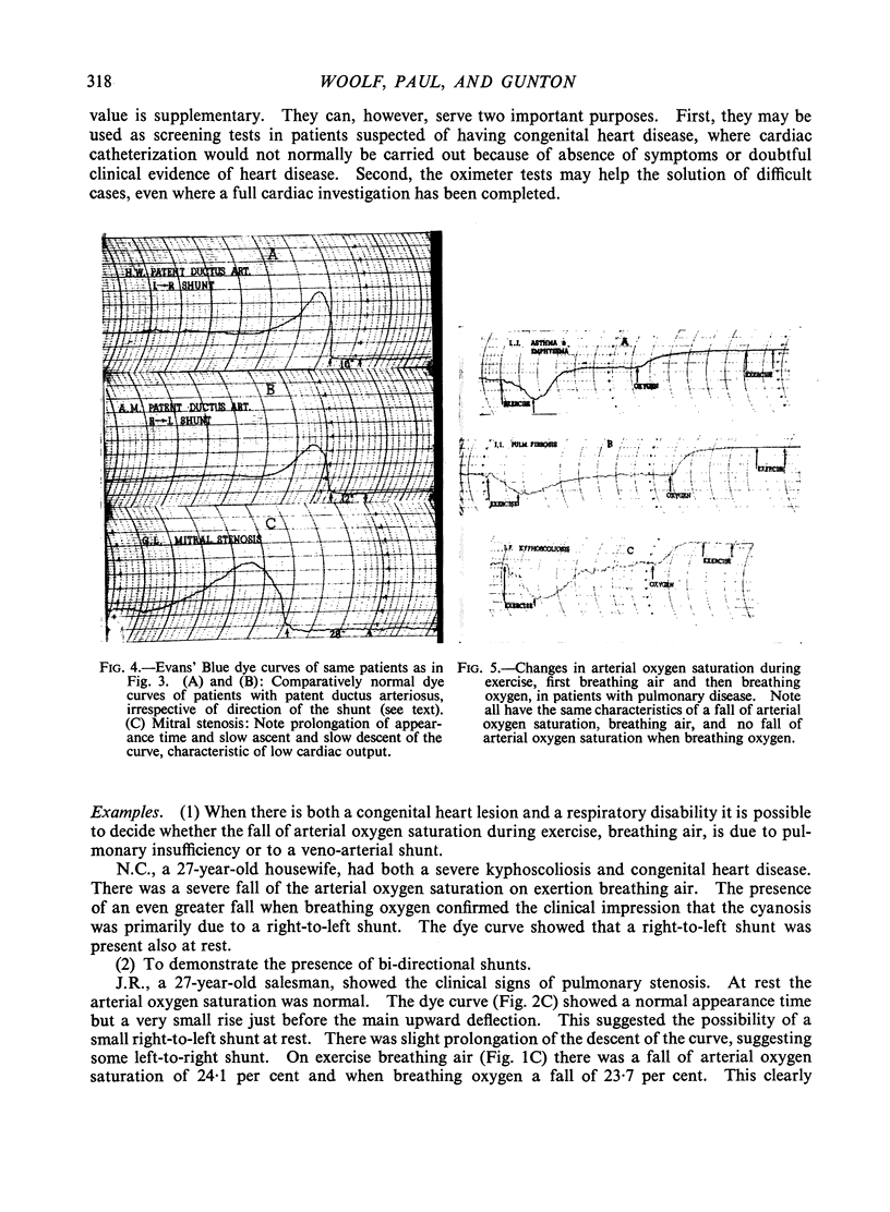
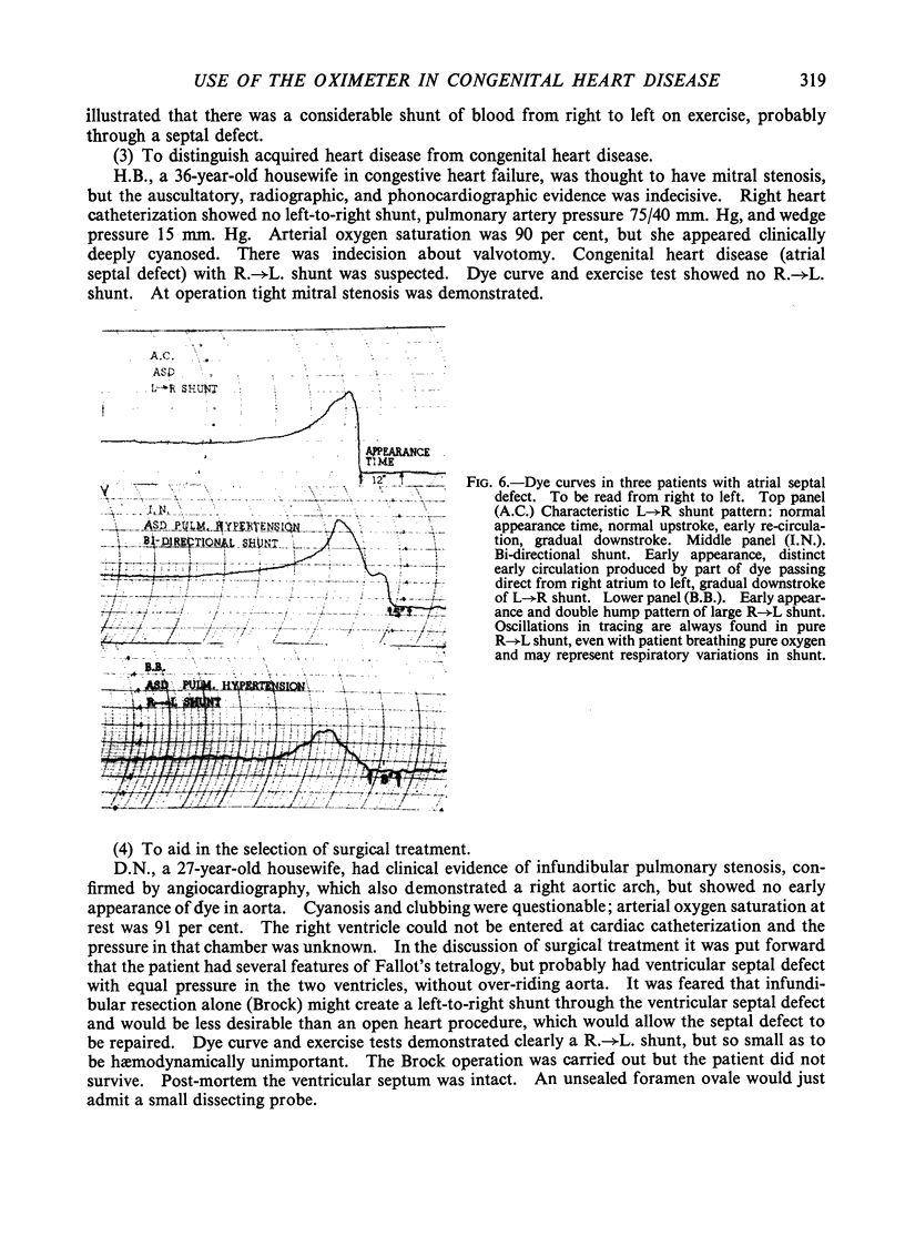
Images in this article
Selected References
These references are in PubMed. This may not be the complete list of references from this article.
- BROADBENT J. C., WOOD E. H. Indicator-dilution curves in acyanotic congenital heart disease. Circulation. 1954 Jun;9(6):890–902. doi: 10.1161/01.cir.9.6.890. [DOI] [PubMed] [Google Scholar]
- BURCHELL H. B., TAYLOR B. E. Circulatory adjustments to the hypoxemia of congenital heart disease of the cyanotic type. Circulation. 1950 Mar;1(3):404–414. doi: 10.1161/01.cir.1.3.404. [DOI] [PubMed] [Google Scholar]
- CALAZEL P., GERARD R., DALEY R., DRAPER A., FOSTER J., BING R. J. Physiological studies in congenital heart disease. XI. A comparison of the right and left auricular, capillary and pulmonary artery pressures in nine patients with auricular septal defect. Bull Johns Hopkins Hosp. 1951 Jan;88(1):20–37. [PubMed] [Google Scholar]
- GILMORE H. R., HAMILTON M., KOPELMAN H., SOMMER L. S. The ear oximeter; its use clinically and in the determination of cardiac output. Br Heart J. 1954 Jul;16(3):301–310. doi: 10.1136/hrt.16.3.301. [DOI] [PMC free article] [PubMed] [Google Scholar]
- GUNTON R. W., PAUL W., WOOLF C. R. Simple tests of respiratory function using a direct-writing ear oximeter. Am Rev Tuberc. 1956 Oct;74(4):511–532. doi: 10.1164/artpd.1956.74.4.511. [DOI] [PubMed] [Google Scholar]
- HICKAM J. B. Atrial septal defect; a study of intracardiac shunts, ventricular outputs, and pulmonary pressure gradient. Am Heart J. 1949 Dec;38(6):801–812. doi: 10.1016/0002-8703(49)90882-0. [DOI] [PubMed] [Google Scholar]
- HULTGREN H. N., GERBODE F. Physiologic studies in a patient with a pulmonary arteriovenous fistula. Am J Med. 1954 Jul;17(1):126–133. doi: 10.1016/0002-9343(54)90213-2. [DOI] [PubMed] [Google Scholar]
- NICHOLSON J. W., 3rd, BURCHELL H. B., WOOD E. H. A method for the continuous recording of Evans blue dye curves in arterial blood, and its application to the diagnosis of cardiovascular abnormalities. J Lab Clin Med. 1951 Mar;37(3):353–364. [PubMed] [Google Scholar]
- SWAN H. J., BURCHELL H. B., WOOD E. H. The presence of venoarterial shunts in patients with interatrial communications. Circulation. 1954 Nov;10(5):705–713. doi: 10.1161/01.cir.10.5.705. [DOI] [PubMed] [Google Scholar]
- VAN LINGEN B., WHIDBORNE J. Oximetry in congenital heart disease with special reference to the effects of voluntary hyperventilation. Circulation. 1952 Nov;6(5):740–748. doi: 10.1161/01.cir.6.5.740. [DOI] [PubMed] [Google Scholar]


