

Abstract
Objective: To evaluate the results from arthroscopic treatment in patients with calcific tendonitis of the shoulder. Methods: Between September 2001 and June 2006, 55 patients with calcific tendonitis of the shoulder that was resistant to conservative treatment were evaluated, with follow-up of 12 to 70 months. The mean age was 42 years, ranging from 30 to 64 years; 44 patients were female (80%). There were 37 right shoulders, and 63.63% of the cases were on the dominant side. Pain was the main symptom, and the mean time between onset of symptoms and arthroscopy was 38 months (range: five to 120 months). The tendon affected was the supraspinatus in 42 cases, the infraspinatus in 11 cases and an association between these in two cases. Acromioplasty was carried out in 12 patients (21.82%) and subacromial bursectomy was performed in all cases. Results: According to the UCLA criteria, 46 cases were excellent and six were good, making a total of 52 satisfactory results (94.54%). Conclusion: Arthroscopic treatment of calcific tendonitis of the shoulder appears to be an effective method, with high rates of satisfactory results. Associated acromioplasty is not necessary.
Keywords: Shoulder Fractures, Fracture Fixation, Internal, Bone Plates
INTRODUCTION
Calcific tendonitis is one of the most common pathological conditions of the rotator cuff, especially affecting patients aged between 30 and 50 years, with higher incidence among females1, 2. Its etiology still remains unknown.
The calcium deposits most frequently originate in the portion of the cuff that is subject to higher-intensity forces and that most frequently presents degenerative alteration, i.e. the supraspinatus tendon(3). Codman named this region the critical zone of the rotator cuff: a location where the blood supply is poor and where pressure is highest, with an impact on the anterior elevation of the arm. Therefore, the most common location for calcification is on the bursal face of the supraspinatus tendon(4), near to its insertion into the great tuberosity. For this reason, when surgical procedures are necessary, they are performed in the subacromial region. Other additional localities are the infraspinatus and subscapularis tendons, and there may be an association between them3, 4.
Bosworth(5) performed a study on a non-selected group of 6,061 workers in a life insurance company, with radiographs on both shoulders, and found that the incidence of calcium deposits was 2.7%. Of these, 35% of the patients had previously presented symptoms. Thus, it is possible for calcification to be present in asymptomatic individuals. Similar conclusions can be drawn from the studies of Welfling et al(6) and Sandstrom and Wahlgreen(7), who found 7.5% and 20%, respectively. There is no evidence correlating calcium deposits in the shoulder with the presence of calculi in organs such as the kidneys and gallbladder, or with osteometabolic diseases (1).
The calcium deposit is normally intratendinous1, 3, 4 and evolves with a defined progression, according to Uhthoff and Sarkar(8), going through a pre-calcifying phase, formation phase and reabsorption phase (Figure 1 and 2), and subsequently remodeling to a normal tendon. It is believed that the duration of this remodeling is the true variable in this process.
Figure 1.

Formative phase, according to Uhthoff
Figure 2.

Calcific tendonitis in the reabsorption phase, according to Uhthoff
Knowledge of the chronological sequence of these three phases is important in therapeutic planning. Since it is self-limited, calcific tendonitis should initially be treated conservatively, with anti-inflammatory drugs and rehabilitation. Symptom progression and absence of pain improvement are indications for other types of treatment: percutaneous aspiration(3), shock waves(9), open surgical removal13, 8 or arthroscopic removal2, 10, 11, 12, 13, 14, 15.
Arthroscopic treatment allows complete joint evaluation and early rehabilitation, and avoids the reported complications of open surgery, such as problems with the deltoid muscle insertion, infection and joint rigidness1, 2, 10. This method aims to remove the calcification associated with bursectomy, with promising results.
The objective of the present study was to evaluate the results obtained from arthroscopic treatment of patients with refractory calcific tendonitis of the shoulder and correlate them with associated acromioplasty.
METHODS
This was a cross-sectional study conducted between September 2001 and June 2006, in which 55 patients with calcific tendonitis of the shoulder that was resistant to conservative treatment underwent resection of the calcification via arthroscopy. All the patients were operated by the same surgeon. The length of follow-up ranged from 12 to 70 months (mean of 31 months).
The patients' mean age was 42 years, ranging from 30 to 64 years; 44 patients were female (80%). There were 37 shoulders were of the right side (67.27%), and the dominant shoulder was affected in 63.63% of the cases. See clinical demographic characteristics in Table 1.
Table 1.
Clinical demographic characteristics
| Case | Gender | Dom | Side | Age(years) | LF | Acrom | Te | Bosw | Gart | SutureRC | Follow-upmonths | UCLA | Compl |
|---|---|---|---|---|---|---|---|---|---|---|---|---|---|
| 1 | M | L | 50 | SS | medium-sized | I | 70 | 26 | Residual Pain | ||||
| 2 | F | + | R | 48 | + | IS | small | II | 69 | 31 | |||
| 3 | F | + | R | 47 | + | SS | medium-sized | I | 67 | 30 | |||
| 4 | F | + | R | 39 | IS | small | I | 66 | 35 | ||||
| 5 | M | + | R | 49 | + | SS | large | I | 61 | 35 | |||
| 6 | F | + | R | 37 | + | SS | small | I | 52 | 31 | |||
| 7 | M | + | R | 45 | + | + | SS | medium sized | III | 50 | 35 | ||
| 8 | M | + | R | 40 | + | + | SS | large | I | + | 49 | 34 | |
| 9 | M | + | R | 30 | + | SS | small | II | 47 | 35 | |||
| 10 | M | L | 49 | + | + | SS | medium-sized | I | 45 | 35 | |||
| 11 | F | + | R | 38 | + | SS | medium-sized | I | 43 | 35 | |||
| 12 | F | L | 51 | + | + | SS | medium-sized | I | 42 | 35 | |||
| 13 | M | L | 64 | + | SS | large | II | 41 | 35 | ||||
| 14 | M | + | R | 35 | + | SS | medium sized | I | 41 | 35 | |||
| 15 | F | + | R | 42 | + | + | IS | medium-sized | I | 40 | 31 | Capsulitis | |
| 16 | F | + | R | 40 | SS | medium-sized | I | 40 | 35 | ||||
| 17 | F | + | R | 38 | + | + | SS | medium-sized | I | 38 | 35 | ||
| 18 | F | + | R | 37 | + | SS | medium-sized | I | 38 | 35 | |||
| 19 | F | L | 30 | + | IS + SS | medium-sized | I | 36 | 35 | ||||
| 20 | F | + | R | 32 | SS | large | I | 35 | 35 | ||||
| 21 | M | + | R | 35 | + | SS | medium-sized | I | 33 | 35 | |||
| 22 | F | + | R | 50 | + | SS | medium-sized | I | 33 | 35 | |||
| 23 | F | + | R | 50 | + | SS | large | II | 33 | 31 | Capsulitis | ||
| 24 | F | L | 58 | + | SS | small | II | 32 | 35 | ||||
| 25 | M | L | 42 | + | SS | small | I | 31 | 35 | Capsulitis | |||
| 26 | M | + | R | 40 | + | SS | large | I | 30 | 35 | |||
| 27 | F | + | R | 38 | + | IS | large | I | 30 | 35 | Capsulitis | ||
| 28 | F | + | R | 48 | IS | small | I | 30 | 35 | ||||
| 29 | F | L | 30 | + | SS | medium-sized | II | 29 | 35 | ||||
| 30 | F | + | R | 32 | + | SS | medium-sized | I | 28 | 35 | |||
| 31 | F | L | 33 | + | SS | medium-sized | III | 27 | 35 | ||||
| 32 | F | + | R | 32 | SS | small | II | 26 | 35 | ||||
| 33 | F | L | 55 | SS | medium-sized | I | 25 | 35 | Capsulitis | ||||
| 34 | F | + | R | 37 | IS | medium-sized | I | 25 | 35 | ||||
| 35 | F | L | 52 | + | SS | medium-sized | I | 25 | 35 | ||||
| 36 | F | + | R | 45 | + | SS | medium-sized | I | 24 | 35 | |||
| 37 | F | L | 60 | SS | medium-sized | II | 24 | 35 | |||||
| 38 | F | + | R | 32 | + | IS | small | II | 23 | 35 | |||
| 39 | F | + | R | 58 | + | IS | small | II | 22 | 35 | |||
| 40 | F | L | 58 | + | SS | small | II | 22 | 32 | Capsulitis | |||
| 41 | F | + | R | 40 | + | SS | large | I | 20 | 35 | |||
| 42 | F | L | 57 | SS | small | III | 20 | 35 | |||||
| 43 | F | + | R | 45 | + | SS | small | I | 20 | 35 | |||
| 44 | F | + | R | 47 | IS | medium-sized | I | 20 | 35 | ||||
| 45 | F | + | R | 50 | + | SS | medium-sized | I | 18 | 35 | |||
| 46 | F | + | R | 45 | SS | medium-sized | II | 15 | 34 | ||||
| 47 | F | L | 34 | SS | medium-sized | I | 13 | 35 | |||||
| 48 | F | + | R | 50 | + | SS | medium-sized | I | 12 | 35 | |||
| 49 | F | + | R | 50 | IS | small | I | 12 | 26 | Residual Pain | |||
| 50 | F | + | R | 40 | + | SS | small | I | 12 | 35 | |||
| 51 | F | + | R | 46 | + | IS + SS | medium-sized | I | 12 | 25 | Residual Pain | ||
| 52 | F | L | 50 | SS | large | I | 12 | 35 | |||||
| 53 | F | + | R | 42 | SS | large | II | 12 | 35 | ||||
| 54 | F | L | 37 | IS | medium-sized | I | 12 | 35 | |||||
| 55 | F | L | 38 | + | SS | medium-sized | I | 12 | 35 |
Legend: Dom – Dominant side, M- Male, F – Female, L – Left, R – Right, LM – raspberry lesion, Acrom – Acromioplasty, Te – tendon affected, Bosw – Bosworth classification, Gart- Gartner classification, RC – Rotator cuff, Compl- Complications, SS – Supraspinatus, IS - Infraspinatus
Source: Medical archives of the HOG
Pain was the main symptom; the time elapsed from the beginning of symptoms to arthroscopy was 38 months on average, with a range from five to 120 months. The “raspberry lesion” described by Snyder(4), which is a vascular stain (hyperemia) on the joint side of the supraspinatus tendon, was found in 56% of the patients (Table 1).
The tendon affected was the supraspinatus in 42 cases, the infraspinatus in 11 cases and an association between these in two cases (Figure 3). Acromioplasty was performed because of the presence of subacromial friction, as shown by fibrillation on the anteroinferior surface of the acromion or when no calcification was found during the operation (Figure 4). On the other hand, subacromial bursectomy was performed in all cases.
Figure 3.

Scapular profile showing supraspinatus calcifications (white arrow) and infraspinatus calcifications (black arrow).
Figure 4.

Acromioplasty.
Radiographic evaluation was performed in the anteroposterior view with internal and external rotation, scapular view with 10° caudal inclinations and axillary view(1). Indices using Bosworth's classification(5), which is based on the size of the calcification, and Gartner's classification(16), which is related to density, are expressed in Figures 5 and 6. Among the patients operated, the defective tendon was sutured after resection of the calcification in only one case (1.81%), which was insufficient sample for statistical analysis. In the series studied, there were no SLAP lesions(4) or associated rupture of the rotator cuff.
Figure 5.

Rates according to Bosworth classification.
Figure 6.

Rates according to Gartner classification.
The arthroscopic surgery was performed in lateral decubitus, with posterior, lateral and anterior ports. In the last 35 operations, the anterior port was not used (Figure 7); in these cases, irrigation was provided through the lateral portal, along with instrumentation. It is important to mark out the joint surface of the supraspinatus tendon in the hyperemia zone using monofilament suture (Figure 8), when present, since this may correspond to the calcification area on the bursal surface(4). Complete removal of calcification without causing iatrogenic damage to the affected tendon is fundamental (Figure 9), along with exhaustive irrigation of the subacromial space, because calcium residues (Figure 10) may cause pain after the operation(2). No image intensifier was used during the surgery. The patients were evaluated using the system proposed by the University of California at Los Angeles (UCLA)(17).
Figure 7.
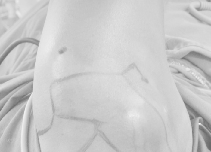
Posterior and lateral arthroscopic ports in lateral decubitus for the right shoulder.
Figure 8.

Guide wire on joint surface of the supraspinatus tendon of the right shoulder, seen through posterior port.
Figure 9.
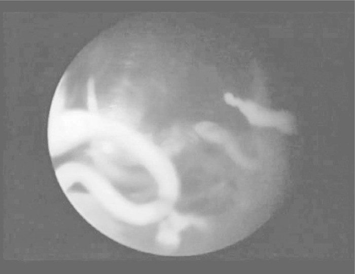
Removal of calcification, seen through posterior port.
Figure 10.

Presence of calcareous granules in the bursal space, seen through posterior port.
The data were analyzed using the SPSS 11.5 software (Statistical Package for the Social Sciences). Variance analysis was performed to investigate whether there were any differences in acromioplasty regarding the means for the continuous variable of age. Fisher's test was used to compare discrete 2×2 variables and the chi-square test was used to investigate whether there were any differences in the discreet variables with more than two groups. A confidence level of 95% was used, i.e. results were significant if p < 0.05.
RESULTS
According to the UCLA criteria, there were 46 excellent results and six good results, thus totaling 52 satisfactory results (94.54%). The three unsatisfactory cases were classified as regular because of postoperative residual pain, even after rehabilitation, although without joint limitation. Six patients evolved with adhesive capsulitis during the early postoperative period, and these cases were all resolved through serial blockage of the suprascapularis nerve.
Calcification was not found in four patients: in three of these, acromioplasty was performed and in one case, it was not.
No cases of infection, nerve lesions or acromioclavicular pain were found.
There was no significant difference between the patients who underwent acromioplasty and those who did not, in relation to age (p = 0.851) (Table 2), gender (p = 0.133), presence or absence of “raspberry lesion” (p = 0.209), dominance (p = 0.231) or side (0.231). There was also no relationship between size/type of calcification and the discrete variable of acromioplasty (p = 0.219 and p = 0.193, respectively).
Table 2.
Correlation between the variables of acromioplasty and age
| Acromioplasty |
|||
|---|---|---|---|
| Variable | Yes | No | p |
| Mean ± SD | Mean ± SD | ||
| Age (years) | 43.46 ± 8.55 | 44.00 ± 9.18 | 0.851 |
Test: Variance analysis
From Table 3, it can be seen that there was no significant difference in the satisfactoriness of the results according to the UCLA rates, with regard to whether or not acromioplasty was performed (p = 0.086). The correlation between the Bosworth/Gartner classification and UCLA evaluation is shown in Table 4.
Table 3.
Distribution of patients regarding UCLA versus acromioplasty
| UCLA |
Acromioplasty |
|
|---|---|---|
| No | Yes | |
| Excellent | 38 | 8 |
| Good | 3 | 3 |
| Regular | 2 | 1 |
| Total | 43 | 12 |
P = 0.086 / Fisher's Test
Table 4.
UCLA versus Bosworth/UCLA versus Gartner
| UCLA |
||||||
|---|---|---|---|---|---|---|
| Classification | Good | Excellent | Regular | |||
| n | % | n | % | n | % | |
| Bosworth | ||||||
| Small | 2 | 33.3 | 11 | 23.9 | 2 | 66.7 |
| Medium sized | 3 | 50.0 | 26 | 56.5 | 1 | 33.3 |
| Large | 1 | 16.7 | 9 | 19.6 | − | 0.0 |
| Gartner | ||||||
| I | 2 | 33.3 | 33 | 71.7 | 2 | 66.7 |
| II | 4 | 66.7 | 10 | 21.7 | 1 | 33.3 |
| III | − | 0.0 | 3 | 6.5 | − | 0.0 |
DISCUSSION
Calcific tendonitis is a self-limiting disease1, 3, 5. It is important to understand that calcification reabsorption will occur, but its timing is unclear. This may be the true variable of the whole process. Conservative treatment is always recommended initially, before considering surgical treatment. In our sample, the time elapsed between the beginning of symptoms and arthroscopy was 38 months on average, with a minimum of five months. Great care needs to be taken in relation to the high spontaneous absorption rate that this disease presents.
Much has been published about the use of arthroscopy for treating shoulder lesions. This technique has already become established, and has been shown to be efficient for treating calcific tendonitis of the shoulder that is refractory to conservative approaches. It presents lower morbidity and allows patients to return earlier to their daily activities2, 4, 10, 11, 12, 13, 14, 15. On the other hand, open surgery presents problems regarding the deltoid muscle insertion, infection, greater joint rigidity and difficulty in viewing the joint3, 8.
In our study the proportion of satisfactory results amounted to 94.54%, which coincides with results in the literature. Godinho et al obtained excellent and good results in 94%(2), Bassini et al(18) in 88.9%, Ark et al(12) in 91% and Molé et al(11) in 89%.
Concordant with other orthopedic papers, we found higher incidence of calcific tendonitis among the female patients, with the majority of such cases in the age group between 30 and 50 years and predominance on the dominant side)2, 3, 10, 12. Radiographic evaluation is important for classifying and locating calcifications, and this needs to be done immediately before the operation, in order to verify that no reabsorption of the calcification has occurred between indication and surgery1, 2.
According to Godinho et al(2), Ark et al(12) and Rupp et al(19), the satisfactoriness of the results from arthroscopic treatment is not influenced by whether the resection of the calcification is partial or total. Our view is that the resection should always be as complete as possible, in agreement with Checchia et al(10) and Jerosch et al(15), although such evaluations are not always easy to perform intraoperatively(18). Porcellini et al(20) observed that the presence of calcium residues after the operation significantly decreased the pain score of the Constant-Murley index, in relation to individuals with complete removal. In the present study, we did not use an image intensifier during the operation, contrary to Checchia et al, who checked whether calcium resection had been completed(10). Additionally, if there is any doubt regarding the removal of the calcification, radiography can be performed on the surgical table(4).
Some authors have advocated not suturing the tendon opening, after resection of the calcification8, 20. Whenever possible, the aim is not to make incisions in the tendon, but to use a percutaneous needle laterally to the acromion, in order to perforate it, with the aid of an arthroscopic curette. The shaver blade must not be passed over the tendon, in order to avoid iatrogenic damage, but instead, it should be used for vacuum suction of the calcification, preferentially using synovial resector blades of small diameter1(4).
In our series, only one cuff suture was performed (1.81%), because we did not have any patients who, after resection, remained with large partial or complete lesions. Among the 54 shoulders in which the defects were not sutured, 94% attained satisfactory results. Our sample of a single patient with a suture was insufficient for statistical analysis. Porcellini et al(20) reported that no tendon sutures were performed in cases of partial longitudinal lesion shorter than 1 cm, which corresponded to 38.1%, and that during the follow-up, there was no rupture of the rotator cuff, either in the patients who underwent suturing or in those who did not. These authors also stated that they did not perform suturing when the removal was incomplete. In the series of 71 shoulders of Checchia et al(10), tendon defects were sutured in 17 cases (23.9%). Among the 54 cases that did not receive suturing, 51 presented satisfactory results, corresponding to 94.44%. It is possible to suggest that this procedure is only necessary in situations of major defects. Whenever tendon incision is needed, it should always be performed longitudinally, along the tendon fibers, and not transversally, in order to avoid tension in the suture, which would compromise rehabilitation.
The question of whether acromioplasty is necessary has arisen. In 1990, Neer(21) stated that it was not necessary, because calcific tendonitis was not associated with subacromial impact. Ellman et al(17) recommended performing it only if there was evidence of friction. In a multicenter French study, 112 patients with calcific tendonitis underwent arthroscopic procedures either with or without subacromial decompression, and it was concluded that there were no statistically significant differences between the two groups(11). In another randomized prospective study on 74 patients either with or without acromioplasty, Mold et al(22) stated that this variable did not improve the final result. Jerosch et al(15) also did not find any benefit from acromioplasty and Snyder only performed concomitant decompression on approximately 20% of the patients(4).
Checchia et al(10) performed acromioplasty on 90.2% of acromion type II and III cases(21) and in the presence of impact that was seen radiographically or during the operation, in the belief that it would diminish the postoperative pain. Godinho et al(2) performed it on 89.1% of the patients with subacromial clamping, long evolution, acromion III and partial resection, and concluded that acromioplasty was unnecessary. In our series, subacromial decompression was not performed on 43 patients, because the acromion was seen to be smooth and shiny during the operation, in the majority of the cases, thus showing the absence of subacromial friction. Acromioplasty was necessary when impaction was present or when calcification was not found. In 43 cases without acromioplasty, 42 were shown to be satisfactory, which may suggest that acromioplasty should not be a routine part of the surgical technique (p = 0.086). There was no relationship between this variable and the size and type of calcification (p = 0.219 and p = 0.193). We did not systematically release the coracoacromial ligament, because this is important for containment of the humeral head and is not a prognostic factor for success in the technique.
Residual pain was present in three patients after the operation. These were treated with rehabilitation and all of them evolved to regular results. The most common complication was adhesive capsulitis, which was also present in the studies of Godinho et al(2) and Checchia et al(10). Six cases were identified and were treated with blockage of the suprascapularis nerve; all of them evolved with satisfactory results, according to the UCLA rate. No patients presented migration of the calcareous deposit to a location within bones(23), and there were also no cases of pain in the acromioclavicular joint(10).
In the last 35 arthroscopies of the series, we used only two ports: the posterior and lateral ports. Because this procedure can be performed quickly, we did not create an anterior port for irrigation, since its absence improved the postoperative cosmetic appearance and did not make the surgery difficult. Irrigation was thus provided through the lateral port, along with instrumentation.
The question that remain are whether the removal of the calcification should be partial or complete; whether acromioplasty should be performed or not; and whether rotator cuff tendon defect should be sutured or not. These continue to be greatly discussed variables in the literature, both in descriptive studies and in randomized clinical trials on the subject of the surgical treatment of calcific tendonitis of the shoulder.
CONCLUSION
Arthroscopic treatment of calcific tendonitis of the shoulder seems to present a high rate of satisfactory results and it can therefore be considered an effective method. Acromioplasty is not necessary and does not improve the results.
Footnotes
Work performed in the Shoulder and Elbow Group, Orthopedic Hospital of Goiania
REFERENCES
- 1.Zoppi AF. Tendinite calcária. In: Godinho GG, editor. Clínica ortopédica - Atualização em cirurgia do ombro. Medsi; Rio de Janeiro: 2000. pp. 111–116. [Google Scholar]
- 2.Godinho GG, Freitas JMA, Vieira AW, Antunes LC, Castanheira EW. Tratamento artroscópico da tendinite calcária do ombro. Rev Bras Ortop. 1997;32(9):669–674. [Google Scholar]
- 3.De Palma AF. Surgery of the shoulder. 3rd ed. Lippincott; Philadelphia: 1983. Calcareous tendinitis; pp. 277–285. [Google Scholar]
- 4.Snyder SJ. Shoulder arthroscopy. McGraw Hill; New York: 1993. [Google Scholar]
- 5.Bosworth BM. Examination of the shoulder for calcium deposits. Technique of fluoroscopy and spot film roentgenography. J Bone Joint Surg. 1941;23:567–577. [Google Scholar]
- 6.Welfling J, Kahn MF, Desroy M, Paolaggi JB, de Sèze S. Les calcifications de l'epaule II. La maladie des calcifications tendineuses multiples. Rev Rhum Mal Osteoartic. 1965;32(6):325–334. [PubMed] [Google Scholar]
- 7.Sandstrom C, Wahlgren F. Peritendinitis calcarea: common disease of middle life: its diagnosis, pathology and treatment. AJR. 1938;40:1–21. [Google Scholar]
- 8.Uhthoff HK, Sarkar K. Calcifying tendonitis. In: Rockwood CA Jr, Matsen FA 3rd, editors. The shoulder. Saunders; Philadelphia: 1990. pp. 147–149. [Google Scholar]
- 9.Rompe JD, Zoellner J, Nafe B. Shock wave therapy versus conventional surgery in the treatment of calcifying tendinitis of the shoulder. Clin Orthop Relat Res. 2001;(387):387–389. doi: 10.1097/00003086-200106000-00010. [DOI] [PubMed] [Google Scholar]
- 10.Checchia SL, Miyazaki AN, Fregoneze M, Santos PD, Silva LA, Mazza LFF. Tratamento artroscópico da tendinite calcária do ombro. Rev Bras Ortop. 2007;42(6):161–168. [Google Scholar]
- 11.Molé D, Kempf JF, Gleyze P, Rio B, Bonnomet F, Walch G. Results of endoscopic treatment of non-broken tendinopathies of the rotator cuff. Rev Chir Orthop Reparatrice Appar Mot. 1993;79(7):532–541. [PubMed] [Google Scholar]
- 12.Ark JW, Flock TJ, Flatow EL, Bigliani LU. Arthroscopic treatment of calcific tendinitis of the shoulder. Arthroscopy. 1992;8(2):183–188. doi: 10.1016/0749-8063(92)90034-9. [DOI] [PubMed] [Google Scholar]
- 13.Tilander BM, Norlin RO. Change of calcifications after arthroscopic subacromial decompression. J Shoulder Elbow Surg. 1998;7(3):213–217. doi: 10.1016/s1058-2746(98)90047-7. [DOI] [PubMed] [Google Scholar]
- 14.Ozkoc G, Akpinar S, Hersekli MA, Ozalay M, Tandogan RN. Arthroscopic treatment of rotator cuff calcifying tendinitis. Acta Orthop Traumatol Turc. 2002;36(5):13–16. [PubMed] [Google Scholar]
- 15.Jerosch J, Strauss JM, Schmiel S. Arthroscopic treatment of calcific tendinitis of the shoulder. J Shoulder Elbow Surg. 1998;7(1):30–37. doi: 10.1016/s1058-2746(98)90180-x. [DOI] [PubMed] [Google Scholar]
- 16.Gartner J. Tendinosis calcarea: results of treatment with needling. Z Orthop Ihre Grenzgeb. 1993;131(5):461–469. doi: 10.1055/s-2008-1040055. [DOI] [PubMed] [Google Scholar]
- 17.Ellman H, Hanker G, Bayer M. Repair of the rotator cuff. End result study of factors influencing reconstruction. J Bone Joint Surg Am. 1986;68(8):1136–1144. [PubMed] [Google Scholar]
- 18.Bassini F, Rescazzi R, Murena PF. Arthroscopic treatment of the calcifying tendinitis of the rotator cuff. J Shoulder Elbow Surg. 1995;4(1):526. [Google Scholar]
- 19.Rupp S, Khon D, Litzenburger H, Seil R. Arthroscopic treatment of chronically painful calcifying tendinitis of the supraespinatus tendon. Arthroscopy. 2006;22(5):521–527. doi: 10.1016/j.arthro.2006.01.012. [DOI] [PubMed] [Google Scholar]
- 20.Porcellini G, Paladini P, Campi F, Paganelli M. Artroscopic treatment of calcifying tendinitis of the shoulder. Clinical and ultrasonografic follow-up findings at two to five years. J Shoulder Elbow Surg. 2004;13(5):503–508. doi: 10.1016/j.jse.2004.04.001. [DOI] [PubMed] [Google Scholar]
- 21.Neer CS. Shoulder reconstruction. WB Saunders; Philadelphia: 1990. Calcium deposit; pp. 427–429. [Google Scholar]
- 22.Mold D, Gonzalvez M, Rio B. Arthroscopic treatment of calcifying tendinitis: is acromioplasty useful? J Shoulder Elbow Surg. 1995;4(1):519. [Google Scholar]
- 23.Seyahi A, Demirhan M. Arthroscopic removal of intraosseous and intratendinous deposits in calcifying tendinitis of the rotator cuff. Arthroscopy. 2009;25(6):590–596. doi: 10.1016/j.arthro.2008.12.024. [DOI] [PubMed] [Google Scholar]