

Abstract
Objective: To evaluate a series of cases of traumatic posterior dislocations in children, with the treatment and results, and to review issues relating to the epidemiology, clinical and radiographic diagnosis, treatment, complications and prognosis of such cases. Methods: Five patients with traumatic hip dislocation, with a mean age of 4.6 ± 0.9 years and a follow-up period of 19.8 ± 7.0 months, were evaluated retrospectively. The time elapsed between dislocation and reduction, the type of treatment, the associated injuries and the subsequent complications were evaluated. Results: The initial treatment was closed reduction, which was performed on average 5.2 ± 3.6 hours after the initial trauma. All the patients underwent the reduction under anesthesia. The complementary treatment included plaster cast immobilization and traction. No need for additional surgery was observed and there were no long-term side effects. Conclusion: Traumatic dislocation of the hip should be treated quickly by means of closed reduction, with appropriate control over the reduction and rigorous observation in order to diagnose and treat late complications.
Keywords: Hip dislocation/epidemiology, Hip dislocation/diagnosis, Hip dislocation/radiography, Hip dislocation/complications, Child
INTRODUCTION
Posterior traumatic dislocation of the hip in children is a rarely seen injury and constitutes an orthopedic emergency(1). During childhood, it may result from low-energy trauma caused by acetabular plasticity and ligament weakness(1).
The treatment should be performed as early as possible. Closed reduction under anesthesia is the method of choice for the initial approach towards these lesions, in order to minimize the complications. However, there continues to be no consensus regarding the treatment after achieving reduction(1).
The most common early complications are associated fractures and neurological lesions. The late complications include episodes of renewed dislocation, chondrolysis, avascular necrosis and degenerative arthritis, usually secondary to delayed reduction. The following factors are related to worse prognosis: dislocation that is not corrected for more than six hours, advanced skeletal maturity, severe joint lesions and the presence of multiple associated trauma(1).
The aim of the present study was to evaluate a series of cases of posterior traumatic dislocation in children, with the treatment and results, and to review the issues relating to the epidemiology, clinical diagnosis, radiographic diagnosis, treatment, complications and prognosis of such cases.
PATIENTS AND METHOD
Five cases of posterior traumatic dislocation of the hip in patients with immature skeletons were evaluated between September 1999 and December 2003. The patients were attended at Hospital da Baleia, Hospital São Camilo and the Minas Gerais Institute of Orthopedics. The mean age was 4.6 ± 0.9 years (ranging from 3.9 to 6 years). Four children (80%) were male and one (20%) was female. The injury mechanism was direct trauma in one case (20%), fall from the patients' own height in three cases (60%) and fall from a high platform in one case (20%). The left side was affected in four cases (80%) and the right side in one case (20%).
In all the cases, the diagnosis was based on physical examinations and radiographic findings from anteroposterior views of the hip. On examination, all five children were unable to walk and presented typical deformities regarding flexion, adduction and medial rotation of the hip that was affected (Figures 1 and 2A). All the patients underwent closed reduction under venous anesthesia, and monitoring after the reduction was performed using conventional radiographs (Figure 2B), Computed tomography was used if joint incongruence or the possibility of occult lesions was suspected. The patients were followed up every week for the first two months, every month over the first two years and every six months from the third year onwards. The control examinations consisted of clinical assessment of the range of motion, gait pattern, discrepancy between the limbs and radiographic evaluations in anteroposterior and batráquio views. The mean length of follow-up was 19.8 ± 7.0 months. The following were evaluated: severity of the trauma, initial time elapsed between the trauma and the reduction, the associated lesions, the functional result in terms of the limitation on joint mobility and residual claudication, the radiographic result in terms of analysis on the sphericity of the femoral head and abnormalities of acetabular development, and new episodes of dislocation. The data were analyzed with the aid of the Epi-Info software, version 6.04.
Figure 1.

Clinical appearance characteristic of posterior dislocation of the hip
Figure 2A.
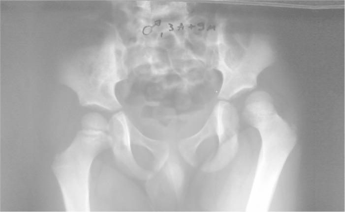
Radiographic appearance of traumatic posterior dislocation of the hip
Figure 2B.

After reduction of the dislocation and without signs of necrosis, seven months later
This study was approved by the Ethics Committees of the respective hospitals, and the parents responsible for these children gave their consent through a free and informed consent statement.
RESULTS
The trauma that resulted in the dislocation was low-energy in four cases (80%). Only one case was high-energy, caused by a fall from a high platform (approximately 2.5 m) (Table 1). The initial treatment consisted of closed reduction under anesthesia, which was performed on average 5.2 ± 3.6 hours after the initial trauma (median of 3.5 hours). Epiphysiolysis of the proximal femur was observed in one case, but after the reduction, the radiographic and tomographic examinations showed spontaneous reduction that remained stable in radioscopic control examinations. It was decided to treat it conservatively, with a plaster cast from the pelvis to the foot. None of the cases presented neurological abnormalities as complications. In the evaluation after the reduction, joint congruence was observed in all the cases, and there were no interpositions or occult associated lesions in the radiographic examinations.
Table 1.
Complementary treatment following closed reduction, among the patients with posterior dislocation of the hip.
| Patient | Gender | Complementary treatment | Duration of immobilization (weeks) | Time taken to restore weight loading(weeks) |
|---|---|---|---|---|
| DBL | M | PCPT | 3 | 4 |
| FFL | F | PCPT | 4 | 8 |
| ASR | F | ST + PCPT | 4 | 4 |
| BCMM | F | ST + PCPT | 4 | 4 |
| EML | F | PCPT | 4 | 4 |
Legend: PCPT – plaster cast from pelvis to foot, ST – skeletal traction, M – male, F – female
Source: SAME – Hospital da Baleia, Hospital Sao Camilo and IMOT
After the complementary treatment, all the children were released for walking without restrictions. Duringthe follow-up, no clinical abnormalities or episodes of renewed dislocation were observed. All the patients presented symmetrical ranges of motion in relation to the contralateral hip, three months after the trauma, and none of them presented residual claudication. The radiographic evolution was satisfactory, with maintenance of the sphericity of the femoral head, acetabular orientation and joint congruence. No cases of chondrolysis, avascular necrosis or joint degeneration were observed during the follow-up. In none of the cases was complementary surgical treatment necessary.
DISCUSSION
Traumatic dislocation of normal hips in children under the age of 16 years(2) is a rare injury, with an incidence of 0.8 cases per million/year, and posterior dislocation accounts for 80% of such cases2, 3, 4, 5, 6, 7, 8, 9, 10, 11. It is more common in boys (4:1)8, 9 and may occur at any age among children, although the peak incidence occurs between four and seven years of age and between 11 and 15 years of age3, 8, 9.
In younger children, the acetabulum is very flexible, loose and cartilaginous1, 2, 3, 4, thus allowing trivial trauma to result in dislocation1, 3, 4, 5, 7, 8, 9, 10, 12. With growth, the cartilage calcifies and the ligament weakness diminishes, such that greater energy is required to dislocate the hip1, 2, 10, 11, 12. Thus, associations between traumatic dislocation of the hip and femoral fractures in children should always be borne in mind(11), especially among older children and adolescents1, 2, 3, 4, 10.
The cases described here presented characteristics similar to those in the literature. We observed that male patients and a mean age of 4.6 years predominated, with lower-energy trauma in most cases, with the exception of one patient who suffered a fall from a height and presented associated proximal epiphysiolysis of the femur.
The clinical diagnosis for this lesion is based on the history of trauma, pain in the hip and inability to walk(2). The typical deformity varied according to the type of dislocation. In posterior dislocation, the leg will be flexed, adducted and internally rotated1, 2, 3, 4, 9. The functioning of the sciatic nerve should be registered before and after the hip reduction3, 4. There is also the possibility of spontaneous but partial reduction of the dislocation, through interposition of soft tissues(13). This situation may not be recognized, thus leading to permanent joint damage1, 12, 14, 15.
Good-quality radiographic evaluation is essential for confirming the diagnosis, revealing the type of dislocation and discarding the hypothesis of associated fractures(2). Fragments from acetabular or femoral fractures are often seen better on radiographs produced before the reduction3, 4. In our case of femoral fracture, the lesion was only evident on the initial radiograph, in agreement with Canale(3).
Traumatic dislocation of the hip in children is an orthopedic emergency7, 11. There is a consensus that the reduction should be performed immediately, preferably using a closed procedure and under general anesthesia9, 16, 17 or with relaxants3, 11, using the same maneuvers as used for reductions in adults (Stimson, Allis and Bigelow)2, 3, 9, 10. After the reduction, the joint congruence should be evaluated, comparing the joint space, lateralization of the head(14) and breakage of Shenton's line(9) with the contralateral side.
In cases of incongruent reduction, computed tomography (CT) and magnetic resonance imaging (MRI) are useful for determining any presence of fragments or interposed tissue(14). If the presence of interposition is confirmed, a second attempt at reduction is made3, 4, or open reduction using a posterior approach is performed3, 4, 10. The indications for open reduction are weakness of the closed reduction, injury to the sciatic nerve with indication for exploration2, 3 and fractures of the acetabulum, femoral neck or femoral head that require surgical treatment(2).
There continues to be no consensus regarding the treatment to be followed after achieving reduction(1). Price et al(12) and Tachdjian(9) recommended using a plaster cast from the pelvis to the foot (PCPF) for four to six weeks, in order to allow the capsule to heal. Gianom et al(6) indicated resting in bed until achieving pain relief, following by walking using crutches and protection of the support for four weeks. Blaster and Hughes(10) proposed resting until the pain improved, followed by a return to walking. Canale(3) and Hebert(2) used cutaneous traction for one week, followed by protection of the support for four to six weeks.
When there is a need for open reduction, skeletal traction (ST) or PCPF is recommended for up to six weeks2, 3, 10. For cases of dislocation that evolved over more than 24 hours, ST prior to reduction is recommended, followed by traction for two to three weeks after the reduction2, 3.
The five cases analyzed here were subjected to closed reduction under anesthesia. Radiographic evaluation to verify joint congruence was performed on all of them. In cases of dislocation associated with femoral fracture, the congruence is confirmed using CT.
Many complications are associated with traumatic dislocation of the hip in children. Nerve injuries occur in around 5% of such children2, 9, and the fibular branch of the sciatic nerve is the one most affected in cases of posterior dislocation due to direct compression(13). Absence of improvement after four to six weeks is an indication for surgical exploration following neurodiagnostic studies(9). If there is loss of function of the sciatic nerve after the reduction, the nerve should be explored surgically(10). Avascular necrosis has an incidence of 8% to 10%, and delays in performing the reduction, high-energy trauma and age greater than five years are factors associated with higher frequency of this complication1, 2, 3, 7. The main risk factor seems to be the time elapsed since the dislocation. Reduction performed after more than six hours of evolution presents a risk of avascular necrosis that is 20 times greater(18). According to Mehlman et al (18), scintigraphic evaluation is unnecessary as routine follow-up. According to Blaster and Hughes(10), it is recommendable for the hip to be evaluated using serial radiographs, at least two years after the dislocation. These authors also did not recommend scintigraphic evaluation or MRI as routine control examinations until reaching skeletal maturity(19). Renewed dislocation is rare and is associated with children younger than eight years of age(10) and children with ligament looseness, especially those with Down syndrome3, 10. Its treatment consists of a new reduction, followed by PCPF for six weeks or capsuloplasty2, 10. Among older children, it may be necessary to associate this with bone procedures such as Salter's operation3, 10 or varus osteotomy(3). Chondrolysis has been reported following traumatic dislocation of the hip, in 6% of the children. It probably results from the joint lesion at the time of dislocation. The treatment should be symptomatic. If joint reconstitution does not occur, arthrodesis or reconstruction should be considered(10). Coxa magna seems to occur as a result of post-traumatic hyperemia. In most of these children, this condition is asymptomatic and does not require any treatment. Ossifying myositis(3) and degenerative arthritis1, 7, 8, 9 are potential sequelae.
The five cases reported here did not present any complications up to the time of the last orthopedic evaluation. Most of the children had good long-term evolution and better results than shown by adults10, 20. A large proportion of such patients (78%) can undertake high-demand activities such as soccer or basketball(18).
CONCLUSION
Traumatic dislocation of the hip occurred in younger children in our case series, with lower-energy trauma and good late evaluation. Despite the satisfactory evolution of our five patients, long-term follow-up needs to be maintained.
Footnotes
Work performed in the Pediatric Orthopedics Group of Belo Horizonte, at Hospital da Baleia (Prof. Matta Machado service), Hospital das Clínicas (UFMG) and Hospital Sao Camilo, Belo Horizonte, and at the Minas Gerais Institute of Orthopedics and Traumatology, Belo Horizonte.
REFERENCES
- 1.Kutty S, Thornes B, Curtin WA, Gilmore MF. Traumatic posterior dislocation of hip in children. Pediatr Emerg Care. 2001;17(1):32–35. doi: 10.1097/00006565-200102000-00009. [DOI] [PubMed] [Google Scholar]
- 2.Hebert S. Fraturas e luxaçies do quadril na criança e no adolescente. In: Hebert S, Xavier R, Pardini Jonior AG, Barros Filho TEP, editors. Ortopedia e Traumatologia – Principios e Prática. 3a. ed. Artmed; Porto Alegre: 2003. pp. 1231–1237. [Google Scholar]
- 3.Canale ST. Luxaçes traumáticas do quadril em crianças. In: Crenshaw AH, editor. Cirurgia Ortopédica de Campbell. 8a. ed. Manole; Sao Paulo: 1996. pp. 1222–1225. [Google Scholar]
- 4.Canale ST, King RE. Luxaçes traumáticas do quadril. In: Rockwood CA Jr, Wilkins KE, King RE, editors. Fraturas em crianças. Tradução de Vilma Ribeiro de Souza Varga. 3a. ed. Manole; São Paulo: 1993. pp. 1061–1089. [Google Scholar]
- 5.Guarniero R, Peixinho M. Luxação traumática do quadril na criança. Rev Bras Ortop. 1990;25(4):93–96. [Google Scholar]
- 6.Gianom D, Kronberger G, Sacher P. Long-term follow-up of traumatic hip dislocation in childhood. Helv Chir Acta. 1994;60(4):623–627. [PubMed] [Google Scholar]
- 7.Hughes MJ, D'Agostino J. Posterior hip dislocation in a five-year-old boy: a case report, review of the literature and current recommendations. J Emerg Med. 1996;14(5):585–590. doi: 10.1016/s0736-4679(96)00131-x. [DOI] [PubMed] [Google Scholar]
- 8.Petrie SG, Harris MB, Willis RB. Traumatic hip dislocation during childhood. A case report and review of the literature. Am J Orthop. 1996;25(9):645–649. [PubMed] [Google Scholar]
- 9.Tachdjian MO. Luxação traumática do quadril. In: Tachdjian MO, editor. Ortopedia pediátrica. Tradução de José Aparecido Lopes. 2a. ed. Manole; Sao Paulo: 1995. pp. 3222–3240. [Google Scholar]
- 10.Blaster RD, Hughes LO. Fractures and traumatic dislocation of the hip in children. In: Beaty JH, Kasser JR, editors. Rockwood & Wilkins Fractures in children. 5th edition. Lippincott Willians & Wilkins; Philadelphia: 2001. pp. 930–938. [Google Scholar]
- 11.Macnicol MF. The Scottish incidence of traumatic dislocation of the hip in childhood. J Pediatr Orthop B. 2000;9(2):122–124. doi: 10.1097/01202412-200004000-00009. [DOI] [PubMed] [Google Scholar]
- 12.Price CT, Phillips JH, Devito DP. Management of fractures. In: Morrisy RT, Weinstein SL, editors. Lovell & Winter's pediatric orthopaedics. 5th edition. Lippincott Willians & Wilkins; Philadelphia: 2001. pp. 1372–1373. [Google Scholar]
- 13.Price CT, Pyevich MT, Knapp DR, Phillips JH, Hawker JJ. Traumatic hip dislocation with spontaneous incomplete reduction: a diagnostic trap. J Orthop Trauma. 2002;16(10):730–735. doi: 10.1097/00005131-200211000-00008. [DOI] [PubMed] [Google Scholar]
- 14.Banskota AK, Spiegel DA, Shrestha S, Shrestha OP, Rajbhandary T. Open reduction for neglected traumatic hip dislocation in children and adolescents. X-ray transparency interpositions after reduction of traumatic dislocations of the hip in children. J Pediatr Orthop. 2007;27(2):187–191. doi: 10.1097/BPO.0b013e31802c547e. [DOI] [PubMed] [Google Scholar]
- 15.Olsson O, Landin LA, Johansson A. Traumatic hip dislocation with spontaneous reduction and capsular interposition. A report of 2 cases. Acta Orthop Scand. 1994;65(4):476–479. doi: 10.3109/17453679408995496. [DOI] [PubMed] [Google Scholar]
- 16.Burgos J, Gonzales-Herrans P, Ocete G. Traumatic hip dislocation with incomplete reduction due to soft tissue interposition in a 4-year-old boy. J Pediatr Orthop B. 1995;4(2):216–218. doi: 10.1097/01202412-199504020-00017. [DOI] [PubMed] [Google Scholar]
- 17.Rieger H, Pennig D, Klein W, Grunert J. Traumatic dislocation of the hip in young children. Arch Orthop Trauma Surg. 1991;110(2):114–117. doi: 10.1007/BF00393886. [DOI] [PubMed] [Google Scholar]
- 18.Mehlman CT, Hubbard GW, Crawford AH, Roy DR, Wall EJ. Traumatic hip dislocation in children. Long-term followup of 42 patients. Clin Orthop Relat Res. 2000;(376):376–379. [PubMed] [Google Scholar]
- 19.Pinheiro PCMS. Uma lesão traumática no quadril da criança. Relato de um caso. Rev Bras Ortop. 1994;29(1/2):44–46. [Google Scholar]
- 20.Salisbury RD, Eastwood DM. Traumatic dislocation of the hip in chlidren. Clin Orthop Relat Res. 2000;(377):377–378. doi: 10.1097/00003086-200008000-00015. [DOI] [PubMed] [Google Scholar]