

Abstract
The authors report the case of a patient with synovial osteochondromatosis of the knee, who had previously been a soccer player.
Keywords: Chondromatosis, Synovial, Athlete, Knee
INTRODUCTION
Synovial osteochondromatosis is an unusual pathological condition characterized by formation of cartilage with synovial metaplasia.
We report the case of a former soccer player who presented osteochondromatosis in his left knee that mimicked a second patella.
CASE REPORT
A 40-year-old white man who was a former soccer player and was born and was living in São Paulo, sought the services of the Sports Traumatology Center (CETE) of the Department of Orthopedics and Traumatology, Federal University of São Paulo (UNIFESP), with a complaint of pain in his left knee that he had had for around six months.
The patient reported that the pain had started around six months earlier and that it had progressed from sporadic pain to become continuous, with high intensity.
For the last two months, the patient had had great difficulty in extending and flexing his left knee.
On physical examination, the patient presented, on inspection, increased volume in the anterior region of the knee and edema (++/4+), pain on palpation of a mass measuring 5.0 cm x 4.0 cm in the infrapatellar region, diminished range of motion and diminished muscle strength.
Radiographs of the knee were requested, and these showed an image of an osteoblastic infrapatellar mass measuring 3.0 x 3.5 cm.
For greater elucidation of the diagnosis, magnetic resonance imaging of the left knee was requested. This showed that there was a well-delimited mass in the infrapatellar region that did not have a malignant appearance.
The patient was treated surgically, with total excision of the mass, which was sent for anatomopathological examination.
We left a splint on the malleolar thigh for analgesia, for one week, and the patient was released for physiotherapy after the splint had been removed.
Two months after the operation, the patient's range of motion in this left knee had returned to normal.
The result from the anatomopathological examination was that this was a case of synovial chondromatosis.
DISCUSSION
Osteochondromatosis is an unusual condition of unknown cause, characterized by formation of cartilage in the synovial membranes.
Synovial osteochondromatosis may be idiopathic or secondary to diseases such as osteoarthrosis, osteochondritis dissecans, chondral fractures and neuropathic arthropathy, among others(1, 2, 3). It generally occurs in a single joint, affecting predominantly the knee, hip or elbow(3, 4, 5).
It affects males twice as often as females, and generally occurs between the ages of 20 and 40 years(6, 7). The patients present pain, edema and limitation of their range of motion.
This is a pathological condition of progressive nature, although rare cases of spontaneous regression have been reported(8).
The treatment is surgical and consists of excision of any intra-articular body and resection of the synovial membranes involved(1, 9, 10).
The histological findings include synovial hyperplasia with foci of cartilaginous metaplasia(1, 4, 10).
In this report, we described a case of synovial chondromatosis of the knee in a former soccer player that mimicked a second patella. The case was diagnosed and treated in accordance with the literature (Figure 1, Figure 2, Figure 3, Figure 4).
Figure 1.
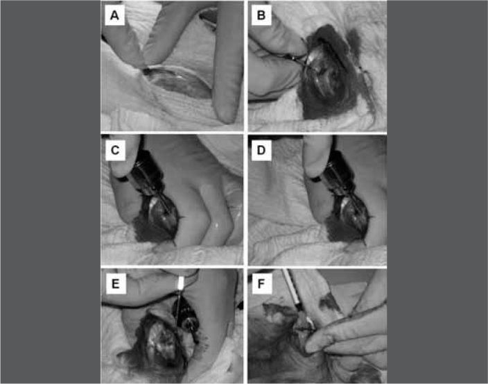
Clinical appearance.
Figure 2.
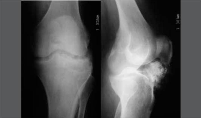
Radiographs (anteroposterior and lateral views).
Figure 3.
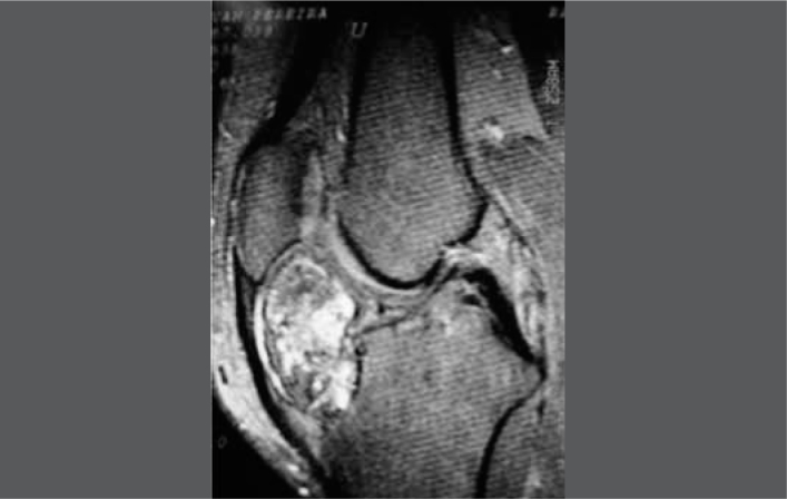
Magnetic resonance imaging.
Figure 4.
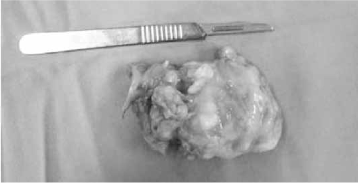
Excised tumor.
Footnotes
The authors declare that there was no conflict of interest in conducting this work
This article is available online in Portuguese and English at the websites:www.rbo.org.brandwww.scielo.br/rbort
REFERENCES
- 1.Koyama J, Ito J, Hayashi T, Kobayashi F. Synovial chondromatosis in the temporomandibular joint complicated by displacement and calcification of the articular disk: report of two cases. AJNR Am J Neuroradiol. 2001;22(6):1203–1206. [PMC free article] [PubMed] [Google Scholar]
- 2.Kramer J, Recht M, Deely DM, Schweitzer M, Pathria MN, Gentili A, Greenway G, Resnick D. MR appearance of idiopathic synovial osteochondromatosis. J Comput Assist Tomogr. 1993;17(5):772–776. doi: 10.1097/00004728-199309000-00020. [DOI] [PubMed] [Google Scholar]
- 3.Murphy FP, Dahlin DC, Sullivan R. Articular synovial chondromatosis. J. Bone Joint Surg Am. 1962;44(1):77–86. [Google Scholar]
- 4.Milgram JW. Synovial osteochondromatosis: a histopathological study of thirty cases. J Bone Joint Surg Am. 1977;59(6):792–801. [PubMed] [Google Scholar]
- 5.McIvor RR, King D. Osteochondromatosis of the hip Joint. J Bone Joint Surg Am. 1962;44(1):87–97. [Google Scholar]
- 6.Resnick S, Niwayama G. Diagnosis of bone and joint disorders. 2nd ed. Sauders; Philadelphia: 1988. [Google Scholar]
- 7.Sekosky M, Lefkowitz H, Steiner I. Osteochondromatosis of the ankle. J Foot Surg. 1990;29(4):330–333. [PubMed] [Google Scholar]
- 8.Swan EF, Owens WF., Jr Synovial chondrometaplasia: a case report with spontaneous regression and a review of the literature. South Med J. 1972;65(12):1496–1500. [PubMed] [Google Scholar]
- 9.Shih WJ, Ryo UY. Synovial osteochondroma of the knee in Tc-99m HMDP bone imaging. Clin Nucl Med. 1988;13(8):617–618. doi: 10.1097/00003072-198808000-00018. [DOI] [PubMed] [Google Scholar]
- 10.Smith R, Hulsey JM. Bone scintigraphic demonstration of synovial chondromatosis. Clin Nucl Med. 1987;12(2):120–122. doi: 10.1097/00003072-198702000-00009. [DOI] [PubMed] [Google Scholar]