

Abstract
Purpose:
Identification of human body or remains after death is a forensic procedure, which is difficult to perform and is mandatory by law and in compliance with social norms. Sexing the recovered human remains is an integral part of the identification process. Maxillary sinus can be used for gender determination as it remains intact even when the skull and other bones may be badly damaged in casualties where the body is incinerated. Computed tomography (CT) provides an excellent method for examining maxillary sinuses.
Materials and Methods:
CT images were used to measure the mediolateral, superoinferior, and anteroposterior dimensions and the volume of the maxillary sinuses in 30 patients (15 males and 15 females) to investigate whether these parameters could be used to determine the gender of an individual for forensic identification. The t-test for independent samples was used to compare these values in males and females and the data were subjected to discriminative analysis using SPSS software.
Results:
Our method was able to predict the gender with an accuracy of 80.0% in males and 86.7% in females, with an overall accuracy rate of 83.3%.
Conclusion:
The accuracy rate in this study was comparable, if not higher than many other methods that have been used to predict the gender of an individual from skeletal remains. The length, width, height, and volume of the maxillary sinuses together with other bones could be used for gender determination with a fair degree of accuracy when the whole skeleton is not available.
Keywords: Forensic dentistry, gender identity, maxillary sinus, computed tomography, X-ray
Introduction
The identity of a human being should be preserved even after death, as there are consequences, often financial, that lead to the court of law, which demands that the identity of the deceased be established before a verdict is passed. Forensic science based evidence is accepted in a judicial setting by the court and plays a major role in the identification of individuals who cannot be identified visually or by other simple means.
Post-mortem identification is a forensic procedure. However, it is difficult to perform after marked post-mortem changes such as decomposition and skeletonization have taken place due to environmental factors such as humidity, temperature, and exposure to microorganisms.[1,2] Nevertheless, a post-mortem is obligatory in terms of the law and social norms.
The identification process involves anthropological analysis for sexing skeletal material, which provides relatively fast and accurate data that help the police investigator narrow down his field of search to a limited geographic area or within a gender.
Gender has been determined from pelvis, skull, and long bones, with assessment of epiphysis and metaphysis in unknown skeletons. The distance between the basion and the prosthion, the circumference of the head, the length of the supraorbital ridge, mastoid process, and mandibular ramus, the shape and length of palate, the circumference of occipital condyle, the sizes of teeth, the length and height of head, the distance between the basion and nasion, the height of the mandibular symphysis, the foramen magnum, the sphenoidal sinus, the sella turcica, and the frontal sinuses have also been used recently for gender determination in unknown remains.[3,4,5,6,7]
It has been reported that maxillary sinuses remain intact despite the skull and other bones getting badly disfigured in victims who are incinerated. Therefore, maxillary sinuses can be used for identification.[8] Jovinic stated that maxillary sinuses reach their mature sizes at about the age of 20 years.[9] During adulthood, their shapes and sizes change, especially due to loss of teeth. The size of the maxillary sinus can be affected due to environmental factors, genetic diseases, or post infections.[10]
Computed tomography (CT) provides an excellent method for examining maxillary sinuses.[11] As the image represents a contiguous series of cross-sections and three-dimensional information and the machine is available in most of the hospitals, CT has been applied in the study of fossil skulls.[11,12,13]
CT scan can be used for determination of gender by measurement of maxillary sinus when other methods are inconclusive, though this method is not error-free.[8]
The aim of the study was to determine the size [mediolateral (ML), superoinferior (SI), and anteroposterior (AP) linear dimensions] and the volume of the maxillary sinuses using CT scanning, and investigate whether these parameters can be used to determine the gender of an individual for forensic identification.
Materials and Methods
The study was designed to measure the ML, SI, and AP dimensions along with the volumes (V) of both the right and the left maxillary sinuses of an individual using CT.
The study was carried out on a small regional population of patients presenting to the imaging center of Institute of Dental Studies and Technologies, Modinagar, Western Uttar Pradesh, India, between December 2009 and August 2011. Patients were selected on the basis of strict inclusion and exclusion criteria that favored intact maxillary sinuses without any pathological, physiological, or surgical deformity. Thirty patients (15 male and 15 female) were included in the study, who were between 20 and 50 years of age, had retained all their permanent teeth, and were referred to the imaging center to have a CT scan of the paranasal sinuses (CT PNS) for various reasons, in which no pathological findings were detected on CT images. Patients who were found to have maxillary sinus pathology on CT scan, facial deformities involving the maxilla, clinical facial asymmetries, or had previously undergone a surgery of the maxillary sinus were excluded from the study.
Non-contrast coronal CT scan was performed on all patients to visualize the maxillary sinuses using GE CT/e Dual Slice CT scanner (GE Healthcare Technologies, Waukesha, WI, USA). Prior to the scan, the patient was instructed to remove all metallic objects including hair pins, jewelry, and so on from the head and neck region and positioned on the CT table in prone position. The patient's neck was hyper-extended with the chin resting on a pad. For stabilization, pads were inserted on both sides of the head. The gantry was angulated to make it perpendicular to the hard palate.
Sections of 3 mm thickness were planned on the preliminary scout view extending from the anterior margin of the frontal sinus to the posterior margin of the sphenoid sinus, with a reconstruction matrix size of 512 × 512 at 120 kV, 100 mA. Coronal CT was performed after instructing the patient to remain steady during the entire procedure. For the purpose of standardization, all scans were performed by the same operator on the same machine using similar exposure parameters. The CT image stack thus acquired was transferred to the Advantage Workstation™ 5 (GE Healthcare Technologies) for post processing.
The ML and SI measurements were made where the maxillary sinus was in its widest position, with the help of the on-screen linear measurement tool on the CT workstation. The linear distance between the two points was annotated on the screen [Figure 1]. To measure the AP dimension of the maxillary sinus, the first and the last appearance of the sinus was noted in the sequential coronal CT sections, and the number of sections between them was determined. Finally, the number of sections obtained was multiplied by 3 (thickness of a single section) to find the AP dimension of the sinus.
Figure 1.

Linear measurement of mediolateral and superoinferior dimensions of maxillary sinus
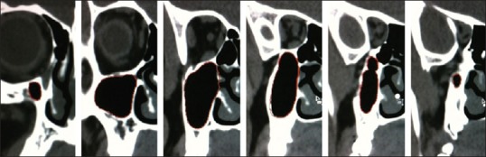
Maxillary sinus volume (V) was calculated using the “Paint on slices” tool on the workstation. To define a volume, the outline of the sinus was traced manually on each slice of the image stack using the on-screen mouse pointer in the coronal plane [Figure 2]. Once the tracing was complete, the workstation automatically segmented the entire volume of the sinus from the surrounding structures and the segmented portion could be visualized and manipulated in 3D [Figure 3].
Figure 2.
“Paint on slice” tool
Figure 3.
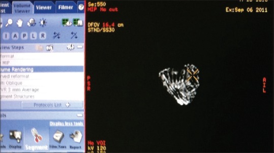
Sinus volume in 3D
At this point, switching to the “Histogram” view on the workstation [Figure 4] automatically reflected the volume of the sinus in cubic centimeters (cc). The entire procedure was repeated for the right and the left maxillary sinuses separately for every patient.
Figure 4.

Workstation showing the sinus volume
The t-test for independent samples was used to compare these values in two groups. Discriminative analysis was used to detect the gender by using the data obtained from CT scans. The analyses were performed using the SPSS ver. 16.0 (SPSS Inc., Chicago, IL, USA).
Results
The study included 30 adult patients consisting of 15 males and 15 females. The patients were distributed into three age groups starting from 20 to 50 years with a class interval of 10 years.
Maxillary sinus dimensions
The mean, along with the standard deviation was calculated for all the dimensions of the right and the left maxillary sinuses, namely ML, SI, and AP, for both males and females [Table 1].
Table 1.
Distribution of maxillary sinus dimensions measured on CT and their standard deviation

For the right maxillary sinus, the mean value of the ML dimension was found to be 27.53 ± 4.26 mm in males and 25.12 ± 6.75 mm in females. The mean of SI dimension was 38.21 ± 5.77 mm in males and 33.34 ± 6.57 mm in females. Also, the mean of AP dimension was 42.60 ± 3.79 mm in males and 36.00 ± 4.09 mm in females. For the left maxillary sinus, the mean value of the ML dimension was found to be 27.01 ± 5.04 mm in males and 23.22 ± 6.21 mm in females. The mean value of the SI dimension was 36.99 ± 4.45 mm in males and 33.11 ± 6.71 mm in females. Also, the mean value of AP was 40.80 ± 2.73 mm in males and 37.20 ± 2.96 mm in females.
A statistically significant (P < 0.05) difference was found in the right SI dimension, left SI dimension, and the left AP dimension of the left maxillary sinuses between males and females. A significant (P < 0.01) difference was found in the right AP dimension of the maxillary sinus between males and females. The other maxillary sinus dimensions showed a pattern of being larger in males than in females; however, the difference was statistically insignificant.
A comparison was made between the dimensions of the right and left maxillary sinus within males and females separately [Table 2]. On non-statistical comparison in males, the right maxillary sinus gave an impression of being slightly larger than the left maxillary sinus in its overall dimensions. Similarly, in females, the right maxillary sinus was marginally larger in dimensions than the left maxillary sinus, with the exception of the AP dimension. Aberrantly, the AP dimension of right maxillary sinus was found to be less than that of the left maxillary sinus in females. However, these intra-gender findings were statistically insignificant (P > 0.05).
Table 2.
Distribution of right and left maxillary sinus dimensions in males (n=15) and females (n=15)

Maxillary sinus volume
The mean with standard deviation was calculated for the volume of the right (VR) and the left (VL) maxillary sinuses for both males and females [Table 3]. For the right maxillary sinus, the mean volume was found to be 16.63 ± 4.54 cc in males and 11.61 ± 5.15 cc in females. For the left maxillary sinus, the mean volume was found to be 15.19 ± 3.94 cc in males and 10.95 ± 4.98 cc in females.
Table 3.
Distribution of maxillary sinus volume measured on CT and its standard deviation
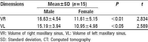
The volume of the maxillary sinuses in males was overall larger than in females. A significant (P < 0.01) statistical difference was found in the right maxillary sinus volume between males and females. A significant (P < 0.05) difference was found in the left maxillary sinus volume between males and females. The maxillary sinuses in males had significantly larger volumes than in females.
A comparison was made between the volume of the right and left maxillary sinus within males and females separately [Table 4]. Overall, both in males and females, the right maxillary sinus volume was found to be larger than the left maxillary sinus volume. However, this intra-gender volume difference was statistically insignificant (P > 0.05).
Table 4.
Distribution of right and left maxillary sinus volume in males and females

Gender determination
A discriminative analysis was performed using the SPSS software to determine whether these linear measurements and volume could be used for gender determination.
The following formula could be used for gender determination from measurements of the right maxillary sinus:
Gender = −5.116 - 0.159×MLR - 0.014 × SIR + 0.171 × APR + 0.218 × VR
where MLR is the ML dimension of the right maxillary sinus, SIR is the SI dimension of the right maxillary sinus, and APR is the AP dimension of the right maxillary sinus.
The accuracy of gender predicted using the linear measurements and volume of the right maxillary sinus was found to be 80.0% in both males and females. Therefore, the overall accuracy of gender prediction was 80.0%.
The following formula can be used for gender determination from measurements of the left maxillary sinus:
Gender = −0.720 - 0.258 × MLL - 0.146 × SIL + 0.120 × APL + 0.586 × VL
where MLL is the ML dimension of the left maxillary sinus, SIL is the SI dimension of the left maxillary sinus, and APL is the AP dimension of the left maxillary sinus.
The accuracy of gender predicted from the linear measurements and volume of the left maxillary sinus was found to be 66.7% in males and 80.0% in females, with an overall gender prediction accuracy of 73.3%.
The following formula can be used for gender determination from measurements of the right and the left maxillary sinuses together:
Gender = 4.033 - 0.101 × MLR - 0.21 × SIR + 0.397 × APR + 0.118 × VR - 0.23 × MLL - 0.014 × SIL - 0.417 × APL + 0.358 × VL
The accuracy of gender predicted from the linear measurements and volume of the right and the left maxillary sinuses together was found to be 80.0% in males and 86.7% in females. The overall accuracy of gender prediction was 83.3% [Table 5].
Table 5.
Gender determination from measurements of right and left maxillary sinuses

When the variables in the formulae derived above are substituted with the desired values, a numeric value is obtained for the “Gender.” When this value is positive, the gender is predicted to be male. A negative value is predicted to be female.
The accuracy of gender prediction, however, was variable. It was observed that the accuracy of gender prediction from the measurements of the right maxillary sinus (80.0%) was more than that of the left maxillary sinus (73.3%). When the measurements of the right and the left maxillary sinuses were accounted together for predicting the gender, the accuracy rate increased to 83.3%.
Discussion
Determination of gender is an important aspect of forensic investigation as it narrows down the investigator's field of search. Various methods which have been discussed below have been used to identify the gender of the human remains from a crime scene or a site of mass disaster.[3,4,5,6,7,8,14,15,16] It has been reported that 100% accuracy can be achieved in gender determination from skeleton, 98.0% from both pelvis and skull, 95.0% from pelvis only or the pelvis and long bones, 90.0–95.0% from both the skull and the long bones, and 80.0–90.0% from the long bones only.[5,17] Morphometric parameters such as mesio-distal and bucco-lingual dimensions of the right permanent teeth were shown to predict the gender with an accuracy of 87.0%.[18] Other parameters such as odontometric differences, root length, and crown diameter have been used to determine the gender with varying degrees of accuracy.[19] The circumference and area of the foramen magnum were used to differentiate the gender with an accuracy of 67.0% and 69.3%, respectively.[16] Nascimento Correia Lima et al., in their study of the linear measurement of palatal bone and skull base showed significant sexual dimorphism, with reliability rates of 63.0% and 65.0%, respectively.[20] A study based on radius and ulna for gender determination showed an accuracy for forearm ranging between 76.0% and 86.0%,[21] whereas Osman showed a sex determination accuracy as high as 96.0%.[22] Eshak in his study of gender determination showed that metacarpals, proximal phalanges, and distal phalanges are sexually dimorphic with an accuracy of 80.0%, 76.6%, and 80.0%, respectively.[23] Three-dimensional volume-rendering reconstructed image of metacarpal gave more accurate result (92.0%). Measurements of hand length and phalanges of the fingers and dimensions of the palm have been used for gender determination with varying degrees of accuracy.[24,25] Various methods involving the soft tissues, such as cheiloscopy (lip prints),[26,27] radiographic methods for gender determination such as distance between the crest of alveolar ridge and the superior margin of mental foramina on orthopantomogram of edentulous individuals, and radiograph of the calcaneus, have been used.[28,29] Torwalt and Robert were able to predict the gender using width of the fourth rib and sternal area on chest radiographs with an accuracy of 95.8% for males and 90.3% for females.[30] Cephalometric analysis on lateral cephalograms seems to be a viable method for gender determination.[31,32] Sex could be determined with an accuracy of 95.6% by making cephalometric plots on lateral teleradiography with an orthodontic software.[31] Hsiao et al., attempted to develop a method to determine sex from lateral cephalometric and discriminant functional analysis and determined sex with 100% accuracy in a random sample of 100 Taiwanese adults.[32]
The studies discussed above for gender determination used plain radiographs, which included lateral cephalograms, and panoramic view for mental foramen, gonial angle, and mandibular ramus, along with ankle and hand–wrist radiographs showed an accuracy levels ranging between 69.0 and 100%.[33,34,35] It seems that plain radiographs have a higher average degree of accuracy in predicting the gender when compared to CT used in our study. However, plain radiographs, along with other conventional methods discussed above can only be used when the bones, skull, or the skeleton are found intact. When human skeleton is found in fragmented or incomplete state, it is necessary to look for denser bones, such as the maxillary sinus, which has a higher possibility of remaining intact by resisting decomposition, fracture, and incineration.
CT has been reported to be a robust method in the estimation of different dimensions of the maxillary sinus.[36] Previous studies have shown that the dimensions of maxillary sinuses from measurements of human skulls were similar to those obtained by CT scans[37] and the consistency of measurements of the paranasal sinuses using CT images have been evaluated in the last decade.[38,39] It may, therefore, be reasonably assumed that CT is a reliable method for measuring the dimensions of the maxillary sinuses. In the literature, the mean values of maxillary sinus measurements were 32, 25, and 35 mm in length, width, and height, respectively.[40]
Our study measured the ML, SI, and AP dimensions of the maxillary sinus on CT in 30 patients including both males (n = 15) and females (n = 15). The right maxillary sinus showed an average size of 27.53 × 38.21 × 42.60 mm in males and 25.12 × 33.34 × 36 mm in females. The left maxillary sinus showed an average size of 27.01 × 36.99 × 40.80 mm in males and 23.22 × 33.11 × 37.20 mm in females.
We found that the overall size of the maxillary sinus was larger in males than in females. These findings were in agreement with those of authors like Fernandes,[41] Teke et al.,[1] and Sahlstrand-Johnason et al.,[36] who have separately reported that the overall size of the maxillary sinus is larger in males than in females. Also, the right maxillary sinus gave an impression of being larger than the left maxillary sinus in overall dimensions in both males and females, but the difference was found to be statistically insignificant.
The volume of the maxillary sinus has been previously measured using CT. Sahlstrand-Johnson et al., reported that the volume of the maxillary sinus can also be accurately estimated by using a simple formula: (ML dimension × SI dimension × AP dimension) divided by 2.[36] Such an estimation of volume is debatable and was not considered for use in this study; however, this tool might be beneficial in clinical practice for approximate estimation of the maxillary sinus volume, where volume measurement applications are not available or precision is not critical. In a study about gender determination, it was found that the volume of maxillary sinuses was larger and wider in males than in females in Europe but narrower in males than in females in Zululand.[42]
Our study used the CT workstation to measure the volume of the right and left maxillary sinuses in males and females by tracing the outline of the sinus manually on each slice of the CT image stack using the on-screen pointer in the coronal plane. The software module on the workstation then automatically calculated the maxillary sinus volume in cubic centimeters. We found that for the right maxillary sinus, the average volume was 16.63 cc in males and 11.61 cc in females. For the left maxillary sinus, the volume was calculated to be 15.19 cc in males and 10.95 cc in females. Therefore, it could be concluded that males have significantly larger maxillary sinus volumes than females. Also, the volume of the right maxillary sinus is slightly larger than the left maxillary sinus in both males and females.
Sahlstrand-Johnson et al.,[36] measured the dimensions of 120 maxillary sinuses from head CTs and found the volume to be larger in males than in females with a mean value of 15.7 ± 5.3 cm3. These authors reported differences in the volume of the maxillary sinus between males and females, and this finding is in accordance with the findings in our study. But they did not find any significant difference between the volumes of the right and the left maxillary sinuses, which is in contrast to our study, as we found that the volume of the right maxillary sinus was larger than that of the left maxillary sinus, though the difference was statistically insignificant.
Teke et al., employed CT to measure the length, width, and height of the maxillary sinus and predicted the gender with an accuracy of 69.4% in females and 69.2% in males and an overall mean accuracy of 69.3%.[1]
Uthman et al., studied the accuracy and reliability of maxillary sinus dimensions measurement in gender classification through the use of reconstructed helical CT images and reported an accuracy of correctly sexing 74.4% of male and 73.3% of female sinuses.[43]
Amin and Hassan investigated the possibility of estimation of sex from some radiologic measurements of the maxillary sinus using multi-detector CT among a known cross-section of Egyptian population, which gave a predictive accuracy of 70.8% in males and 62.5% in females.[44]
Improvising on the CT-based studies, which did not include the volume of the maxillary sinuses, our study measured the volume of the maxillary sinus along with its three other linear dimensions (ML, SI, AP) to predict the gender of an individual. It was observed that if the volume is also included as a variable in the discriminative analysis along with the other three dimensions, the accuracy of gender prediction increases to 80.0% in males and 86.7% in females, with an overall accuracy rate of 83.3%.
Conclusion
Gender determination is an important step in the identification in forensic medicine. The maxillary sinuses remain intact although the skull and other bones may be badly disfigured in victims who are incinerated, and therefore, maxillary sinuses can be used for identification.
CT provides an excellent method for examining maxillary sinuses. CT measurements of maxillary sinuses may be useful to support gender determination in forensic medicine. This study, building upon similar previous studies, demonstrates that the length, width, and height of the maxillary sinus, along with its volume can be used to predict the gender of an individual with a fair degree of accuracy. The accuracy rate in this study is comparable, if not higher than many other methods that have been used to predict the gender of an individual from skeletal remains. However, the result of the study does not prove this method to be infallible. Further studies with larger sample size are required to make this procedure conclusive and achieve standardization.
Footnotes
Source of Support: Nil
Conflict of Interest: None declared
References
- 1.Teke HY, Duran S, Canturk N, Canturk G. Determination of gender by measuring the size of the maxillary sinuses in computerized tomography scans. Surg Radiol Anat. 2007;29:9–13. doi: 10.1007/s00276-006-0157-1. [DOI] [PubMed] [Google Scholar]
- 2.Riepert T, Ulmcke D, Schweden F, Nafe B. Identification of unknown dead bodies by X-ray image comparison of the skull using the X-ray simulation program FoXSIS. Forensic Sci Int. 2001;117:89–98. doi: 10.1016/s0379-0738(00)00452-7. [DOI] [PubMed] [Google Scholar]
- 3.Cameriere R, Ferrante L, Mirtella D, Rollo FU, Cingolani M. Frontal sinuses for identification: Quality of classifications, possible error and potential corrections. J Forensic Sci. 2005;50:770–3. [PubMed] [Google Scholar]
- 4.Günay Y, Altinkök M, Çagdir S, Kirangil B. Gender determination with skull measurements (in Turkish) J Forensic Med. 1997;13:13–9. [Google Scholar]
- 5.Günay Y, Altinkök M, Çagdir S, Sari H. Is foremen magnum size useful for gender determination (in Turkish) Bull Legal Med. 1998;3:41–5. [Google Scholar]
- 6.Rogers TL. Determining the sex of human remains through cranial morphology. J Forensic Sci. 2005;50:493–500. [PubMed] [Google Scholar]
- 7.Quatrehomme G, Fronty P, Sapanet M, Grévin G, Bailet P, Ollier A. Identification by frontal sinus pattern in forensic anthropology. Forensic Sci Int. 1996;83:147–53. doi: 10.1016/s0379-0738(96)02033-6. [DOI] [PubMed] [Google Scholar]
- 8.Lerno P. Identification par le sinus maxillaire. Odontol Leg. 1983;216:39. [PubMed] [Google Scholar]
- 9.Jovanović S, Jelicić N, Kargovska-Klisarova A. Postnatal development and anatomical relationship of the maxillary sinus. Acta Anat (Basel) 1984;118:122–8. [PubMed] [Google Scholar]
- 10.Karakas S, Kavakli A. Morphometric examination of the paranasal sinuses and mastoid air cells using computed tomography. Ann Saudi Med. 2005;25:41–5. doi: 10.5144/0256-4947.2005.41. [DOI] [PMC free article] [PubMed] [Google Scholar]
- 11.Reichs KJ. Quantified comparison of frontal sinus patterns by means of computed tomography. Forensic Sci Int. 1993;61:141–68. doi: 10.1016/0379-0738(93)90222-v. [DOI] [PubMed] [Google Scholar]
- 12.Wind J. Computerized x-ray tomography of fosil hominid skulls. Am J Phys Anthropol. 1984;63:265–82. doi: 10.1002/ajpa.1330630303. [DOI] [PubMed] [Google Scholar]
- 13.Wind J, Zonneveld FW. Computed tomography of an Australopithecus skull (Mrs Ples): A new technique. Naturwissenschaften. 1989;76:325–7. doi: 10.1007/BF00368433. [DOI] [PubMed] [Google Scholar]
- 14.Krishan K, Kanchan T, Sharma A. Sex determination from hand and foot dimensions in North Indian population. J Forensic Sci. 2011;56:453–9. doi: 10.1111/j.1556-4029.2010.01652.x. [DOI] [PubMed] [Google Scholar]
- 15.Gülekon IN, Turgut HB. The external occipital protuberance: Can it be used as a criterion in the determination of sex? J Forensic Sci. 2003;48:513–6. [PubMed] [Google Scholar]
- 16.Uthman AT, Al-Rawi NH, Al-Timimi JF. Evaluation of foramen magnum in gender determination using helical CT scanning. Dentomaxillofac Radiol. 2012;41:197–202. doi: 10.1259/dmfr/21276789. [DOI] [PMC free article] [PubMed] [Google Scholar]
- 17.Krogman WM, Iscan MY. 2nd ed. Springfield, IL: Charles Thomas Publisher; 1986. The Human Skeleton in Forensic Medicine; pp. 189–243. [Google Scholar]
- 18.Garn SM, Cole PE, Wainwright RL, Guire KE. Sex discriminatory effectiveness using combinations of permanent teeth. J Dent Res. 1977;56:697. doi: 10.1177/00220345770560062601. [DOI] [PubMed] [Google Scholar]
- 19.Tak J, Gupta N. An adjunct in sex determination. Guident. 2011;5:94–5. [Google Scholar]
- 20.Nascimento Correia Lima N, Fortes de Oliveira O, Sassi C, Picapedra A, Francesquini L, Jr, Daruge E., Jr Sex determination by linear measurements of palatal bones and skull base. J Forensic Odontostomatol. 2012;30:38–44. [PMC free article] [PubMed] [Google Scholar]
- 21.Barrier IL, L’Abbé EN. Sex determination from the radius and ulna in modern South African sample. Forensic Sci Int. 2008;179:85.e1–7. doi: 10.1016/j.forsciint.2008.04.012. [DOI] [PubMed] [Google Scholar]
- 22.Celbis O, Agritmis H. Estimation of stature and determination of sex from radial and ulna bone lengths in a Turkish corpse sample. Forensic Sci Int. 2006;158:135–9. doi: 10.1016/j.forsciint.2005.05.016. [DOI] [PubMed] [Google Scholar]
- 23.Eshak GA, Ahmed HM, Abdel Gawad EA. Gender determination from hand bones length and volume using multidetector computed tomography: A study in Egyptian people. J Forensic Leg Med. 2011;18:246–52. doi: 10.1016/j.jflm.2011.04.005. [DOI] [PubMed] [Google Scholar]
- 24.Özaslan A, İşcan MY, Özaslan İ, Tuğcu H, Koç S. El Ölçülerinden Cinsiyet Tespiti (Sex determination from the hand) In: Kitabi Cantürk G, Agritmis H., editors. Yillik Adli Toplantilari. Istanbul, Adli Tip Kurumu: Annual Forensic Medicine Meeting Proceeding; 2002. pp. 114–8. [Google Scholar]
- 25.Kanchan T, Rastogi P. Sex determination from hand dimensions of North and South Indians. J Forensic Sci. 2009;54:546–50. doi: 10.1111/j.1556-4029.2009.01018.x. [DOI] [PubMed] [Google Scholar]
- 26.Sharma P, Saxena S, Rathod V. Cheiloscopy: The study of lip prints in sex identification. J Forensic Dent Sci. 2009;1:24–7. [Google Scholar]
- 27.Gondivkar SM, Indurkar A, Degwekar S, Bhowate R. Cheiloscopy for sex determination. J Forensic Dent Sci. 2009;1:56–60. [Google Scholar]
- 28.Rai B. Possible identification marker in orthopantomograms: Edentulous. Middle East J Sci Res. 2007;2:82–3. [Google Scholar]
- 29.Riepert T, Drechsler T, Schild H, Nafe B, Mattern R. Estimation of sex on the basis of radiographs of the calcaneus. Forensic Sci Int. 1996;77:133–40. doi: 10.1016/0379-0738(95)01832-8. [DOI] [PubMed] [Google Scholar]
- 30.Torwalt CR, Hoppa RD. A test of sex determination from measurements of chest radiographs. J Forensic Sci. 2005;50:785–90. [PubMed] [Google Scholar]
- 31.Veyre-Goulet SA, Mercier C, Robin O, Guérin C. Recent human sexual dimorphism study using cephalometric plots on lateral teleradiography and discriminant function analysis. J Forensic Sci. 2008;53:786–9. doi: 10.1111/j.1556-4029.2008.00759.x. [DOI] [PubMed] [Google Scholar]
- 32.Hsiao TH, Chang HP, Liu KM. Sex determination by discriminant function analysis of lateral radiographic cephalometry. J Forensic Sci. 1996;41:792–5. [PubMed] [Google Scholar]
- 33.Upadhyay RB, Upadhyay J, Agarwal P, Rao NN. Analysis of gonial angle in relation to age, gender, and dentition status by radiological and anthropometric methods. J Forensic Dent Sci. 2012;4:29–33. doi: 10.4103/0975-1475.99160. [DOI] [PMC free article] [PubMed] [Google Scholar]
- 34.Chandra A, Singh A, Badni M, Jaiswal R, Agnihotri A. Determination of sex by radiographic analysis of mental foramen in North Indian population. J Forensic Dent Sci. 2013;5:52–5. doi: 10.4103/0975-1475.114556. [DOI] [PMC free article] [PubMed] [Google Scholar]
- 35.Indira AP, Markande A, David MP. Mandibular ramus: An indicator for sex determination-A digital radiographic study. J Forensic Dent sci. 2012;4:58–62. doi: 10.4103/0975-1475.109885. [DOI] [PMC free article] [PubMed] [Google Scholar]
- 36.Sahlstrand-Johnson P, Jannert M, Strömbeck A, Abul-Kasim K. Computed tomography measurements of different dimensions of maxillary and frontal sinuses. BMC Med Imaging. 2011;11:8. doi: 10.1186/1471-2342-11-8. [DOI] [PMC free article] [PubMed] [Google Scholar]
- 37.Ariji Y, Ariji E, Yoshiura K, Kanda S. Computed tomographic indices for maxillary sinus size in comparison with the sinus volume. Dentomaxillofac Radiol. 1996;25:19–24. doi: 10.1259/dmfr.25.1.9084281. [DOI] [PubMed] [Google Scholar]
- 38.Kawarai Y, Fukushima K, Ogawa T, Nishizaki K, Gunduz M, Fujimoto M, et al. Volume quantification of healthy paranasal cavity by three-dimensional CT imaging. Acta Otolaryngol Suppl. 1999;540:45–9. [PubMed] [Google Scholar]
- 39.Pirner S, Tingelhoff K, Wagner I, Westphal R, Rilk M, Wahl FM, et al. CT-based manual segmentation and evaluation of paranasal sinuses. Eur Arch Otorhinolaryngol. 2009;266:507–18. doi: 10.1007/s00405-008-0777-7. [DOI] [PubMed] [Google Scholar]
- 40.Williams PL, Bannister LH, Berry MM, Collins P, Dyson M, Dussek JE, et al. 38th ed. Edinburgh: Churchill Livingstone; 1995. Gray's Anatomy; p. 1637. [Google Scholar]
- 41.Fernandes CL. Volumetric analysis of maxillary sinuses of Zulu and European crania by helical, multislice computed tomography. J Laryngol Otol. 2004;118:877–81. doi: 10.1258/0022215042703705. [DOI] [PubMed] [Google Scholar]
- 42.Fernandes CL. Forensic ethnic identification of crania: The role of the maxillary sinus-A new approach. Am J Forensic Med Pathol. 2004;25:302–13. doi: 10.1097/01.paf.0000146379.85804.da. [DOI] [PubMed] [Google Scholar]
- 43.Uthman AT, Al-Rawi NH, Al-Naaimi AS, Al-Timimi JF. Evaluation of maxillary sinus dimensions in gender determination using helical CT scanning. J Forensic Sci. 2011;56:403–8. doi: 10.1111/j.1556-4029.2010.01642.x. [DOI] [PubMed] [Google Scholar]
- 44.Amin MF, Hassan EI. Sex identification in Egyptian population using Multidetector Computed Tomography of the maxillary sinus. J Forensic Leg Med. 2012;19:65–9. doi: 10.1016/j.jflm.2011.10.005. [DOI] [PubMed] [Google Scholar]