INTRODUCTION
The ear is a high-risk location for skin cancer due to frequent sun exposure. The ear’s anatomical features, such as thin skin overlying cartilage, minimal subcutaneous tissue, and close proximity to subcutaneous lymphatic channels, confer increased risk of invasion and metastasis. Cutaneous neoplasms of the ear are common, and the auricle is the third most common site for basal cell carcinomas (BCCs). Squamous cell carcinomas (SCCs) also commonly develop on the ear, and they may be more prevalent than BCCs.1 They tend to behave aggressively with high rates of recurrence and metastasis.2,3 Due to the occult location of crevices of the external ear and the potential for a lesion to develop on the posterior aspect, late diagnosis is common. The purpose of this study was to review skin malignancies of the ear in our institution over a 5-year period looking at tumor characteristics, demographics, excision margins, recurrence, and metastatic spread.
METHODOLOGY
A retrospective review of 228 patients was carried out of all surgically excised primary cutaneous malignancies of the ear in University Hospital Galway from 2009 to 2014. The electronic histopathological database was used to obtain tumor characteristics and recurrence rates.
RESULTS
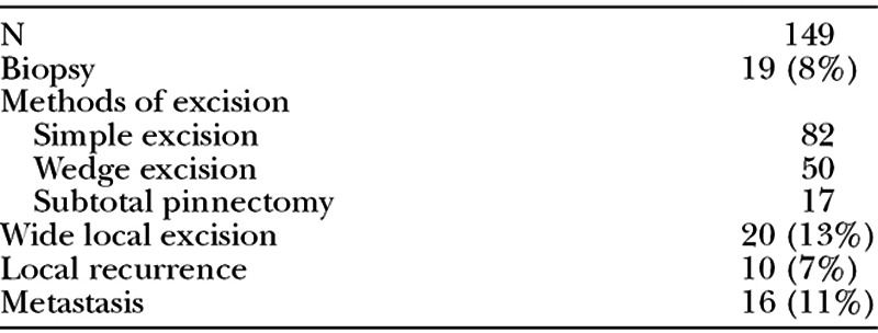
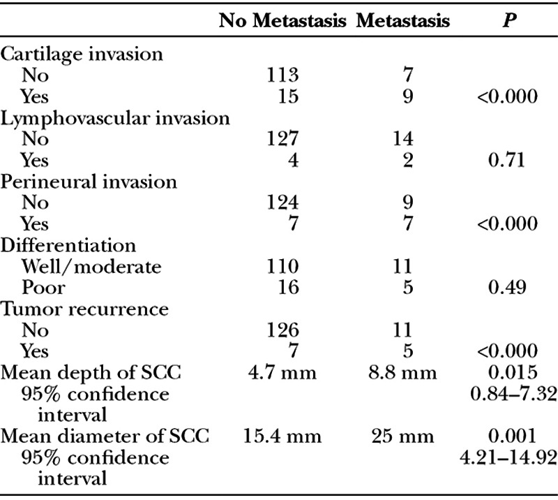
A total of 245 primary cutaneous malignancies of the ear were treated during the 5-year period (94% male and 6% female.) The average age at diagnosis was 75 years. Table 1 details the lesions excised over the period. The most common skin cancer was SCC (61%) followed by BCC (23%). SCCs of the ear were 2.6 times more common than BCCs. Of all lesions excised, 19% required further wide local excision due to incomplete or inadequate margins. Seven percent of patients with SCC and 4% of patients with BCC developed local recurrence, and 11% of patients with SCC and 1 patient with melanoma developed metastases. Increased size and depth, as well as cartilage and perineural invasion, were significantly associated with SCC metastasis (P = 0.001, 0.015, <0.001, and <0.001; Tables 2–4).
Table 1.
Malignancies of the Ear

Table 2.
Squamous Cell Carcinomas of the Ear

Table 4.
Basal Cell Carcinomas of the Ear

DISCUSSION
Malignancies of the ear account for a significant portion of skin cancers. We have shown that SCC is the most common skin cancer of the ear. All subtypes of auricular cancer, especially SCCs, behave more aggressively compared with other anatomical locations. In keeping with other studies, high-risk features associated with SCCs include increased tumor size and depth, invasion of cartilage, perineural invasion, and an SCC located on the ear.2 The skin of the ear seems to behave differently. Overall, it is thought that thin skin, lack of subcutaneous adipose tissue, and close underlying cartilage contribute to a wider subclinical extension and horizontal growth along the dermis and perichondrium, often making initial adequate resection difficult by conventional methods.4 It is a high-risk site for incomplete excision.5 Lesions of the auricle and periauricular regions also have a tendency to spread along the embryologic fusion planes of the ear.6 This may increase the rates of incomplete excision. Due to the increased prevalence of subclinical extension, wide margins are required when excising suspicious lesions. They necessitate urgent treatment and may require re-excision or adjuvant treatment.
Table 3.
Factors Associated with Metastasis of Auricular SCC

Footnotes
Presented at the Irish Association of Plastic Surgeons Summer Meeting, May 14-15 2015, Galway, Ireland.
Disclosure: The authors have no financial interest to declare in relation to the content of this article. The Article Processing Charge was paid for by the Irish Association of Plastic Surgeons.
IAPS: Irish Association of Plastic Surgeons (IAPS) Summer Meeting, in Galway, Ireland, May 14-15, 2015.
REFERENCES
- 1.Ahmad I, Das Gupta AR. Epidemiology of basal cell carcinoma and squamous cell carcinoma of the pinna. J Laryngol Otol. 2001;115:85–86. doi: 10.1258/0022215011907497. [DOI] [PubMed] [Google Scholar]
- 2.Rowe DE, Carroll RJ, Day CL., Jr Prognostic factors for local recurrence, metastasis, and survival rates in squamous cell carcinoma of the skin, ear, and lip. Implications for treatment modality selection. J Am Acad Dermatol. 1992;26:976–990. doi: 10.1016/0190-9622(92)70144-5. [DOI] [PubMed] [Google Scholar]
- 3.Byers R, Kesler K, Redmon B, et al. Squamous carcinoma of the external ear. Am J Surg. 1983;146:447–450. doi: 10.1016/0002-9610(83)90228-3. [DOI] [PubMed] [Google Scholar]
- 4.Gal TJ, Futran ND, Bartels LJ, et al. Auricular carcinoma with temporal bone invasion: outcome analysis. Otolaryngol Head Neck Surg. 1999;121:62–65. doi: 10.1016/s0194-5998(99)70126-9. [DOI] [PubMed] [Google Scholar]
- 5.Bogdanov-Berezovsky A, Cohen AD, Glesinger R, et al. Risk factors for incomplete excision of basal cell carcinomas. Acta Derm Venereol. 2004;84:44–47. doi: 10.1080/00015550310020585. [DOI] [PubMed] [Google Scholar]
- 6.Mohs F, Larson P, Iriondo M. Micrographic surgery for the microscopically controlled excision of carcinoma of the external ear. J Am Acad Dermatol. 1988;19:729–737. doi: 10.1016/s0190-9622(88)70229-7. [DOI] [PubMed] [Google Scholar]


