
Figure 2.
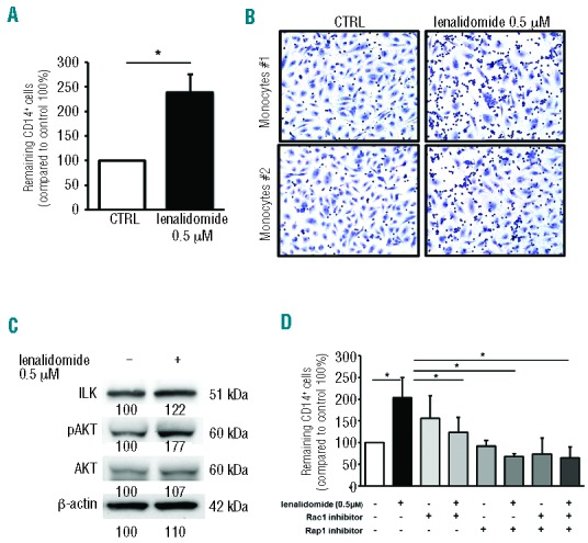
Lenalidomide promotes CLL monocytes adhesion to endothelium. (A) CD14+ monocytes from CLL patients (n=10) were allowed to adhere to HUVEC layer for 2 h and then treated with lenalidomide 0.5 μM or vehicle (DMSO). Histograms represent the mean relative adhesion of lenalidomidetreated monocytes compared to the untreated control. Data represent 8 independent experiments. Columns and error bars represent mean±SEM (Student t-test, *P<0.05). (B) Representative May-Grunwald Giemsa staining shows CLL monocytes adhesion to HUVEC in presence or absence of lenalidomide 0.5 μM. (C) Western blot analysis of CLL CD14+ monocytes was performed with ILK, anti-phospho Akt, total Akt and β-actin antibodies after 4 h of treatment with lenalidomide. The immunoblots show ILK and Akt activation in one representative case. Densitometric quantification of bands normalized to the untreated control is shown below the immunoblots. (D) Monocytes from 6 CLL patients were treated with Rac1 inhibitor or Rap1 inhibitor or both for 30 min before stimulation with lenalidomide 0.5 μM for an additional 20 min. Histograms represent the mean relative adhesion to HUVEC. (Student t-test, *P<0.05).