

Abstract
Using site-directed mutagenesis in conjunction with NMR structural data on the adhesion domain of human CD2, we have defined the binding region for CD58. Previous structural studies of rat and human CD2 indicate that this adhesion domain is immunoglobulin-like. Here we report that the CD58 binding site is a well-circumscribed, charged surface area covering approximately 770 A2 on the AGFCC'C" face of the CD2 beta barrel. This site contains beta-strand residues in the carboxyl-terminal half of the F strand (including Lys-82 and Tyr-86), the top of the C strand (Asp-32 and Lys-34), and the C' strand (Gln-46), which are all solvent exposed. In addition, several exposed residues on the FG loop (Gly-90, Lys-91, Asn-92, and Val-93), the CC' loop (Lys-41 and Lys-43), and the C'C" loop (Arg-48 and Lys-51) form this site. In contrast, neither residues on the more peripheral G and C" strands of the same CD2 surface nor residues on B, E, and D strands of the opposite face are involved in CD58 binding. This CD58 binding site is predicted to lie most distal to the T-lymphocyte surface membrane, with ready access to CD58 on the surface of the opposing antigen-presenting cell.
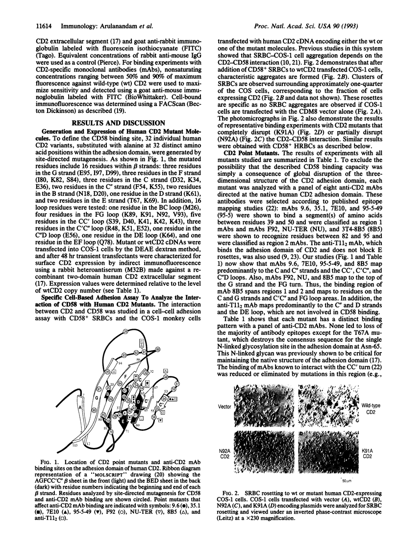
Full text
PDF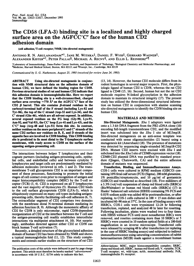
Images in this article

Selected References
These references are in PubMed. This may not be the complete list of references from this article.
- Amit A. G., Mariuzza R. A., Phillips S. E., Poljak R. J. Three-dimensional structure of an antigen-antibody complex at 2.8 A resolution. Science. 1986 Aug 15;233(4765):747–753. doi: 10.1126/science.2426778. [DOI] [PubMed] [Google Scholar]
- Amzel L. M., Poljak R. J. Three-dimensional structure of immunoglobulins. Annu Rev Biochem. 1979;48:961–997. doi: 10.1146/annurev.bi.48.070179.004525. [DOI] [PubMed] [Google Scholar]
- Arulanandam A. R., Koyasu S., Reinherz E. L. T cell receptor-independent CD2 signal transduction in FcR+ cells. J Exp Med. 1991 Apr 1;173(4):859–868. doi: 10.1084/jem.173.4.859. [DOI] [PMC free article] [PubMed] [Google Scholar]
- Arulanandam A. R., Moingeon P., Concino M. F., Recny M. A., Kato K., Yagita H., Koyasu S., Reinherz E. L. A soluble multimeric recombinant CD2 protein identifies CD48 as a low affinity ligand for human CD2: divergence of CD2 ligands during the evolution of humans and mice. J Exp Med. 1993 May 1;177(5):1439–1450. doi: 10.1084/jem.177.5.1439. [DOI] [PMC free article] [PubMed] [Google Scholar]
- Bentley G. A., Boulot G., Riottot M. M., Poljak R. J. Three-dimensional structure of an idiotope-anti-idiotope complex. Nature. 1990 Nov 15;348(6298):254–257. doi: 10.1038/348254a0. [DOI] [PubMed] [Google Scholar]
- Driscoll P. C., Cyster J. G., Campbell I. D., Williams A. F. Structure of domain 1 of rat T lymphocyte CD2 antigen. Nature. 1991 Oct 24;353(6346):762–765. doi: 10.1038/353762a0. [DOI] [PubMed] [Google Scholar]
- Howard F. D., Ledbetter J. A., Wong J., Bieber C. P., Stinson E. B., Herzenberg L. A. A human T lymphocyte differentiation marker defined by monoclonal antibodies that block E-rosette formation. J Immunol. 1981 Jun;126(6):2117–2122. [PubMed] [Google Scholar]
- Jones E. Y., Davis S. J., Williams A. F., Harlos K., Stuart D. I. Crystal structure at 2.8 A resolution of a soluble form of the cell adhesion molecule CD2. Nature. 1992 Nov 19;360(6401):232–239. doi: 10.1038/360232a0. [DOI] [PubMed] [Google Scholar]
- Kato K., Koyanagi M., Okada H., Takanashi T., Wong Y. W., Williams A. F., Okumura K., Yagita H. CD48 is a counter-receptor for mouse CD2 and is involved in T cell activation. J Exp Med. 1992 Nov 1;176(5):1241–1249. doi: 10.1084/jem.176.5.1241. [DOI] [PMC free article] [PubMed] [Google Scholar]
- Koyasu S., Lawton T., Novick D., Recny M. A., Siliciano R. F., Wallner B. P., Reinherz E. L. Role of interaction of CD2 molecules with lymphocyte function-associated antigen 3 in T-cell recognition of nominal antigen. Proc Natl Acad Sci U S A. 1990 Apr;87(7):2603–2607. doi: 10.1073/pnas.87.7.2603. [DOI] [PMC free article] [PubMed] [Google Scholar]
- Krensky A. M., Sanchez-Madrid F., Robbins E., Nagy J. A., Springer T. A., Burakoff S. J. The functional significance, distribution, and structure of LFA-1, LFA-2, and LFA-3: cell surface antigens associated with CTL-target interactions. J Immunol. 1983 Aug;131(2):611–616. [PubMed] [Google Scholar]
- Makgoba M. W., Sanders M. E., Shaw S. The CD2-LFA-3 and LFA-1-ICAM pathways: relevance to T-cell recognition. Immunol Today. 1989 Dec;10(12):417–422. doi: 10.1016/0167-5699(89)90039-X. [DOI] [PubMed] [Google Scholar]
- Meuer S. C., Hussey R. E., Fabbi M., Fox D., Acuto O., Fitzgerald K. A., Hodgdon J. C., Protentis J. P., Schlossman S. F., Reinherz E. L. An alternative pathway of T-cell activation: a functional role for the 50 kd T11 sheep erythrocyte receptor protein. Cell. 1984 Apr;36(4):897–906. doi: 10.1016/0092-8674(84)90039-4. [DOI] [PubMed] [Google Scholar]
- Miller G. T., Hochman P. S., Meier W., Tizard R., Bixler S. A., Rosa M. D., Wallner B. P. Specific interaction of lymphocyte function-associated antigen 3 with CD2 can inhibit T cell responses. J Exp Med. 1993 Jul 1;178(1):211–222. doi: 10.1084/jem.178.1.211. [DOI] [PMC free article] [PubMed] [Google Scholar]
- Moebius U., Clayton L. K., Abraham S., Diener A., Yunis J. J., Harrison S. C., Reinherz E. L. Human immunodeficiency virus gp120 binding C'C" ridge of CD4 domain 1 is also involved in interaction with class II major histocompatibility complex molecules. Proc Natl Acad Sci U S A. 1992 Dec 15;89(24):12008–12012. doi: 10.1073/pnas.89.24.12008. [DOI] [PMC free article] [PubMed] [Google Scholar]
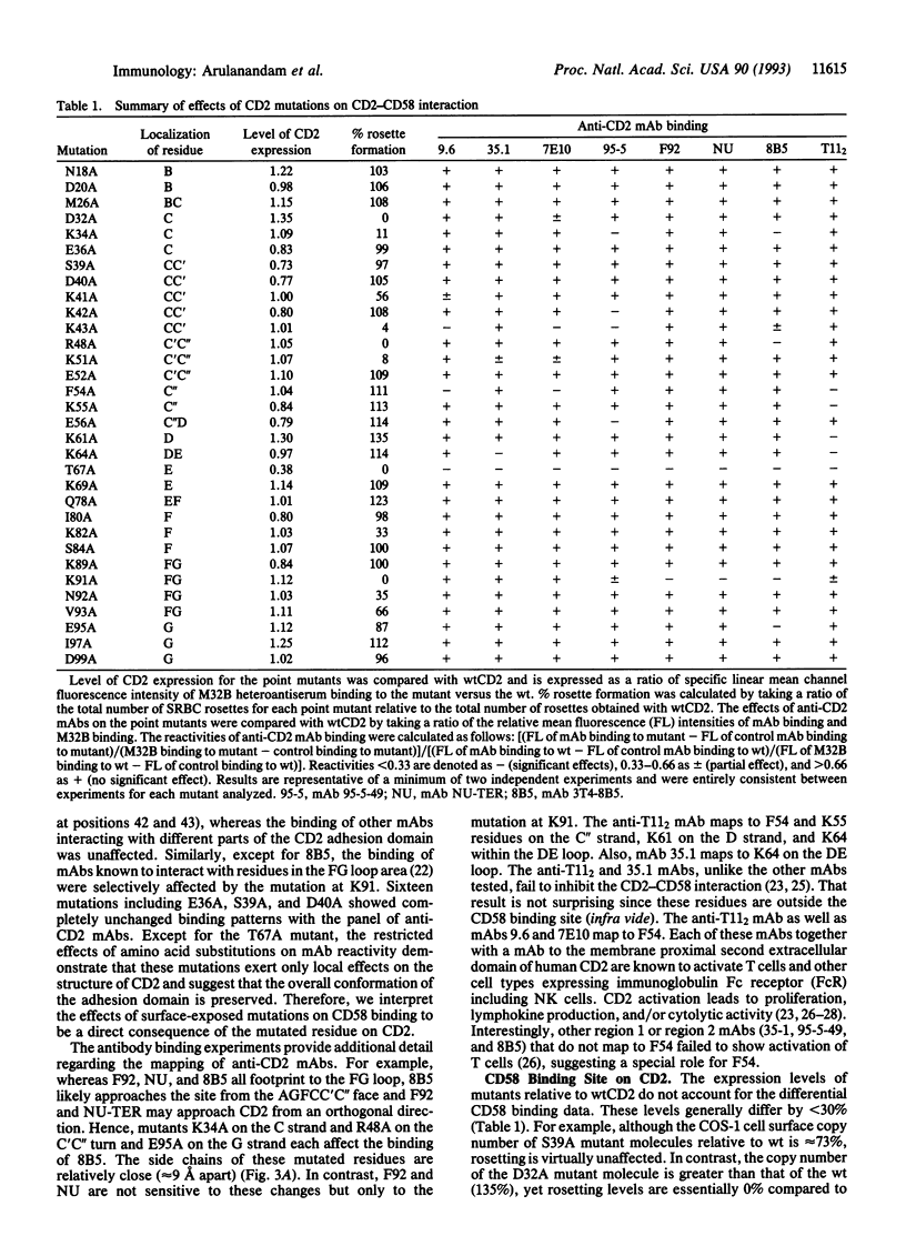
- Moebius U., Pallai P., Harrison S. C., Reinherz E. L. Delineation of an extended surface contact area on human CD4 involved in class II major histocompatibility complex binding. Proc Natl Acad Sci U S A. 1993 Sep 1;90(17):8259–8263. doi: 10.1073/pnas.90.17.8259. [DOI] [PMC free article] [PubMed] [Google Scholar]
- Moingeon P., Chang H. C., Wallner B. P., Stebbins C., Frey A. Z., Reinherz E. L. CD2-mediated adhesion facilitates T lymphocyte antigen recognition function. Nature. 1989 May 25;339(6222):312–314. doi: 10.1038/339312a0. [DOI] [PubMed] [Google Scholar]
- Peterson A., Seed B. Monoclonal antibody and ligand binding sites of the T cell erythrocyte receptor (CD2). 1987 Oct 29-Nov 4Nature. 329(6142):842–846. doi: 10.1038/329842a0. [DOI] [PubMed] [Google Scholar]
- Recny M. A., Luther M. A., Knoppers M. H., Neidhardt E. A., Khandekar S. S., Concino M. F., Schimke P. A., Francis M. A., Moebius U., Reinhold B. B. N-glycosylation is required for human CD2 immunoadhesion functions. J Biol Chem. 1992 Nov 5;267(31):22428–22434. [PubMed] [Google Scholar]
- Recny M. A., Neidhardt E. A., Sayre P. H., Ciardelli T. L., Reinherz E. L. Structural and functional characterization of the CD2 immunoadhesion domain. Evidence for inclusion of CD2 in an alpha-beta protein folding class. J Biol Chem. 1990 May 25;265(15):8542–8549. [PubMed] [Google Scholar]
- Sanders S. K., Fox R. O., Kavathas P. Mutations in CD8 that affect interactions with HLA class I and monoclonal anti-CD8 antibodies. J Exp Med. 1991 Aug 1;174(2):371–379. doi: 10.1084/jem.174.2.371. [DOI] [PMC free article] [PubMed] [Google Scholar]
- Sayre P. H., Chang H. C., Hussey R. E., Brown N. R., Richardson N. E., Spagnoli G., Clayton L. K., Reinherz E. L. Molecular cloning and expression of T11 cDNAs reveal a receptor-like structure on human T lymphocytes. Proc Natl Acad Sci U S A. 1987 May;84(9):2941–2945. doi: 10.1073/pnas.84.9.2941. [DOI] [PMC free article] [PubMed] [Google Scholar]
- Sayre P. H., Hussey R. E., Chang H. C., Ciardelli T. L., Reinherz E. L. Structural and binding analysis of a two domain extracellular CD2 molecule. J Exp Med. 1989 Mar 1;169(3):995–1009. doi: 10.1084/jem.169.3.995. [DOI] [PMC free article] [PubMed] [Google Scholar]
- Seed B. An LFA-3 cDNA encodes a phospholipid-linked membrane protein homologous to its receptor CD2. 1987 Oct 29-Nov 4Nature. 329(6142):840–842. doi: 10.1038/329840a0. [DOI] [PubMed] [Google Scholar]
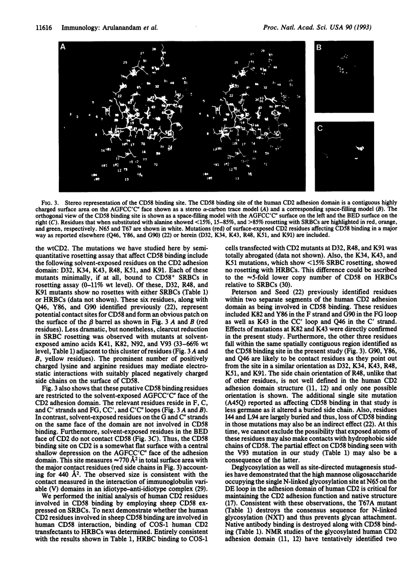
- Seed B., Aruffo A. Molecular cloning of the CD2 antigen, the T-cell erythrocyte receptor, by a rapid immunoselection procedure. Proc Natl Acad Sci U S A. 1987 May;84(10):3365–3369. doi: 10.1073/pnas.84.10.3365. [DOI] [PMC free article] [PubMed] [Google Scholar]
- Selvaraj P., Plunkett M. L., Dustin M., Sanders M. E., Shaw S., Springer T. A. The T lymphocyte glycoprotein CD2 binds the cell surface ligand LFA-3. 1987 Mar 26-Apr 1Nature. 326(6111):400–403. doi: 10.1038/326400a0. [DOI] [PubMed] [Google Scholar]
- Siliciano R. F., Pratt J. C., Schmidt R. E., Ritz J., Reinherz E. L. Activation of cytolytic T lymphocyte and natural killer cell function through the T11 sheep erythrocyte binding protein. Nature. 1985 Oct 3;317(6036):428–430. doi: 10.1038/317428a0. [DOI] [PubMed] [Google Scholar]
- Somoza C., Driscoll P. C., Cyster J. G., Williams A. F. Mutational analysis of the CD2/CD58 interaction: the binding site for CD58 lies on one face of the first domain of human CD2. J Exp Med. 1993 Aug 1;178(2):549–558. doi: 10.1084/jem.178.2.549. [DOI] [PMC free article] [PubMed] [Google Scholar]
- Springer T. A. Adhesion receptors of the immune system. Nature. 1990 Aug 2;346(6283):425–434. doi: 10.1038/346425a0. [DOI] [PubMed] [Google Scholar]
- Staunton D. E., Fisher R. C., LeBeau M. M., Lawrence J. B., Barton D. E., Francke U., Dustin M., Thorley-Lawson D. A. Blast-1 possesses a glycosyl-phosphatidylinositol (GPI) membrane anchor, is related to LFA-3 and OX-45, and maps to chromosome 1q21-23. J Exp Med. 1989 Mar 1;169(3):1087–1099. doi: 10.1084/jem.169.3.1087. [DOI] [PMC free article] [PubMed] [Google Scholar]
- Tiefenthaler G., Dustin M. L., Springer T. A., Hünig T. Serologic cross-reactivity of T11 target structure and lymphocyte function-associated antigen 3. Evidence for structural homology of the sheep and human ligands of CD2. J Immunol. 1987 Oct 15;139(8):2696–2701. [PubMed] [Google Scholar]
- Withka J. M., Wyss D. F., Wagner G., Arulanandam A. R., Reinherz E. L., Recny M. A. Structure of the glycosylated adhesion domain of human T lymphocyte glycoprotein CD2. Structure. 1993 Sep 15;1(1):69–81. doi: 10.1016/0969-2126(93)90009-6. [DOI] [PubMed] [Google Scholar]
- Wyss D. F., Withka J. M., Knoppers M. H., Sterne K. A., Recny M. A., Wagner G. 1H resonance assignments and secondary structure of the 13.6 kDa glycosylated adhesion domain of human CD2. Biochemistry. 1993 Oct 19;32(41):10995–11006. doi: 10.1021/bi00092a008. [DOI] [PubMed] [Google Scholar]
- van der Merwe P. A., McPherson D. C., Brown M. H., Barclay A. N., Cyster J. G., Williams A. F., Davis S. J. The NH2-terminal domain of rat CD2 binds rat CD48 with a low affinity and binding does not require glycosylation of CD2. Eur J Immunol. 1993 Jun;23(6):1373–1377. doi: 10.1002/eji.1830230628. [DOI] [PubMed] [Google Scholar]