Abstract
IL-2 is a lymphocyte growth factor that is an important component of many immune-based cancer therapies. The efficacy of IL-2 is thought to be limited by the expansion of T regulatory cells, which express the high affinity IL-2 receptor subunit, IL-2Rα. IL-15 is under investigation as an alternative to IL-2. Although both cytokines signal through IL-2Rβγ, IL-15 does not bind IL-2Rα and therefore induces less T regulatory cell expansion. However, we found that transferred effector CD8+ T cells induced curative responses in lymphoreplete mice only with IL-2-based therapy. While conventional in vitro assays showed similar effector T cell responsiveness to IL-2 and IL-15, upon removal of free cytokine, IL-2 mediated sustained signaling dependent on IL-2Rα. Mechanistically, IL-2Rα sustained signaling by promoting a cell-surface IL-2 reservoir and recycling of IL-2 back to the cell surface. Our results demonstrate that IL-2Rα endows T cells with the ability to compete temporally for limited IL-2 via mechanisms beyond ligand affinity. These results suggest that strategies to enhance IL-2Rα expression on tumor-reactive lymphocytes may facilitate the development of more effective IL-2-based therapies.
Introduction
The administration of IL-2 is an important component of many cancer immune therapy strategies including adoptive T cell transfer (1–4). Despite its widespread use, the efficacy of IL-2 is limited by short half-life, toxicity, and expansion of IL-2Rαhi T regulatory cells. IL-15 is a promising alternative. Like IL-2, IL-15 signals exclusively through the intermediate affinity IL-2Rβγ subunits (CD122/CD132). However, for high affinity cytokine binding, IL-2 and IL-15 utilize specific IL-2Rα (CD25) and IL-15Rα subunits. This differential α-chain dependence likely dictates the distinct biological outcomes associated with IL-2 and IL-15 (5, 6). In the case of the latter, membrane-bound IL-15Rα can lead to the recycling of IL-15, which sustains cellular signaling and lymphocyte survival (7). However, despite homology with IL-15Rα (8), IL-2Rα is not thought to facilitate sustained signaling or cytokine recycling due to lower affinity for IL-2 (2–4). While briefly expressed on activated lymphocytes, IL-2Rα is constitutively highly expressed on T regulatory cells. For this reason, IL-2 but not IL-15 is essential for T regulatory cell survival and expansion, and mice deficient in IL-2 or IL-2Rα develop T cell-mediated autoimmunity (9, 10). In contrast, mice deficient in IL-15 or IL-15Rα are relatively healthy with reduced frequencies of CD8+ memory-phenotype cells and NK cells (11, 12). Therefore, given the potential undesirable consequences of engaging the IL-2Rα pathway, we hypothesized that IL-15-based therapy would most efficiently augment the efficacy of adoptively transferred tumor-reactive effector CD8+ T cells, particularly in lymphoreplete mice with an intact T regulatory cell population.
Results
IL-2- but not IL-15- therapy mediates anti-tumor immunity after adoptive transfer of activated CD8+ T cells
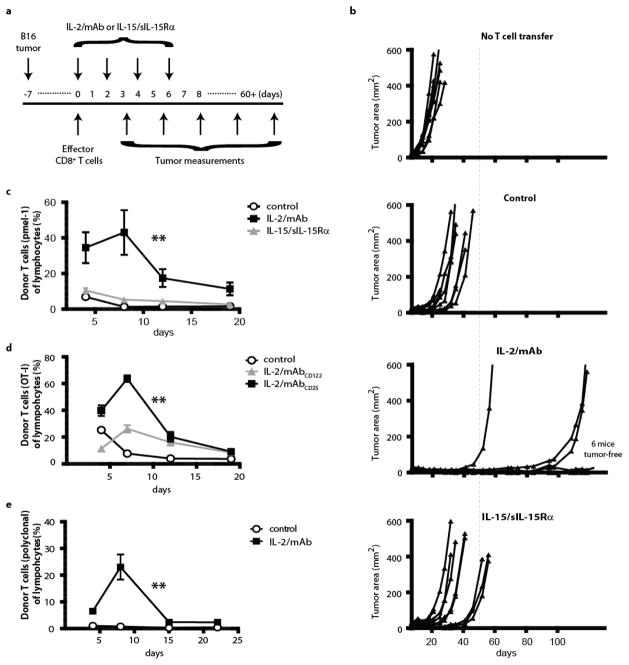
To assess the impact of cytokine therapy on adoptively transferred effector CD8+ T cells, we used IL-2/anti-IL-2 mAb (IL-2/mAb) and IL-15/sIL-15Rα-Fc (IL-15/sIL-15Rα) complexes, in which the antibody or receptor acts as a carrier molecule to improve the half-life and biological activity of free cytokine in vivo (13–15). To test effector T cell responsiveness to cytokines in a clinically relevant model, B6 mice were injected (s.c.) with B16 melanoma tumor cells (Fig. 1a). After the establishment of palpable tumors, unirradiated mice received activated IL-12-conditioned T cells (Tc1) from pmel-1 TCR transgenic mice, from which CD8+ T cells recognize an endogenous B16 tumor antigen (H-2Db-restricted gp10025–33 peptide). We have shown these Tc1 effector cells are highly efficacious against tumor in lymphodepleted mice (16). For the first week after adoptive transfer, IL-15/sIL-15Rα or IL-2/mAb (clone 5355) complexes were administered every 48 hours. While 6 of 9 mice that received IL-2/mAb complexes were cured of established tumor, mice that received either IL-15/sIL-15Rα complexes or no cytokine therapy showed no tumor regression (Fig. 1b). To better understand this differential response, we assessed the persistence of donor Tc1 cells in recipients that received treatment with IL-2/mAb complexes or IL-15/sIL-15Rα complexes. Independent of the presence of tumor, only IL-2/mAb complexes enhanced the persistence of effector CD8+ T cells in a systemic fashion across multiple organs (Fig. 1c and Supplementary Fig. 1a). Notably, without lymphodepletion or vaccination, we routinely achieved sustained donor T cell frequencies of 20% or higher in the peripheral blood. Furthermore, donor Tc1 cells were equally functional across treatment groups as indicated by the ability to produce IFNγ and TNFα (Supplementary Fig. 1b). Finally, as a control, we found that the transfer of tumor-reactive effector CD8+ T cells was necessary for curative therapy. Thus, tumor-bearing mice treated with only IL-2/mAb or IL-15/sIL-15Rα complexes exhibited minimally delayed tumor growth, albeit comparable between cytokine conditions (Supplementary Fig. 2).
Figure 1. IL-2/mAb but not IL-15/sIL-15Rα complexes induce potent effector T cell responses in tumor-bearing mice.
(a) Treatment scheme for B6 mice injected s.c. with B16 melanoma tumor cells 7 days prior to the adoptive transfer of 3x10⁶ pmel-1 Tc1 cells. Mice were then treated with hIL-2/mAb (clone 5355) or hIL-15/sIL-15Rα complexes. (b) Tumor volume from ‘a’ (n=9/group); each line represents one mouse. (*) Based on a log-rank test and time to sacrifice (at 400mm2) for analysis, mice treated with IL-2/mAb complexes had significantly improved outcomes versus each other condition (p<0.001 for each comparison). The average tumor areas when treatment was initiated ranged between 15–20mm2 between the 4 groups. (c) The frequency of donor Tc1 cells in the blood of mice (n=4/group) treated as in ‘a’ but in the absence of tumor. Each point represents the average and bars indicate standard error. (d) The frequency of donor OT-I Tc1 cells in the blood of mice (n=5/group) treated with mIL-2/mAbCD122 (clone S4B6) or mIL-2/mAbCD25 (clone 1A12). Each point represents the average and bars indicate standard error. (e) The frequency of donor polyclonal T cells in the blood of mice (n=5/group) treated with hIL-2/mAb (clone 5355) complexes or vehicle alone. Each point represents the average and bars indicate standard error. For c-e, (**) indicates a significant difference (p<0.001) between indicated and other conditions. Random effects linear regression was used for modeling data and calculating p-values comparing conditions. All results are representative of at least 2 independent experiments.
Donor T cell expression of IL-2Rα is critical for preferential IL-2-mediated responses
The preferential response of effector CD8+ T cells to IL-2/mAb but not IL-15/sIL-15Rα complexes was contrary to our expectation. This response was not dose related as IL-2/mAb and IL-15/sIL-15Rα complexes expanded IL-2Rβγhi cells such as memory-phenotype CD8+ T cells and NK cells to a similar extent in vivo (Supplementary Fig. 3) (17). However, only IL-2/mAb complexes expanded T regulatory cells (Supplementary Fig. 3), which are characterized by their expression of IL-2Rα. As IL-12-conditioned (Tc1) effector CD8+ T cells express very high levels of IL-2Rα (16), our results suggested an unappreciated role for cell surface IL-2Rα on effector T cells in dictating responsiveness to IL-2 therapy. To formally test this, we made use of two anti-IL-2 mAbs with the ability to differentially redirect IL-2 based on lymphocyte cell surface IL-2Rα expression. IL-2/mAbCD25 complexes (clone 1A12) preferentially expand IL-2Rαhi lymphocytes, while IL-2/mAbCD122 complexes (clone S4B6) act in an IL-2Rα-independent manner (15, 18). We tested these two complexes in lymphoreplete mice injected with Tc1 cells. For only this experiment, we generated Tc1 cells from another TCR transgenic mouse, OT-I, to confirm our results with a different TCR. While IL-2/mAbCD122 complexes mediated a minimal increase in persistence, IL-2/mAbCD25 complexes induced donor T cell levels of greater than 60% of total lymphocytes (Fig. 1d). To further confirm that this effect was dependent on IL-2Rα and not on IL-12 conditioning or selective TCR engagement, we stimulated polyclonal T cells from wildtype mice with plate-bound anti-CD3 mAb, a method that generates IL-2Rαhi effector CD8+ T cells. Upon adoptive transfer into lymphoreplete mice, IL-2/mAb complexes (clone 5355) greatly enhanced the persistence of polyclonal T cells (Fig. 1e). Finally, as an additional control, Tc0 cells, which have lower levels of surface IL-2Rα (16), showed limited IL-2/mAb-driven persistence (Supplementary Fig. 4).
IL-2Rα induces sustained IL-2 signaling in effector CD8+ T cells after cytokine withdrawal
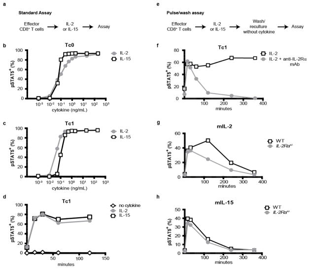
To uncover the mechanism behind the remarkable IL-2Rα-dependent responsiveness of effector Tc1 cells in vivo, we assayed IL-2 and IL-15 activity downstream of IL-2Rβγ using standard in vitro assays quantifying phosphorylation of STAT5 (a proximal signaling event), viability, and proliferation (Fig. 2a). In the context of STAT5 phosphorylation in response to titrated cytokine, we found that Tc1 (IL-2Rαhi) cells exhibited marginally increased sensitivity to IL-2 versus IL-15 when compared to Tc0 effector cells (IL-2Rαmed) (Fig. 2b,c), which is consistent with previous findings (19). The addition of a blocking antibody (anti-IL-2Rα mAb, PC61 clone) also showed a minimal benefit of IL-2Rα engagement on Tc1 cells in comparison between titrated IL-2 and IL-15 (Supplementary Fig. 5). Notably, Tc1 cells responded comparably to IL-2 and IL-15 in standard assays of proliferation and viability (Supplementary Fig. 6). Importantly, there was no difference in the kinetics of STAT5 phosphorylation between cells cultured in IL-2 or IL-15 (Fig. 2d). The mildly enhanced sensitivity of Tc1 cells to IL-2 versus IL-15 seemed unlikely to account for the dramatic difference in activity observed in vivo. Therefore, we hypothesized that IL-2Rα does not simply improve cellular affinity for IL-2, but allows for sustained IL-2 signaling after a T cell transitions from a cytokine-rich to a cytokine-free environment. To test this idea, we used a cytokine pulse assay. Tc1 and Tc0 cells were cultured overnight with a saturating dose of IL-2 or IL-15, washed, and replated without cytokine as shown in Figure 2e. Consistent with our hypothesis, only pre-culture of Tc1 cells with IL-2 led to sustained STAT5 phosphorylation in the absence of additional cytokine (Supplementary Fig. 7). To directly test the role of IL-2Rα in promoting sustained signaling on effector CD8+ T cells, we cultured Tc1 cells for 90 minutes with IL-2 in the absence or presence of blocking anti-IL-2Rα antibody (PC61 clone). This shorter pulse was equally sufficient for inducing sustained signaling as indicated by STAT5 phosphorylation (Fig. 2f). Importantly, blockade of IL-2Rα completely abolished the sustained IL-2 signaling as indicated by STAT5 phosphorylation and proliferation (Fig. 2f and Supplementary Fig. 8 & 9). Polyclonal effector CD8+ T cells activated in the absence of IL-12 also showed sustained IL-2 signaling, and importantly, effector cells generated from IL-2Rα+/− mice showed roughly half the sustained IL-2 signaling (Fig. 2g). To ensure that these cells had similar IL-2Rβγ signaling potential, we pulsed wildtype and IL-2Rα+/− effector CD8+ T cells with IL-15 and found no differences in their response (Fig. 2h). Notably, the ability to induce sustained IL-2 signaling on mouse effector cells was observed with human and mouse IL-2 (Supplementary Fig. 10). Furthermore, culture of human effector T cells with hIL-2 but not hIL-15 led IL-2Rα-dependent sustained STAT5 phosphorylation (Supplementary Fig. 11). Finally, to verify that IL-2/mAb complexes (clone 5355) used in our in vivo experiments were permissive to engagement of IL-2Rα, we repeated the pulse assay with hIL-2 and excess anti-IL-2 mAb. In vitro generated IL-2/mAb complexes induced sustained IL-2 signaling that was dependent on IL-2Rα (Supplementary Fig. 12a). In contrast, IL-2/mAbCD122 complexes (clone S4B6), which do not engage IL-2Rα (15, 18), failed to induce sustained signaling in vitro (Supplementary Fig. 12b).
Figure 2. IL-2Rα mediates sustained signaling in effector CD8+ T cells following withdrawal of IL-2.
(a) Diagram of the standard cytokine assay in which effector cells are assayed after incubation with titrated cytokine. (b,c) Levels of pSTAT5 in Tc1 and Tc0 cells that were cultured with increasing amounts of mIL-2 or mIL-15 for 1 hour. (d) As in ‘b’, except Tc1 cells were incubated as indicated for up to 2 hours with 200ng/ml of cytokine and assayed for pSTAT5. (e) Diagram of the cytokine pulse assay in which effector cells are incubated with saturating amounts of cytokine (200ng/ml). Cells are then washed thoroughly, recultured at 37°C without additional cytokine, and assayed for pSTAT5. (f) Levels of pSTAT5 in Tc1 cells that were pulsed with mIL-2 with or without anti-IL-2Rα mAb (PC61 clone) for 1 hour, washed, and recultured at 37°C for the times indicated. (g,h) Levels of pSTAT5 in polyclonal effector T cells from wildtype (IL-2Rα+/+) or IL-2Rα+/− mice that were pulsed for 1 hour with mIL-2 or mIL-15, and assayed as described in ‘e’. Except for ‘g’ and ‘h’, all effector cells were generated from pmel-1 mice. All results are representative of at least 3 independent experiments.
IL-2Rα facilitates sustained IL-2 signaling through creation of an extracellular reservoir and recycling
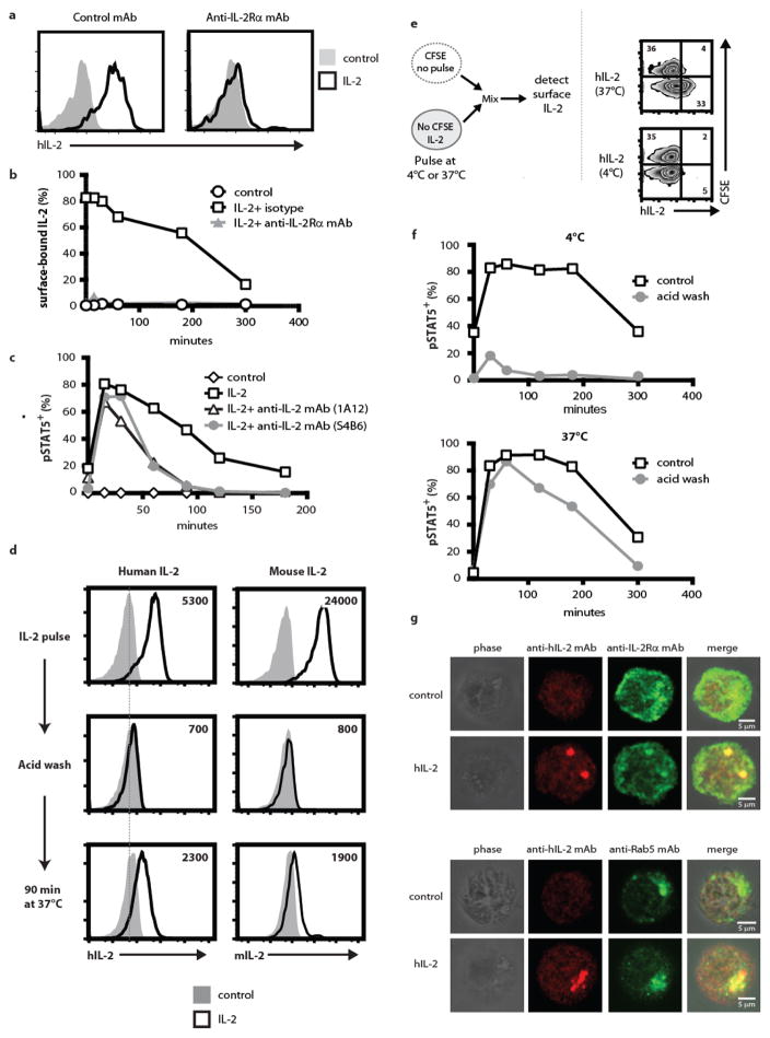
To understand how IL-2Rα promotes sustained IL-2 signaling, we hypothesized two non-mutually exclusive possibilities. First, IL-2Rα may bind IL-2 and create a cell-surface cytokine reservoir due to the high ratio of surface IL-2Rα to IL-2Rβγ, as IL-2/IL-2Rα internalization can only occur in the presence of both IL-2Rβ and γ (20, 21). Such a reservoir of IL-2 bound to IL-2Rα would mediate gradual signaling by continually feeding the rate-limiting, endocytosed IL-2Rβγ. In support of this possibility, we detected high surface levels of IL-2 on effector CD8+ T cells that gradually waned after extended culture, and this cell-surface IL-2 was dependent on available IL-2Rα (Fig. 3a,b). Furthermore, antibodies against IL-2 added after the removal of free cytokine from IL-2 pulsed cells were able to dampen sustained signaling (Fig. 3c). A second possible way in which IL-2Rα might sustain signaling is by promoting recycling of IL-2 from within the cell to the surface, thus allowing for repetitive signaling. To test this hypothesis, Tc1 cells were pulsed with IL-2 at 37°C to allow for cytokine internalization. Cells were then stripped of surface IL-2 using an acid wash. Upon reculture at 37°C, we were able to detect re-appearance of either mIL-2 or hIL-2 on the cell surface (Fig. 3d). Minimal surface IL-2 was observed when cells were pulsed at 4°C or on the surface of mixed bystander Tc1 cells (Fig. 3e). Importantly, the species-specificity of our reagents precluded autocrine production as the source of cell surface IL-2 after acid wash (Supplementary Fig. 13). In additional support of IL-2Rα-mediated recycling, we observed sustained pSTAT5 signaling after acid washing of cells pulsed with hIL-2 at 37°C but not 4°C (Fig. 3f). Because internalization of IL-2Rαβγ does not occur at 4°C, these data provide further support that sustained signaling occurs in part through an IL-2Rα bound pool of internalized IL-2. It is notable that we could not block sustained STAT5 signaling in cells pulsed with mIL-2 at 4°C followed by acid washing, possibly reflecting a higher affinity of mIL-2 for mIL-2Rα compared with that of hIL-2 for mIL-2Rα (18, 22). Finally, confocal microscopy showed discrete punctate structures of either mIL-2 or hIL-2 when cells were incubated with cytokine at 37°C but not 4°C (Supplementary Fig. 14 & 15). These punctate structures colocalized with IL-2Rα, Rab5, and EEA1, but less frequently with LAMP-1, consistent with intracellular IL-2 being accessible to the recycling pathway (Fig. 3g,h and Supplementary Fig. 16) (23, 24). Taken together, these results suggest that IL-2Rα both promotes an extracellular reservoir for IL-2 and mediates recycling of IL-2.
Figure 3. IL-2Rα facilitates sustained IL-2 signaling through creation of an extracellular reservoir and recycling.
(a) Presence of IL-2 on the surface of polyclonal T cells depends on IL-2Rα. Polyclonal effector CD8+ T cells were pulsed for 2 hours with or without mIL-2. Prior to (and during) the pulse, T cells were incubated with anti-IL-2Rα mAb (PC61). Cells were then washed and stained for surface IL-2. (b) Time course of surface IL-2 on polyclonal T cells after reculture at 37°C. (c) Levels of pSTAT5 in Tc1 cells that were pulsed with IL-2, washed, and recultured at 37°C with or without anti-IL-2 mAb (clone S4B6 or 1A12). (d) Recycling of IL-2 on effector T cells. Pmel-1 Tc1 cells were incubated with hIL-2 or mIL-2 at 37°C for 2 hours. As indicated, cells were then acid washed and recultured at 37°C for 90 minutes in the presence of anti-hIL-2 mAb conjugated to Alexa647. Cells were then washed, fixed, and assayed by flow cytometry. (e) Recycling of IL-2 on pulsed cells while mixed with non-pulsed cells. Pmel-1 Tc1 cells were pulsed with hIL-2 at either 4°C or 37°C for 2 hours, and then acid washed. Cells were then mixed with non-pulsed CFSE-labeled Tc1 cells. The mixed cells were recultured at 37°C for 45 minutes in the presence of anti-hIL-2 mAb conjugated to Alexa647. Cells were then washed, fixed, and assayed by flow cytometry. (f) Internalized IL-2 leads to sustained pSTAT5 signaling. Pmel-1 Tc1 cells were pulsed with hIL-2 at either 4°C or 37°C for 2 hours, and then acid washed. Cells were then recultured at 37°C and assayed for pSTAT5. (g). Subcellular localization of hIL-2 and IL-2Rα. Pmel-1 Tc1 cells were pulsed with hIL-2 (or media alone) for 1 hour at 37°C, and stained for hIL-2 and IL-2Rα. Cells were then imaged by confocal microscopy. (h) Subcellular localization of hIL-2 and Rab5. As in ‘g’, except cells were stained for Rab5. Results are representative of 3 independent experiments.
IL-2Rα expression on donor CD8+ T cells provides a competitive advantage to IL-2 therapy in a lymphoreplete but not lymphopenic host environment
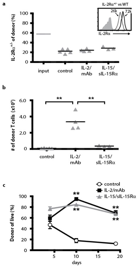
Our results thus far suggest that the differential responsiveness of Tc1 cells to IL-2- and IL-15 therapy in vivo is a consequence of IL-2Rα on donor T cells providing a competitive advantage to accessing cytokine. To formally test this hypothesis, we initially attempted to activate T cells from wildtype and IL-2Ra−/− mice. However, this proved technically not feasible for us as T cells isolated from IL-2Ra−/− mice were resistant to normal activation, likely due to the immune alterations in the absence of IL-2 responsiveness (9). Therefore, we used polyclonal IL-2Rα+/− T cells, as these cells activated comparably to wildtype T cells and had approximately half the expression of IL-2Rα (Fig. 4a). Using the Thy1.1 congenic marker to distinguish between genotypes, these two cell populations were mixed and adoptively transferred into non-irradiated B6(CD45.1) recipient mice. Mice were treated with IL-2/mAb or IL-15/sIL-15Rα for 1 week. We hypothesized that IL-2Rα+/− donor CD8+ T cells would not persist as well as their wildtype counterparts due to loss of one allele. In contrast to our expectations, wildtype and IL-2Rα+/− donor T cells did not show differential responsiveness to treatment with IL-2/mAb or IL-15/sIL-15Rα complexes (Fig. 4a, b). These results suggest a threshold of IL-2Rα in vivo, both in terms of level and durability of expression, that when reached is sufficient for providing donor cells a competitive advantage to IL-2 therapy in a lymphoreplete environment.
Figure 4. IL-2Rα on donor T cells is critical for persistence in lymphoreplete but not lymphodepleted hosts.
(a) Wildtype and IL-2Rα+/− effector CD8+ T cells show similar persistence with or without IL-2 therapy. Effector T cells from wildtype and IL-2Rα+/− mice were activated, mixed, and injected into recipient mice (n=3–5/group). Mice received injections of hIL-2/mAb (clone 5355) complexes, hIL-15/sIL-15Rα complexes, or vehicle alone. The proportion of IL-2Rα+/− T cells among all donor T cells in the spleen was determined pre- and post- transfer. Each triangle represents one mouse and the bars indicate the mean. (b) The total number of donor T cells per spleen for the experiment shown in ‘a’. The bars indicate the mean. The symbol (**) indicates a significant difference (p<0.001) between indicated conditions. (c) In lymphodepleted mice, IL-2 and IL-15 therapy augment the persistence of effector Tc1 cells comparably. Pmel-1 Tc1 cells (107) were injected into mice given total body irradiation (600 rads) 1 day earlier. On days 0, 2, 4, and 6 after adoptive transfer, mice (n = 5 per group) were treated with hIL-2/mAb (clone 5355) or hIL-15/sIL-15Rα complexes, and the frequency of donor cells in blood was assayed at the time points indicated. Each point represents the average and bars indicate standard error. The symbol (**) indicates a significant difference (p<0.001) between control and indicated conditions. All results are representative of 2 independent experiments.
As an alternative means of assessing the role of IL-2Rα on donor T cells in vivo, we compared the responsiveness of IL-2Rαhi donor T cells to IL-2- and IL-15 therapy with the addition of lymphodepletion to destroy host cells. We predicted that the advantage of IL-2Rα-competent cytokine therapy would be lost in the absence of host IL-2Rβγ+ lymphocytes competing for cytokine (Supplementary Fig. 17a). Thus, mice were given total body irradiation (600rad) prior to adoptive transfer of effector Tc1 CD8+ T cells, and then treated for one week with IL-2/mAb and IL-15/sIL-15Rα complexes. Consistent with our prediction, both IL-2 and IL-15 therapy effectively augmented the persistence of donor cells both in the blood and in the spleen, and only in lymphodepleted mice (Fig. 4c and Supplementary Fig. 17b). These results demonstrate a critical role for IL-2Rα on donor T cells in promoting IL-2 responsiveness in a lymphoreplete host environment.
Discussion
Administration of IL-2 is a critical component of many T cell-based strategies for cancer therapy. However, IL-2 has a short half-life and toxicity at therapeutic doses. Furthermore, as IL-2 selectively expands T regulatory cells, it has been proposed that IL-15-based therapies may more effectively support adoptively transferred effector T cells. However, our results show that in the absence of lymphodepletion, only IL-2Rα-competent cytokine therapy led to sustained persistence and anti-tumor immunity of effector CD8+ T cells expressing elevated IL-2Rα. Interestingly, host cells appear to play an important role in this process, as in mice preconditioned with total body irradiation, IL-2Rα-independent (or IL-15-based) cytokine therapy effectively augmented donor T cell responses. Together, these results suggest that IL-2Rα expression on donor T cells allows them to outcompete host cells for limited amounts of IL-2. Our results have important implications for the utilization of cytokine-based therapeutics in human patients as efficacy of IL-2- and IL-15-based therapies will be dictated by donor cell cytokine receptor subunit expression and competition with host lymphocytes.
In addition to our clinically relevant findings, we have uncovered molecular mechanisms which explain how IL-2Rα increases the durability of IL-2 signaling. It has been widely assumed that IL-2Rα improves IL-2 responsiveness primarily by acting as a high affinity receptor. We find, however, that beyond ligand affinity, IL-2Rα is able to promote sustained IL-2 signaling after withdrawal of cytokine, thereby providing a temporal dimension to IL-2 signaling. To enable sustained signaling, we find at least two distinct IL-2Rα-dependent mechanisms, including the creation of a cell-surface cytokine reservoir and recycling of IL-2 back to the cell surface. Because it is known that the IL-2Rα subunit is recycled after cytokine-mediated internalization, instead of being degraded like the IL-2Rβγ subunits (25), our results suggest a mechanism by which IL-2Rα can rescue IL-2 from degradation. These properties may be relevant during early stages of infection when lymphocytes express high levels of IL-2Rα (26–28).
Our results with IL-2Rα are reminiscent of the ability of membrane-bound IL-15Rα to facilitate recycling and sustained cellular responsiveness to IL-15 (7). Interestingly, mutants of IL-2, selected for high affinity binding to IL-2Rα (and thus thought to more resemble IL-15 in terms of high affinity for cognate alpha receptor), do exhibit prolonged cytokine responsiveness when assayed on cell lines and this has been attributed to the creation of both an extracellular reservoir and recycling (29–31). Importantly, our findings are distinct as they show that wildtype IL-2 can induce sustained cytokine signaling on primary mouse and human lymphocytes. Furthermore, we find sustained IL-2-signaling is directly dependent on IL-2Rα as shown by antibody-mediated blockade and genetic targeting.
There are emerging questions from our study that warrant additional investigation. Foremost, our results suggest strategies to enhance IL-2Rα expression on donor T cells may allow for more effective IL-2-based anti-tumor responses. In this context, it will be important to assess the therapeutic potential of strategies that redirect IL-2 toward IL-2Rα, approaches that improve the half-life of IL-2, and also the administration of low-dose IL-2. There also remain important biological questions from our mechanistic findings. For example, how does the presence of IL-2Rα impact the fate of internalized IL-2 through different endocytic compartments? Our ability to image and track IL-2 by confocal microscopy may provide evidence in support of IL-2Rα-mediated endosomal IL-2 recycling. Another question is related to IL-2-responsive T regulatory cells. Do cytokine-stimulated effector T cells have resistance to suppression by such T regulatory cells? Also important is whether IL-2Rα can mediate sustained signaling on T regulatory cells or other cell types. This may depend on the ratio and localization of the different receptor subunits. It may also be relevant that ADP ribosylation can regulate IL-2 binding to IL-2Rα (32).
In conclusion, our findings have important implications for efforts to boost the therapeutic value of IL-2- and IL-15-mediated signaling in patients with cancer as well as infectious disease. Our results suggest that in non-lymphopenic patients, IL-2Rα-competent cytokine therapy will be effective if responding T cells express elevated levels of IL-2Rα. In contrast, in lymphodepleted patients where host cells are destroyed and do not compete for cytokine availability, IL-2Rα-independent signaling will likely be sufficient, and thus either IL-15 or IL-2-based therapies will mediate effective responses.
Materials and Methods
Study design
This was a preclinical study to assess the efficacy of cytokine therapy to augment anti-tumor T cell immune responses. We found that IL-2-based therapies were more efficacious than IL-15-based therapies in our tumor model, and thereafter, focused on understanding the mechanism of this differential response. For in vivo experiments, the numbers of mice are outlined in the figure legends. For all experiments, the number of independent replicates is outlined in each figure legend. Randomization and blinding for tumor experiments was done as described in the tumor methods below. Additional study design details are also included in the statistical analysis section below.
Recombinant proteins and antibodies
Human (h) IL-15, hIL-2, and anti-hCD3 mAb (clone OKT3) were kindly provided by the NCI Biological Resources Branch Preclinical Repository. Mouse (m) IL-2, mIL-12, and mIL-15 were purchased from Shenandoah Biotechnology. Recombinant sIL-15Ra-Fc (551-MR-100), anti-hIL-2 mAb (clone 5355), and anti-hIL-2 mAb (clone 5334) were purchased from R&D systems. Anti-mIL-2 mAb (clone 5B9-2-1) was purchased from Peprotech. Anti-hIL-2 mAbCD25 (clone 5344.111) was obtained from BD Bioscience. Anti-mIL-2 mAbCD122 (clone S4B6) and anti-IL-2Rα (clone PC61) were obtained from Bioxcell. Anti-mIL-2 mAbCD25 (clone JES6-1A12), anti-mCD3 mAb (clone 145-2C11), and anti-mCD28 mAb (clone 37.51) were obtained from the UCSF monoclonal antibody core. Antibodies used for flow cytometric and confocal analysis are described below.
Mice and tumor cells
C57BL/6 (B6), B6.PL (Thy1.1), B6(CD45.1), pmel-1 TCR transgenic, and OT-I TCR transgenic mice were purchased from Jackson Laboratory. All animals were housed under specific pathogen-free conditions in accordance with institutional and federal guidelines. For tumor experiments, B16-F1 cells were obtained from ATCC.
T cell cultures
Mouse Tc1 and Tc0 cells were generated from pmel-1 and OT-I TCR transgenic mice as previously described (16). Briefly, splenocytes were cultured for three days with relevant peptide (for pmel-1, hgp10025–33 peptide (KVPRNQDWL) and for OT-I, OVA257–264 peptide (SIINFEKL)) and cultured with (Tc1) or without (Tc0) mIL-12 (10ng/ml). Polyclonal mouse T cells were generated by culturing B6 splenocytes for three days with plate-bound anti-CD3 mAb (clone 145-2C11, 1ug/ml) unless otherwise stated. Activated human T cells were generated by culturing de-identified human PBMCs (Research Blood Components) from healthy adult donors for two or three days with plate-bound anti-CD3 mAb (clone OKT3, 1ug/ml).
Tumor and persistence studies in mice
For tumor experiments, B6 mice were challenged subcutaneously with 2.5x10⁵ B16-F1 tumor cells. Prior to randomizing mice to treatment groups, some mice were excluded due to abnormal tumor growth. As indicated, mice were treated by adoptive transfer of activated T cells (Tc1 or Tc0) by intravenous tail vein injection. Cytokine complexes were administered by intraperitoneal injection on days 0, 2, 4, and 6 after adoptive transfer unless otherwise indicated. Cytokine complexes used include: hIL-15/sIL-15Rα, hIL-15 (0.5ug)/sIL-15Ra-Fc (2.3ug); hIL-2/mAb, hIL-2 (1.5ug)/anti-IL-2 mAb (7.5ug, clone 5355); hIL-2/mAbCD25, hIL-2 (1.5ug)/anti-IL-2 mAb (7.5ug, clone 5344.111); mIL-2/mAbCD122, mIL-2 (1.5ug)/anti-IL-2 mAb (7.5ug, clone S4B6); and mIL-2/mAbCD25, mIL-2 (1.5ug)/anti-IL-2 mAb (7.5ug, clone JES6-1A12). Tumor growth was measured by caliper every 2–4 days by personnel blinded to the treatment regimen. Tumor surface area (mm2) was calculated as length x width. Mice were sacrificed when tumors reached 400mm2. For persistence studies, mice received adoptive transfer of activated T cells (Tc1 or Tc0). Peripheral blood lymphocytes or indicated organs were stained for CD8 and either Thy1.1 or CD45.1 to identify donor T cells. In experiments with a mixed transfer, we used effector T cells from wildtype (Thy1.1) and IL-2Rα+/− (Thy1.2) mice that were activated with plate-bound anti-CD3/anti-CD28 mAb, mixed, and transferred into B6(CD45.1) mice. Where indicated, mice also received total body irradiation (600rad) one day prior to adoptive T cell transfer. In all adoptive transfer experiments, donor and recipient mice were gender-matched and recipient mice were 6 to 12 weeks of age. All animals were housed under specific pathogen-free conditions in accordance with institutional and federal guidelines.
Flow cytometry
Flow cytometry analysis was performed as previously described (17). The antibodies used in this study include CD8 (53–6.7), CD25 (PC61), CD45.1 (A20), IFNγ (XMG1.2), STAT5 pY694 (47/Stat5(pY694)), Thy1.1 (A20), and TNFα (TN3-19.12).These were purchased from BD Bioscience, Biolegend (San Diego, CA), and eBioscience (San Diego, CA). For analysis of phosphorylation of STAT5, we followed the manufacturer’s protocol using Lyse/Fix and PermIII buffer (BD Bioscience). To examine cellular proliferation, cells were fixed and permeabilized according to the manufacturer’s protocol for Cytofix/Cytoperm (BD Bioscience) and stained with anti-Ki67 mAb (SolA15, eBioscience). Alternatively, BrdU (10μm) was added one hour prior to harvest, and cells were analyzed for BrdU incorporation as previously described (17). For Foxp3 staining, we followed the protocol outlined in the Foxp3 kit (eBioscience). Flow cytometry was performed on BD LSRII and BD FACSAccuri. Data were analyzed using FlowJo software (TreeStar). In all experiments, initial gating of live cells was performed using forward scatter and side scatter parameters, and cells were then gated on live lymphocytes. Isotype and fluorescence minus one (FMO) controls were performed as required. For experiments assessing IL-2, we always included control conditions without IL-2 pulsing.
In vitro experiments
For functional assays, Tc1 or Tc0 cells were incubated with cytokines and assayed for pSTAT5, Ki67, BrdU incorporation, or propidium iodide exclusion. For pulse assays, cells were incubated with or without cytokine at 200ng/ml at either 4°C or 37°C for 90 minutes unless otherwise indicated. Cells were then washed at least three times, replated without cytokine, and assayed for pSTAT5. When added during the pulse step, anti-IL-2Rα mAb was added 15 minutes prior to cytokine addition. Acid wash was performed by washing cells twice for 2 minutes at 4°C with an acid wash buffer consisting of complete media adjusted to pH3.5 or pH3.75 with 1N HCl. For analysis of recycling of IL-2 to the cell surface, acid washed cells were replated in media at 37°C for the indicated amount of time with anti-IL-2 mAb conjugated to Alexa647 (5334 or 5B9-2-1 clone). To assess IFNγ and TNFα production, we added hgp10025–33 (1ug/ml) or PMA (50ng/mL) and ionomycin (1μM) to splenocytes for 5 hours in the presence of monensin (GolgiStop, BD Bioscience).
Confocal Microscopy
Tc1 cells were incubated with hIL-2 (200ng/ml), mIL-2 (200ng/ml), or no cytokine, for 1 hour at either 4°C or 37°C unless otherwise stated. Cells were washed, fixed, and permeabilized using the Cytofix/Cytoperm protocol. To determine the subcellular localization of internalized IL-2 by confocal microscopy, cells were stained with anti-hIL-2 mAb and either anti-IL-2Rα pAb (R&D systems), anti-Rab5 mAb (C8B1, Cell Signaling), anti-LAMP1 mAb (1D4B, Biolegend), or anti-EEA1 mAb (C45B10, Cell Signaling). To detect anti-IL-2Rα, we used an anti-goat IgG conjugated to Alexa488 (R&D systems). To detect EEA-1 and Rab5, we used an anti-rabbit IgG conjugated to Alexa488 (F(ab′)2 fragment, Cell Signaling). After washing, cells were transferred to SuperFrost microscope slides via cytospin. Immunofluorescence staining was visualized with a confocal microscope (Olympus Fluoview FV10i laser scanning confocal microscope system, Olympus) using a 60x water immersion objective (1.2 NA). Image analysis was performed using the FV10-ASW 1.7 software. In all images, IL-2 staining is presented as a red pseudocolor. In all experiments, cells pulsed without IL-2 were used as the primary control.
Statistical Analyses
Before analysis, graphical displays were made of all data vs. conditions to identify the need for transformations to adhere to model assumptions. For experiments comparing outcomes at a fixed point in time, log transforms were taken and comparisons of means performed using two-sample t-tests or linear regression (depending on the number of conditions). Where appropriate, t-tests assumed unequal variance across conditions. Comparisons of conditions where mice were followed over time were made at individual timepoints based on random effects linear regression models (with random effects to account for correlation of data from the same mouse over repeated measures) with the outcome (e.g. % T-cells) log-transformed. Graphical displays were used to assess appropriateness of transformation. Residual plots were inspected to assess assumptions of linear regression models. Time to sacrifice was compared across groups using log-rank tests. Time to sacrifice was compared across groups using log-rank tests. Percent colocalization was compared with log(percent) as the outcome (due to skewness) and main effects of LAMP-1 (vs. EEA-1) and rater. The LAMP-1 effect was evaluated based on the Wald test of the regression coefficient. Model results were exponentiated to provide point estimates for LAMP-1 and EEA-1 colocalization. In the interest of addressing our hypotheses and not over-testing, we did not perform hypothesis tests for every possible comparison in each figure. Instead, we focused on addressing our hypotheses and where comparisons were insignificant (p>0.05) it is stated in the text; where tests were significant, it is stated and/or indicated with asterisks in figures. P-values are reported to two significant digits, except when the p-value is less than 0.001; for p-values smaller than 0.001, it is reported as ‘p<0.001’. P-values are not corrected for multiple comparisons. For all analyses, statistical significance was based on a two-sided alpha level of 0.05. Statistical analyses were performed using Stata/IC (version 12.1) and R statistical software.
Supplementary Material
Fig. S1. IL-2/mAb complexes selectively enhance the persistence of donor T cells
Fig. S2. In the absence of donor T cells, hIL-2/mAb and IL-15/sIL-15Rα complexes mediate comparable anti-tumor immunity
Fig. S3. Treatment with IL-2/mAb, IL-2/mAbCD25, and IL-15/sIL-15Rα complexes induces differential expansion of CD8+ memory-phenotype T cells, NK cells, and T regulatory cells.
Fig. S4. Tc1 but not Tc0 effector CD8+ T cells show preferential responsiveness to IL-2/mAb complexes.
Fig. S5. Blockade of IL-2Rα has minimal impact on Tc1 cells in response to titrated IL-2 or IL-15.
Fig. S6. Tc1 effector CD8+ T cells exhibit comparable functional sensitivity to IL-2 and IL-15 in vitro.
Fig. S7. Tc1 effector CD8+ T cells pulsed with IL-2 mediate sustained cytokine signaling.
Fig. S8. IL-2 mediated sustained cytokine signaling is IL-2Rα-dependent in 11 independent experiments.
Fig. S9. Tc1 effector CD8+ T cells pulsed with IL-2 exhibit IL-2Rα-dependent proliferation after cytokine withdrawal.
Fig. S10. Human IL-2 mediates sustained cytokine signaling on mouse Tc1 effector CD8+ T cells.
Fig. S11. Human effector CD8+ T cells pulsed with IL-2 mediate sustained IL-2Rα-dependent signaling.
Fig. S12. Human IL-2/mAb (clone 5355), but not mouse IL-2/mAbCD122 (clone S4B6) complexes, are permissive to IL-2Rα-dependent sustained signaling in vitro.
Fig. S13. Antibodies for mouse and human IL-2 are species-specific.
Fig. S14. Detection of hIL-2 by confocal microscopy is species-specific and dependent on pulsing cells with cytokine at 37°C.
Fig. S15. Detection of mIL-2 by confocal microscopy.
Fig. S16. Colocalization of hIL-2 with EEA-1 and LAMP-1 by confocal microscopy.
Fig. S17. In lymphodepleted mice, IL-15/sIL-15Rα and hIL-2/mAb mediate comparable engraftment of Tc1 effector CD8+ T cells.
Acknowledgments
We thank Monika Gooz for assistance with confocal microscopy. We thank Ivan Molano and John Zhang for technical assistance with fluorescent labelling of antibodies.
Funding:
Support was provided by the following grants and fellowships from the National Institutes of Health and the National Cancer Institute: P01CA154778-01, R01CA133503, 5R01CA175061, and 5F30CA177208. Flow cytometry and imaging facilities for this research were supported, in part, by Cancer Center Support Grant P30CA138313 to the Hollings Cancer Center, Medical University of South Carolina. We are also grateful to the Melanoma Research Alliance, the Prevent Cancer Foundation, and the Leukemia and Lymphoma Society for research funding.
Footnotes
Author contributions.
EWS and CJM designed research, performed experiments, analyzed data, and wrote the manuscript. SS, CBJ, NS, AP, DJN, and KA designed research, performed experiments, analyzed data, and edited the manuscript. EGM designed experiments, analyzed data, performed statistical analysis, and edited the manuscript. SM, CMP, ALD, AWG, ZL, and DJC designed experiments, analyzed data and edited the manuscript. MPR designed experiments, performed experiments, analyzed data, wrote the manuscript, and supervised the study.
Competing interests.
DJC and MPR are co-inventors on a provisional patent application related to genetically modifying lymphocytes with IL-2 receptor genes. The other authors declare that they have no competing interests.
References and notes
- 1.Restifo NP, Dudley ME, Rosenberg SA. Adoptive immunotherapy for cancer: harnessing the T cell response. Nat Rev Immunol. 2012;12:269–281. doi: 10.1038/nri3191. published online EpubApr (nri3191 [pii] [DOI] [PMC free article] [PubMed] [Google Scholar]
- 2.Waldmann TA. The biology of interleukin-2 and interleukin-15: implications for cancer therapy and vaccine design. Nat Rev Immunol. 2006;6:595–601. doi: 10.1038/nri1901. 10published online EpubAug nri1901 [pii] [DOI] [PubMed] [Google Scholar]
- 3.Ma A, Koka R, Burkett P. Diverse functions of IL-2, IL-15, and IL-7 in lymphoid homeostasis. Annu Rev Immunol. 2006;24:657–679. doi: 10.1146/annurev.immunol.24.021605.090727. doi:1010.1146/annurev.immunol.24.021605.090727. [DOI] [PubMed] [Google Scholar]
- 4.Liao W, Lin JX, Leonard WJ. Interleukin-2 at the crossroads of effector responses, tolerance, and immunotherapy. Immunity. 2013;38:13–25. doi: 10.1016/j.immuni.2013.01.004. published online EpubJan 24 (S1074-7613(13)00011-3 [pii] [DOI] [PMC free article] [PubMed] [Google Scholar]
- 5.Ring AM, Lin JX, Feng D, Mitra S, Rickert M, Bowman GR, Pande VS, Li P, Moraga I, Spolski R, Ozkan E, Leonard WJ, Garcia KC. Mechanistic and structural insight into the functional dichotomy between IL-2 and IL-15. Nat Immunol. 2012;13:1187–1195. doi: 10.1038/ni.2449. published online EpubDec ni.2449 [pii] [DOI] [PMC free article] [PubMed] [Google Scholar]
- 6.Arneja A, Johnson H, Gabrovsek L, Lauffenburger DA, White FM. Qualitatively different T cell phenotypic responses to IL-2 versus IL-15 are unified by identical dependences on receptor signal strength and duration. Journal of immunology. 2014;192:123–135. doi: 10.4049/jimmunol.1302291. published online EpubJan 1. [DOI] [PMC free article] [PubMed] [Google Scholar]
- 7.Dubois S, Mariner J, Waldmann TA, Tagaya Y. IL-15Ralpha recycles and presents IL-15 In trans to neighboring cells. Immunity. 2002;17:537–547. doi: 10.1016/s1074-7613(02)00429-6. published online EpubNov (S1074761302004296 [pii]) [DOI] [PubMed] [Google Scholar]
- 8.Giri JG, Kumaki S, Ahdieh M, Friend DJ, Loomis A, Shanebeck K, DuBose R, Cosman D, Park LS, Anderson DM. Identification and cloning of a novel IL-15 binding protein that is structurally related to the alpha chain of the IL-2 receptor. The EMBO journal. 1995;14:3654–3663. doi: 10.1002/j.1460-2075.1995.tb00035.x. published online EpubAug 1. [DOI] [PMC free article] [PubMed] [Google Scholar]
- 9.Willerford DM, Chen J, Ferry JA, Davidson L, Ma A, Alt FW. Interleukin-2 receptor alpha chain regulates the size and content of the peripheral lymphoid compartment. Immunity. 1995;3:521–530. doi: 10.1016/1074-7613(95)90180-9. published online EpubOct (1074-7613(95)90180-9 [pii]) [DOI] [PubMed] [Google Scholar]
- 10.Sadlack B, Merz H, Schorle H, Schimpl A, Feller AC, Horak I. Ulcerative colitis-like disease in mice with a disrupted interleukin-2 gene. Cell. 1993;75:253–261. doi: 10.1016/0092-8674(93)80067-o. published online EpubOct 22 ( [DOI] [PubMed] [Google Scholar]
- 11.Kennedy MK, Glaccum M, Brown SN, Butz EA, Viney JL, Embers M, Matsuki N, Charrier K, Sedger L, Willis CR, Brasel K, Morrissey PJ, Stocking K, Schuh JC, Joyce S, Peschon JJ. Reversible defects in natural killer and memory CD8 T cell lineages in interleukin. 15-deficient mice. The Journal of experimental medicine. 2000;191:771–780. doi: 10.1084/jem.191.5.771. published online EpubMar 6 ( [DOI] [PMC free article] [PubMed] [Google Scholar]
- 12.Lodolce JP, Boone DL, Chai S, Swain RE, Dassopoulos T, Trettin S, Ma A. IL-15 receptor maintains lymphoid homeostasis by supporting lymphocyte homing and proliferation. Immunity. 1998;9:669–676. doi: 10.1016/s1074-7613(00)80664-0. published online EpubNov ( [DOI] [PubMed] [Google Scholar]
- 13.Rubinstein MP, Kovar M, Purton JF, Cho JH, Boyman O, Surh CD, Sprent J. Converting IL-15 to a superagonist by binding to soluble IL-15R{alpha} Proceedings of the National Academy of Sciences of the United States of America. 2006;103:9166–9171. doi: 10.1073/pnas.0600240103. published online EpubJun 13. [DOI] [PMC free article] [PubMed] [Google Scholar]
- 14.Stoklasek TA, Schluns KS, Lefrancois L. Combined IL-15/IL-15Ralpha immunotherapy maximizes IL-15 activity in vivo. Journal of immunology. 2006;177:6072–6080. doi: 10.4049/jimmunol.177.9.6072. published online EpubNov 1 (177/9/6072 [pii]) [DOI] [PMC free article] [PubMed] [Google Scholar]
- 15.Boyman O, Kovar M, Rubinstein MP, Surh CD, Sprent J. Selective stimulation of T cell subsets with antibody-cytokine immune complexes. Science. 2006;311:1924–1927. doi: 10.1126/science.1122927. published online EpubMar 31. [DOI] [PubMed] [Google Scholar]
- 16.Rubinstein MP, Cloud CA, Garrett TE, Moore CJ, Schwartz KM, Johnson CB, Craig DH, Salem ML, Paulos CM, Cole DJ. Ex vivo interleukin-12-priming during CD8(+) T cell activation dramatically improves adoptive T cell transfer antitumor efficacy in a lymphodepleted host. Journal of the American College of Surgeons. 2012;214:700–707. doi: 10.1016/j.jamcollsurg.2011.12.034. discussion 707–708. published online EpubApr. [DOI] [PMC free article] [PubMed] [Google Scholar]
- 17.Rubinstein MP, Lind NA, Purton JF, Filippou P, Best JA, McGhee PA, Surh CD, Goldrath AW. IL-7 and IL-15 differentially regulate CD8+ T-cell subsets during contraction of the immune response. Blood. 2008;112:3704–3712. doi: 10.1182/blood-2008-06-160945. published online EpubNov 1. [DOI] [PMC free article] [PubMed] [Google Scholar]
- 18.Spangler JB, Tomala J, Luca VC, Jude KM, Dong S, Ring AM, Votavova P, Pepper M, Kovar M, Garcia KC. Antibodies to Interleukin-2 Elicit Selective T Cell Subset Potentiation through Distinct Conformational Mechanisms. Immunity. 2015;42:815–825. doi: 10.1016/j.immuni.2015.04.015. published online EpubMay 19. [DOI] [PMC free article] [PubMed] [Google Scholar]
- 19.Lisiero DN, Soto H, Liau LM, Prins RM. Enhanced sensitivity to IL-2 signaling regulates the clinical responsiveness of IL-12-primed CD8(+) T cells in a melanoma model. Journal of immunology. 2011;186:5068–5077. doi: 10.4049/jimmunol.1003317. published online EpubMay 1. [DOI] [PMC free article] [PubMed] [Google Scholar]
- 20.Robb RJ, Greene WC. Internalization of interleukin. 2 is mediated by the beta chain of the high-affinity interleukin 2 receptor. The Journal of experimental medicine. 1987;165:1201–1206. doi: 10.1084/jem.165.4.1201. published online EpubApr 1. [DOI] [PMC free article] [PubMed] [Google Scholar]
- 21.Takeshita T, Asao H, Ohtani K, Ishii N, Kumaki S, Tanaka N, Munakata H, Nakamura M, Sugamura K. Cloning of the gamma chain of the human IL-2 receptor. Science. 1992;257:379–382. doi: 10.1126/science.1631559. published online EpubJul 17 ( [DOI] [PubMed] [Google Scholar]
- 22.Liu KD, Greene WC, Goldsmith MA. The alpha chain of the IL-2 receptor determines the species specificity of high-affinity IL-2 binding. Cytokine. 1996;8:613–621. doi: 10.1006/cyto.1996.0082. published online EpubAug. [DOI] [PubMed] [Google Scholar]
- 23.Grant BD, Donaldson JG. Pathways and mechanisms of endocytic recycling. Nature reviews Molecular cell biology. 2009;10:597–608. doi: 10.1038/nrm2755. published online EpubSep. [DOI] [PMC free article] [PubMed] [Google Scholar]
- 24.Mu FT, Callaghan JM, Steele-Mortimer O, Stenmark H, Parton RG, Campbell PL, McCluskey J, Yeo JP, Tock EP, Toh BH. EEA1, an early endosome-associated protein. EEA1 is a conserved alpha-helical peripheral membrane protein flanked by cysteine “fingers” and contains a calmodulin-binding IQ motif. J Biol Chem. 1995;270:13503–13511. doi: 10.1074/jbc.270.22.13503. published online EpubJun 2 ( [DOI] [PubMed] [Google Scholar]
- 25.Hemar A, Subtil A, Lieb M, Morelon E, Hellio R, Dautry-Varsat A. Endocytosis of interleukin 2 receptors in human T lymphocytes: distinct intracellular localization and fate of the receptor alpha, beta, and gamma chains. The Journal of cell biology. 1995;129:55–64. doi: 10.1083/jcb.129.1.55. published online EpubApr ( [DOI] [PMC free article] [PubMed] [Google Scholar]
- 26.Obar JJ, Molloy MJ, Jellison ER, Stoklasek TA, Zhang W, Usherwood EJ, Lefrancois L. CD4+ T cell regulation of CD25 expression controls development of short-lived effector CD8+ T cells in primary and secondary responses. Proceedings of the National Academy of Sciences of the United States of America. 2010;107:193–198. doi: 10.1073/pnas.0909945107. published online EpubJan 5. [DOI] [PMC free article] [PubMed] [Google Scholar]
- 27.Cousens LP, Orange JS, Biron CA. Endogenous IL-2 contributes to T cell expansion and IFN-gamma production during lymphocytic choriomeningitis virus infection. Journal of immunology. 1995;155:5690–5699. published online EpubDec 15. [PubMed] [Google Scholar]
- 28.Andersson EC, Christensen JP, Marker O, Thomsen AR. Changes in cell adhesion molecule expression on T cells associated with systemic virus infection. Journal of immunology. 1994;152:1237–1245. published online EpubFeb 1. [PubMed] [Google Scholar]
- 29.Rao BM, Driver I, Lauffenburger DA, Wittrup KD. High-affinity CD25-binding IL-2 mutants potently stimulate persistent T cell growth. Biochemistry. 2005;44:10696–10701. doi: 10.1021/bi050436x. published online EpubAug 9. [DOI] [PubMed] [Google Scholar]
- 30.Fallon EM, Liparoto SF, Lee KJ, Ciardelli TL, Lauffenburger DA. Increased endosomal sorting of ligand to recycling enhances potency of an interleukin-2 analog. J Biol Chem. 2000;275:6790–6797. doi: 10.1074/jbc.275.10.6790. published online EpubMar 10. [DOI] [PubMed] [Google Scholar]
- 31.Rao BM, Driver I, Lauffenburger DA, Wittrup KD. Interleukin. 2 (IL-2) variants engineered for increased IL-2 receptor alpha-subunit affinity exhibit increased potency arising from a cell surface ligand reservoir effect. Molecular pharmacology. 2004;66:864–869. doi: 10.1124/mol.66.4. published online EpubOct. [DOI] [PubMed] [Google Scholar]
- 32.Teege S, Hann A, Miksiewicz M, MacMillan C, Rissiek B, Buck F, Menzel S, Nissen M, Bannas P, Haag F, Boyer O, Seman M, Adriouch S, Koch-Nolte F. Tuning IL-2 signaling by ADP-ribosylation of CD25. Scientific reports. 2015;5:8959. doi: 10.1038/srep08959. [DOI] [PMC free article] [PubMed] [Google Scholar]
Associated Data
This section collects any data citations, data availability statements, or supplementary materials included in this article.
Supplementary Materials
Fig. S1. IL-2/mAb complexes selectively enhance the persistence of donor T cells
Fig. S2. In the absence of donor T cells, hIL-2/mAb and IL-15/sIL-15Rα complexes mediate comparable anti-tumor immunity
Fig. S3. Treatment with IL-2/mAb, IL-2/mAbCD25, and IL-15/sIL-15Rα complexes induces differential expansion of CD8+ memory-phenotype T cells, NK cells, and T regulatory cells.
Fig. S4. Tc1 but not Tc0 effector CD8+ T cells show preferential responsiveness to IL-2/mAb complexes.
Fig. S5. Blockade of IL-2Rα has minimal impact on Tc1 cells in response to titrated IL-2 or IL-15.
Fig. S6. Tc1 effector CD8+ T cells exhibit comparable functional sensitivity to IL-2 and IL-15 in vitro.
Fig. S7. Tc1 effector CD8+ T cells pulsed with IL-2 mediate sustained cytokine signaling.
Fig. S8. IL-2 mediated sustained cytokine signaling is IL-2Rα-dependent in 11 independent experiments.
Fig. S9. Tc1 effector CD8+ T cells pulsed with IL-2 exhibit IL-2Rα-dependent proliferation after cytokine withdrawal.
Fig. S10. Human IL-2 mediates sustained cytokine signaling on mouse Tc1 effector CD8+ T cells.
Fig. S11. Human effector CD8+ T cells pulsed with IL-2 mediate sustained IL-2Rα-dependent signaling.
Fig. S12. Human IL-2/mAb (clone 5355), but not mouse IL-2/mAbCD122 (clone S4B6) complexes, are permissive to IL-2Rα-dependent sustained signaling in vitro.
Fig. S13. Antibodies for mouse and human IL-2 are species-specific.
Fig. S14. Detection of hIL-2 by confocal microscopy is species-specific and dependent on pulsing cells with cytokine at 37°C.
Fig. S15. Detection of mIL-2 by confocal microscopy.
Fig. S16. Colocalization of hIL-2 with EEA-1 and LAMP-1 by confocal microscopy.
Fig. S17. In lymphodepleted mice, IL-15/sIL-15Rα and hIL-2/mAb mediate comparable engraftment of Tc1 effector CD8+ T cells.


