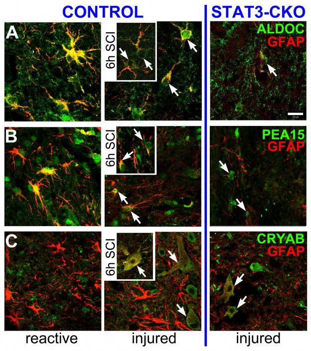
Figure 13. ALDOC, PEA15 and CRYAB distinguish reactive and injured astrocytes acutely after spinal cord crush injury.
Shown are gray matter astrocytes one day (main) and 6 hr (inserts) post-SCI in Control (left) and STAT3-CKO (right) spinal cords. A, ALDOC and GFAP were robustly co-expressed in territorial, reactive astrocytes in the penumbra around the lesion (left). STAT3-CKO cords at same time post-SCI were missing reactive astrocytes (not shown). Areas near or within the lesion contained injured astrocytes showing ALDOC depletion (6h post-SCI, arrows) or attenuated signals (1d post-SCI, center, arrows). ALDOC and GFAP signals were faint in lesioned areas of STAT3-CKO astrocytes (right, arrow). B, At one day post-injury, reactive astrocytes co-expressed PEA15 and GFAP (yellow) in their cell bodies that were found in the lesion penumbra of CON cords (left). Injured CON spinal tissue had few PEA-15 expressing astrocytes with faint (1d post-SCI, center) to marginal (6 h, insert) PEA15 signals (green, arrows), while still showing GFAP (red, center and insert). Right: STAT3-CKO lesioned tissues had marginal astroglial PEA15 signals that was surrounded by faintly stained GFAP-positive processes (red, arrows). C, CRYAB was absent in strongly GFAP-positive reactive CON astrocytes (left, red), but was expressed in injured CON astrocytes with swollen cell bodies and thin processes where it was colocalized with weak GFAP signals (yellow, 6h insert, arrow). Similarly, injured STAT3-CKO astrocytes showing alike morphologies also expressed CRYAB colocalized with GFAP (right, arrows). Bar = 20 μm.

