Abstract
Abstract
Three new highly oxidative cembranoids, sarcophytrols D–F (1–3), were obtained from the South China Sea soft coral Sarcophyton trocheliophorum, along with two known related ones (4 and 5). Their structures were elucidated by extensive spectroscopic analyses and by comparison with literature data. The discovery of these new secondary metabolites enriched the family of cembranoids deduced from the title animal.
Graphical Abstract
Keywords: Soft coral, Sarcophyton trocheliophorum, Cembranoids, Sarcophytrol, Structure elucidation
Introduction
Soft corals (phylum Cnidaria, class Anthozoa, subclass Octocorallia, order Alcyonacea) often equal or exceed the total coverage of scleractinian corals in coral reef ecosystems [1–4]. The family Alcyoniidae within the order Alcyonacea contains the productive genus Sarcophyton [5, 6]. Till now, nearly 16 species of soft corals of the genus Sarcophyton, from various geographical areas, have been chemically investigated, being reported to comprise diverse diterpenes with different skeletons (cembrane, sarsolenane, capnosane, etc.) [5–9], biscembranoids [10–12], polyhydroxylated sterols [13, 14], and other related metabolites [15, 16].
Sarcophyton species are prolific in the South China Sea. In the course of our search for bioactive substances from Chinese marine organisms, Sarcophyton trocheliophorum of the coast of Yalong Bay, Hainan Province, has been collected and chemically investigated, which were found to encompass numerous cembranoids with a variety of oxidation and cyclization patterns, including two unprecedented structures, methyl sarcotroates A and B [7–9, 12, 17]. In addition, many of them exhibited significant inhibitory activities against human PTP1B enzyme [7, 8, 17]. Recently, in order to find more chemically appealing and biologically active cembrane-based metabolites, S. trocheliophorum was re-collected from the same location while in a different growing period, and a further chemical investigation yielded three new highly oxidative compounds named sarcophytrols D–F (1–3), along with two known ones (4 and 5). Details of the isolation, structure elucidation and biological study of these secondary metabolites are reported herein.
Results and discussion
Samples of S. trocheliophorum (400 g, dry weight) were extracted exhaustively with acetone, and the extract was partitioned between H2O and Et2O. The Et2O soluble fraction was subjected to silica gel chromatography (light petroleum ether/acetone gradient). The lower polar fractions were subsequently purified on repeated column chromatography (silica gel, Sephadex LH-20, reversed phase-C18 silica gel and semi preparative-HPLC) to afford five pure metabolites, compounds 1–5. A preliminary NMR analysis revealed that all the new molecules shared the same cembrane skeleton. Among them, two known compounds were readily identified as 11,12-epoxy-1(E),3(E),7(E)-cembratrien-15-ol (4) [18] and sinugibberol (5) [19] by comparison of their spectral data and [α]D values with those reported in the literature.
Sarcophytrol D (1) was obtained as colorless oil. The molecular formula was established as C20H30O3 by HRESIMS (m/z 343.2248 [M + Na]+), sixteen mass units more than that of compound 4 [18]. In fact, their NMR spectra were similar, except for signals appeared in the downfield area. Differed from 4, the 1H and 13C NMR (Tables 1 and 2) data of 1 showed the presence of a trans-disubstituted double bond (δH 5.75, d, J = 15.7 Hz; δH 5.68, ddd, J = 15.7, 7.4, 5.4 Hz). In its 1H–1H COSY, the cross-peaks between H-5 (δH 2.83, 2.75) and H-6 (δH 5.68), between H-6 and H-7 (δH 5.75) indicated the linkage of C-5 to the trans-double bond. Furthermore, HMBC correlations from Me-19 (δH 1.40) to C-7 (δC 140.4) and from Me-18 (δH 1.78) to C-5 (δC 41.47) confirmed the position of the double bond. In addition, Me-19 of compound 1 has been upfield shifted (from δH 1.67 in 4 to 1.40 in 1). Based on these NMR and MS variations, the hydroxylation at C-8 accompanying the double-bond migration from Δ7(8) to Δ6(7) in the structure of 1 were supported. Assignments of 1H and 13C NMR signals of 1 were made by the application of detailed 2D NMR analysis.
Table 1.
1H NMR data [δ H (mult., J in Hz)] for compounds 1–3 a
| No. | 1 | 2 | 3 |
|---|---|---|---|
| 2 | 6.30, d, (11.4) | 6.32, d (11.3) | 5.34, d (6.4) |
| 3 | 5.86, d, (11.4) | 5.73, d (11.3) | 3.36, d (6.4) |
| 5 | 2.83, dd (17.7, 7.4) 2.75, dd (17.7, 5.4) |
2.84, dd (17.7, 9.2) 2.78, dd (17.7, 4.6) |
2.07, m 1.61, m |
| 6 | 5.68, ddd (15.7, 7.4, 5.4) | 5.81, ddd (15.6, 9.2, 4.6) | 2.22, m 2.02, m |
| 7 | 5.75, d (15.7) | 5.71, dd (15.6, 1.5) | 5.26, t (5.8) |
| 9 | 2.00, ddd (12.7, 8.3, 1.9) 1.70, ddd (12.7, 11.5, 9.1) |
1.96, m 1.79, m |
2.44, dd (6.3, 13.7) 2.30, dd (3.4, 13.7) |
| 10 | 1.92, m 1.63, m |
2.03, m 1.52, m |
3.70, ddd (8.7, 7.7, 4.0) |
| 11 | 3.08, t (6.2) | 3.22, dd (7.4, 4.3) | 2.90, d (8.7) |
| 13 | 1.94, m 1.10, dd (12.9, 6.6) |
2.22, m 0.79, m |
2.11, m 1.44, dd (2.3, 12.5) |
| 14 | 2.33, ddd (13.2, 11.1, 6.8) 2.22, dd (11.1, 3.0) |
2.22, m | 2.21, m 2.16, m |
| 16 | 1.36, s | 1.36, s | 1.36, s |
| 17 | 1.36, s | 1.36, s | 1.36, s |
| 18 | 1.78, s | 1.78, s | 1.25, s |
| 19 | 1.40, s | 1.36, s | 1.76, s |
| 20 | 1.25, s | 1.24, s | 1.30, s |
aBruker-DRX-500 spectrometer (500 MHz) in CDCl3; chemical shifts (ppm) referred to CHCl3 (δ H 7.26); assignments were deduced from analysis of 1D and 2D NMR spectra
Table 2.
13C NMR data (δ C, mult.) for compounds 1–3 a and 5 b
| No. | 1 | 2 | 3 | 5 |
|---|---|---|---|---|
| 1 | 147.6, s | 147.3, s | 150.9, s | 150.9, s |
| 2 | 117.4, d | 117.6, d | 121.0, d | 125.8, d |
| 3 | 119.8, d | 119.6, d | 58.7, d | 59.3, d |
| 4 | 138.2, s | 138.4, s | 61.9, s | 61.7, s |
| 5 | 41.4, t | 41.4, t | 37.3, t | 36.8, t |
| 6 | 125.0, d | 123.6, d | 22.1, t | 22.3, t |
| 7 | 140.4, d | 141.0, d | 128.6, d | 121.2, d |
| 8 | 72.6, s | 72.8, s | 133.1, s | 134.9, s |
| 9 | 40.1, t | 38.9, t | 43.8, t | 40.2, t |
| 10 | 24.0, t | 23.8, t | 69.1, d | 26.0, d |
| 11 | 64.4, d | 63.6, d | 65.1, d | 62.1, d |
| 12 | 62.4, s | 61.4, s | 62.4, s | 61.2, s |
| 13 | 41.5, t | 42.0, t | 39.2, t | 38.0, t |
| 14 | 24.6, t | 25.3, t | 24.7, t | 24.5, t |
| 15 | 73.8, s | 73.8, s | 73.4, s | 73.4, s |
| 16 | 29.2, q | 29.3, q | 29.7, q | 29.7, q |
| 17 | 29.2, q | 29.7, q | 29.7, q | 29.7, q |
| 18 | 18.5, q | 18.5, q | 18.5, q | 18.0, q |
| 19 | 28.8, q | 31.3, q | 17.9, q | 14.7, q |
| 20 | 15.8, q | 15.7, q | 18.0, q | 16.1, q |
aBruker-DRX-500 spectrometer (125 MHz) in CDCl3; chemical shifts (ppm) referred to CHCl3 (δ C 77.0); assignments were deduced from analysis of 1D and 2D spectra
bData reported in the literature [19]
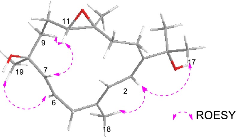
As for the relative configuration of compound 1, according to the ROESY correlations between H-2 (δH 6.30)/Me-17 (δH 1.36), H-2/Me-18 (δH 1.78) and H-3 (δH 5.86)/H-14b (δH 2.22) (Fig. 1), the olefinic geometries were assigned to 1E and 3E. The 13C NMR chemical shift of Me-20 (δC < 20 ppm) and the absence of the ROESY correlations of H-11 (δH 3.08)/Me-20 (δH 1.25) indicated the trans-configuration of the epoxy group at C-11/C-12 in 1, which was the same as that in 4. Additional ROESY interactions of H-11/H-7, H-7/H-9b (δH 1.70), H-9b/H-11 suggested these three protons were co-facial, assigned tentatively as β-orientation. Thus, Me-19 was accordingly β-oriented due to the diagnostic cross-peak of Me-19 (δH 1.40) with H-7, allowing the determination of the structure of 1 as showed in Fig. 2, which was the Δ6(7)-8α-hydroxyl derivative of 4.
Fig. 1.
Key COSY, HMBC and ROESY correlations for compound 1
Fig. 2.
Chemical structures of compounds 1–5
Sarcophytrol E (2) was also obtained as colorless oil. The molecular formula, C20H30O3, established by HRESIMS (m/z 343.2243 [M + Na]+), was identical to that of 1. A detailed 2D NMR analysis of 2 and careful comparison with the NMR data of 1 (Tables 1 and 2) revealed that their structures were almost the same. In fact, the only differences between 2 and 1 were the C-19 signal downfield shifted in the 13C NMR spetrum (δC 28.8 in 1 and δC 31.3 in 2), while the C-6 and C-9 signals upfield shifted (δC 125.0, 40.1 in 1 and δC 123.6, 38.9 in 2, respectively). The observed differences can be rationalized when the two compounds are C-8 epimers, which was confirmed by the ROESY interaction of H-6 (δH 5.81)/Me-19 (δH 1.36) (Fig. 3). Since the hydroxyl group at C-8 of 1 was α-oriented, the opposite configuration at this center is therefore tentatively suggested for 2.
Fig. 3.
Key ROESY correlations for compound 2
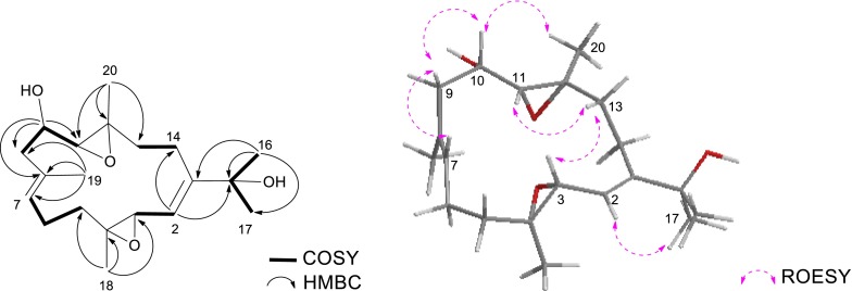
The HRESIMS of sarcophytrol F (3) established the molecular formula C20H32O4 (m/z 359.2188 [M + Na]+), 16 mass units more than that of sinugibberol (5) [19]. The 1H and 13C NMR data of 3 showed great similarity as those of 5 (Tables 1 and 2), some minor differences were observed in relation to the functional group. The presence of a secondary hydroxyl group in the molecule was readily recognized by a signal resonating at δH 3.70 (1H, ddd, J = 8.7, 7.7, 4.0 Hz) in its 1H NMR spectrum, and by a carbon signal at δC 69.1 (CH) in the 13C NMR and DEPT spectra. The oxygenated methine proton was secured at C-10 by a COSY cross-peak between H-9 (δH 2.44, 2.30) and H-11 (δH 3.57), and the HMBC correlations with C-8, C-9 and C-11 (Fig. 4). Due to the presence of the 10-OH, 13C NMR chemical shifts of C-9 to C-12 were all reasonably downfield shifted with respect to those of 5.
Fig. 4.
Key COSY, HMBC and ROESY correlations for compound 3
Finally, the relative configuration of 3 was determined by ROESY experiment and by comparison of the NMR data with 5 (Fig. 4). The 13C NMR chemical shift of Me-18 and Me-20 (δC < 20 ppm) and the absence of ROESY correlations of Me-18 (δH 1.25)/H-3 (δH 3.36), and Me-20 (δH 1.30)/H-11 (δH 2.90), indicated the trans-configurations of these two epoxy groups at C-3/C-4 and C-11/C-12 in 3, which were the same as those in 5. The similar 13C NMR data of C-3 and C-4 in 3 and 5 further confirmed the same stereochemistry of this epoxy group. Furthermore, the ROESY correlations of H-11/H-13a (δH 1.44), H-3/H-13a, suggested that H-11 and H-3 were co-facial, leading to the determination of the relative configurations at C-11 and C-12 where the other epoxy group resided on. In addition, the ROESY correlations of H-10/Me-20 indicated that 10-OH and H-11 were co-facial. Due to the scarcity of material, the modified Mosher’s method could not be able to apply for the determination of the absolute configuration in C-10 position in 3 at this moment. Thus the structure of sarcophytrol F (3) was tentatively determined as 10-hydroxyl derivative of 5.
All the compounds were tested for the cytotoxic activities and inhibitory activities against human protein tyrosine phosphatase 1B (PTP1B), a key target for the treatment of type-II diabetes and obesity [20]. Unfortunately, none of them showed inhibitory effects toward the above bioassays. Further study should be conducted to understand the real biological/ecological role of these metabolites in the life cycle of the animal, as well as to carry out other biological evaluations such as antioxidant, anti-inflammatory, anti-fouling activities, etc.
Experimental Section
General Experimental Procedures
Optical rotations were measured on a Perkin-Elmer 341 polarimeter. HRESIMS spectra were recorded on a Waters-Micromass Q-TOF Ultima Global electrospray mass spectrometer. NMR spectra were measured on a Bruker-DRX-500 spectrometer with the residual CHCl3 (δH 7.26 ppm, δC 77.0 ppm) as internal standard. Chemical shifts are expressed in δ (ppm) and coupling constants (J) in Hz. 1H and 13C NMR assignments were supported by 1H-1H COSY, HSQC, and HMBC experiments. Commercial silica gel (Qing Dao Hai Yang Chemical Group Co., 200–300 and 400–600 mesh), C18 reversed-phase silica gel (150–200 mesh, Merck) and Sephadex LH-20 (Amersham Biosciences) were used for column chromatography. Semi-preparative HPLC (Agilent 1100 series liquid chromatography using a VWDG1314A detector at 210 nm and a semi-preparative ODS-HG-5 [5 μm, 10 mm (i.d.) × 25 cm] column was also employed. Pre-coated silica gel GF254 plates (Qing Dao Hai Yang Chemical Group Co. Ltd. Qingdao, People’s Republic of China) were used for analytical thin-layer chromatography (TLC). All solvents used were of analytical grade (Shanghai Chemical Reagents Company, Ltd.).
Animal Material
The soft corals S. trocheliophorum were collected by scuba at Yalong Bay, Hainan Province, China, in February 26, 2006, at a depth of −15 to −20 m, and identified by Professor R.-L. Zhou of South China Sea Institute of Oceanology, Chinese Academy of Sciences. The voucher sample is deposited at the Shanghai Institute of Materia Medica, CAS, under registration No. YAL-4.
Extraction and Isolation
The lyophilized bodies of S. trocheliophorum (400 g, dry weight) were minced into pieces and exhaustively extracted with Me2CO at room temperature (3 × 1 L). The solvent-free Me2CO extract was partitioned between Et2O and H2O. The organic phase was evaporated under reduced pressure to give a dark brown residue (10 g), which was subjected to silica gel column chromatography (CC) and eluted with petroleum ether (PE) in Me2CO (0–100 %, gradient) to yield 14 fractions (A–M).
Fraction D was chromatographed over silica gel (PE/Et2O, 1:1) to give pure 4 (32.1 mg). Fraction F was subjected to Sephadex LH-20 CC (PE/CH2Cl2/MeOH, 2:1:1), followed by repeated silica gel CC (PE/CH2Cl2, 20:1) to yield 6 sub-fractions (F1-F6). F2 gave compound 5 (2.3 mg) after silica gel CC (PE/CH2Cl2, 10:1). Fraction G was chromatographed over Sephadex LH-20 (CHCl3/MeOH, 1:1), followed by ODS (MeOH/H2O, 50:50–90:10) to yield 10 sub-fractions (G1-G10). After purification by RP-HPLC (MeOH/H2O, 88:12, 2.0 mL/min), G5 yielded pure 1 (2.4 mg, tR 6.3 min). Fraction H gave compound 3 (2.3 mg, tR 12.5 min) after Sephadex LH-20 CC (PE/CHCl3/MeOH, 2:1:1), CC on ODS (MeOH/H2O, 45:55–90:10) and RP-HPLC (MeOH/H2O, 73:27, 2.0 mL/min). Fraction I was subjected to Sephadex LH-20 CC (PE/CHCl3/MeOH, 2:1:1) to give five sub-fractions (I1-I5). Fraction I4 was first split by CC on ODS (MeOH/H2O, 50:50–90:10) and then purified by silica gel (CHCl3/MeOH, 50:1) to afford pure 2 (3.1 mg).
Sarcophytrol D (1): colorless oil; [α] +18.6 (c 0.07, MeOH); 1H and 13C NMR data, see Tables 1 and 2; HRESIMS m/z 343.2248 [M + Na]+ (calcd for C20H32O3Na, 343.2249).
Sarcophytrol E (2): colorless oil; [α] +48.6 (c 0.10, MeOH); 1H and 13C NMR data, see Tables 1 and 2; HRESIMS m/z 343.2243 [M + Na]+ (calcd for C20H32O3Na, 343.2249).
Sarcophytrol F (3): colorless oil; [α] −12.7 (c 0.20, MeOH); 1H and 13C NMR data, see Tables 1 and 2; HRESIMS m/z 359.2188 [M + Na]+ (calcd for C20H32O4Na, 359.2188).
Acknowledgments
This research work was financially supported by the National Marine “863” Projects (Nos. 2013AA092902 and 2012AA092105), the Natural Science Foundation of China (Nos. 81520108028, 81273430, 41506187, 41306130, 41476063), NSFC Shangdong Joint Fund for Marine Science Research Centers (Grant No. U1406402), SCTSM Project (No. 14431901100 and 15431901000), the SKLDR/SIMM Projects (SIMM1203ZZ-03 and 1501ZZ-03), and was partially funded by the EU 7th Framework Programme-IRSES Project (No. 246987).
Compliance with Ethical Standards
Conflict of Interest
All authors declare no conflict of interest.
Footnotes
Wen-Ting Chen and Lin-Fu Liang have contributed equally.
Contributor Information
Wei Xiao, Phone: +86 21 50805813, Email: wzhzh-nj@163.com.
Yue-Wei Guo, Phone: +86 21 50805813, Email: ywguo@mail.shcnc.ac.cn, Email: ywguo@simm.ac.cn.
References
- 1.Dinesen ZD. Coral Reefs. 1983;1:229–236. doi: 10.1007/BF00304420. [DOI] [Google Scholar]
- 2.Fabricius KE. Coral Reefs. 1997;16:159–167. doi: 10.1007/s003380050070. [DOI] [Google Scholar]
- 3.Riegl B, Schleyer MH, Cook PJ, Branch GM. Bull. Mar. Sci. 1995;56:676–691. [Google Scholar]
- 4.Tursch B, Tursch A. Mar. Biol. 1982;68:321–332. doi: 10.1007/BF00409597. [DOI] [Google Scholar]
- 5.Liang LF, Guo YW. Chem. Biodivers. 2013;10:2161–2196. doi: 10.1002/cbdv.201200122. [DOI] [PubMed] [Google Scholar]
- 6.Anjaneyulu ASR, Rao GV. J. Indian Chem. Soc. 1997;74:272–278. [Google Scholar]
- 7.Liang LF, Kurtan T, Mandi A, Yao LG, Li J, Zhang W, Guo YW. Org. Lett. 2013;15:274–277. doi: 10.1021/ol303110d. [DOI] [PubMed] [Google Scholar]
- 8.Liang LF, Kurtan T, Mandi A, Yao LG, Li J, Zhang W, Guo YW. Eur. J. Org. Chem. 2014;2014:1841–1847. doi: 10.1002/ejoc.201301683. [DOI] [Google Scholar]
- 9.Chen WT, Yao LG, Li XW, Guo YW. Tetrahedron Lett. 2015;56:1348–1352. doi: 10.1016/j.tetlet.2015.01.157. [DOI] [Google Scholar]
- 10.Yan XH, Gavagnin M, Cimino G, Guo YW. Tetrahedron Lett. 2007;48:5313–5316. doi: 10.1016/j.tetlet.2007.05.096. [DOI] [Google Scholar]
- 11.Jia R, Kurtán T, Mándi A, Yan X-H, Zhang W, Guo Y-W. J. Org. Chem. 2013;78:3113–3119. doi: 10.1021/jo400069n. [DOI] [PubMed] [Google Scholar]
- 12.Liang LF, Lan LF, Taglialatela-Scafati O, Guo YW. Tetrahedron. 2013;69:7186–7381. [Google Scholar]
- 13.Dong H, Gou YL, Kini RM, Xu HX, Chen SX, Teo SLM, But PPH. Chem. Pharm. Bull. 2000;48:1087–1089. doi: 10.1248/cpb.48.1087. [DOI] [PubMed] [Google Scholar]
- 14.Chen WT, Liu HL, Yao LG, Guo YW. Steroids. 2014;92:56–61. doi: 10.1016/j.steroids.2014.08.027. [DOI] [PubMed] [Google Scholar]
- 15.Cheng ZB, Deng YL, Fan CQ, Han QH, Lin SL, Tang GH, Luo HB, Yin S. J. Nat. Prod. 2014;77:1928–1936. doi: 10.1021/np500394d. [DOI] [PubMed] [Google Scholar]
- 16.Řezanka T, Dembitsky VM. Tetrahedron. 2001;57:8743–8749. doi: 10.1016/S0040-4020(01)00853-5. [DOI] [Google Scholar]
- 17.Liang LF, Gao LX, Li J, Taglialatela-Scafati O, Guo Y-W. Bioorg. Med. Chem. 2013;21:5076–5080. doi: 10.1016/j.bmc.2013.06.043. [DOI] [PubMed] [Google Scholar]
- 18.Duh CY, Hou RS. J. Nat. Prod. 1996;59:595–598. doi: 10.1021/np960174n. [DOI] [Google Scholar]
- 19.Hou RS, Duh CY, Chiang MY, Lin CN. J. Nat. Prod. 1995;58:1126–1130. doi: 10.1021/np50121a026. [DOI] [PubMed] [Google Scholar]
- 20.Byon JCH, Kusari AB, Kuseti J. Mol. Cell. Biochem. 1998;182:101–108. doi: 10.1023/A:1006868409841. [DOI] [PubMed] [Google Scholar]


