Abstract
Patients with inflammatory bowel disease have an additional risk of developing cancer compared with the general population. This is due to local chronic inflammation that leads to the development of gastrointestinal cancers and the use of thiopurines, associated with a higher risk of lymphoproliferative disorders, skin cancers, or uterine cervical cancers. Similar to the general population, a previous history of cancer in inflammatory bowel disease patients increases the risk of developing a secondary cancer. Large studies have not shown an increased risk of cancer in patients treated with biologics. In this review we discuss the prevention and treatment of cancer in patients with inflammatory bowel disease.
Keywords: Crohn’s disease, ulcerative colitis, anti-TNF, chemotherapy, cancer
Introduction
Inflammatory bowel disease (IBD) is a chronic immune-mediated disease with a relapsing course that affects mainly the gastrointestinal tract. Crohn’s disease (CD) and ulcerative colitis (UC) are the two main disorders included in this definition. CD is characterized by a transmural inflammation that can involve, discontinuously, the entire gastrointestinal tract from mouth to the perianal area, but ileum and colon are preferentially affected. In UC, a continuous mucosal inflammation of the colon, affecting the rectum and a variable extent of the colon in continuity can be observed. IBD is frequently associated with extraintestinal manifestations such as arthritis, eye involvement, skin disorders, primary sclerosing cholangitis (PSC), venous or arterial thromboembolism or pulmonary involvement, and/or concomitant immune-mediated diseases.
The pathogenesis of IBD remains unclear but the emerging hypothesis proposes that inflammation in IBD is the consequence of a dysregulated mucosal immune response to antigens derived from the commensal microbiota in a genetically susceptible host. It means that, although there is a genetic susceptibility for this disease, a diversity of environmental factors can act as a trigger for its clinical presentation.
The goal of medical treatment is to modify the disease course by achieving mucosal healing. Drugs used in the treatment of IBD include 5-aminosalicylic acid (5-ASA), immunomodulators (IMM) and biologic agents. 5-ASA is a bowel-specific aminosalicylate drug that acts locally in the gut and is used for induction and maintenance treatment in patients with UC [1]. As it is rapidly absorbed in the jejunum, allowing only 20% to reach the terminal ileum and colon, a number of 5-ASA compounds have been developed to prevent absorption of 5-ASA in the proximal gastrointestinal tract, thereby increasing delivery to the colon.
IMM, that encompasses azathioprine, 6-mercaptopurine, and methotrexate, have shown their efficacy mainly as maintenance therapy of remission in IBD [1,2]. Azathioprine is a derivative of thioguanine, a purine mimic antimetabolite. It is a pro-drug that is transformed into its principal metabolite, 6-mercaptopurine, via a non-enzymatic nucleophilic attack by sulfhydryl-containing compounds, such as glutathione, present in red blood cells and other tissues. 6-mercaptopurine is then metabolized in the liver and gut by one of three enzymes. Methotrexate is a folic acid analogue that inhibits purine and pyrimidine synthesis, which accounts for its efficacy in the therapy of cancer, and its adenosine-mediated anti-inflammatory effects makes it a good immunosuppressant alternative drug. Methotrexate is a prodrug that becomes active only when polyglutamated within cells.
Among biologic agents, anti-tumor necrosis factor (TNF)-α drugs have been widely used for the treatment of IBD [3-9]. Anti-TNF-α are monoclonal antibodies that block the pro-inflammatory cytokine TNF-α involved in the pathogenesis of IBD. The approved anti-TNF-α agents in Europe are infliximab, a mouse-human chimeric IgG1 antibody, adalimumab, a fully human recombinant IgG1 antibody, and golimumab, a human IgG1 anti-TNF-α. All are approved for induction and maintenance therapy.
More recently, other biologic agents have been introduced, but their safety in patients with cancer will not be discussed in detail in this review given that there is a lack of available data. The anti-integrin agents - natalizumab (humanized IgG4 antibody against α4 integrin) and vedolizumab (humanized IgG1 antibody against α4β7 integrin) - modulate gut lymphocyte trafficking, and ustekinumab is a monoclonal antibody that blocks interleukin (IL)-12 and IL-23 [10-13]. The risk of developing progressive multifocal leukoencephalopathy associated with the John Cunningham virus in patients who receive natalizumab has limited its use. Vedolizumab and ustekinumab have shown beneficial effects in IBD patients in phase III studies respectively.
The main long-term complications of immunosuppressive therapy for IBD are the risk of opportunistic infections and cancer development. Several mechanisms have been proposed as promotors of carcinogenesis, including alteration in DNA, activation of oncogenes, reduction in physiologic immunosurveillance, and facilitation of the action of oncogenic viruses [14-16]. Anti-TNF-α agents have a dual effect on tumor progression, as TNF-α can show anti-tumor effects by initiating cellular apoptosis of malignant cells, but its secretion by most tumors can also help cellular survival and increase tumor proliferation [17,18].
Cancer risk in IBD patients
In order to understand the association of IBD and cancer, three different situations should be considered in patients with IBD: 1) cancer related to the local chronic inflammation (IBD-related cancer); 2) cancer related to the use of immunosuppressive drugs in this population (immunosuppressive treatment-related cancer); and 3) any unrelated cancer that can occur in a patient with IBD.
IBD-related cancer
Colorectal cancer (CRC)
Long-term inflammation of the colon is associated with an increased risk of CRC, both in CD and UC [19-21]. However, recent population-based studies have suggested that the risk may not be as high as it was initially believed, and, moreover, that the risk has actually decreased over time [22-24].
IBD-associated cancers develop in chronically inflamed mucosa, so the duration and the anatomic extent of disease affect the development of IBD-related CRC. According to a population-based study published in 1990, the incidence of CRC increased by 14.8 times in patients with extensive colitis and by three times in patients with left-sided colitis; it was similar to that in the general population in patients with proctitis [25]. A more recent study showed a lower but still significantly increased risk (5.6 times for pancolitis, 2.1 times for Crohn’s colitis, and 1.7 times for proctitis [26]). Compared with sporadic CRC, IBD-related CRC is detected in younger patients, is more often found in the proximal colon, and two or more synchronous CRC are more frequently detected.
Regarding chemoprevention, international guidelines suggest the use of 5-ASA compounds in patients with UC and the use of ursodeoxycholic acid in patients with PSC associated with IBD, as it has been proven that the use of these drugs reduces the incidence of CRC [27-29].
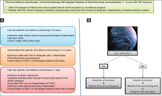
In most cases, CRC in IBD patients develops from dysplasia, so surveillance colonoscopies for patients with UC or colonic CD are recommended to detect dysplastic changes in colonic mucosa [27,28]. According to the European Crohn’s and Colitis Organization (ECCO) guidelines, the first surveillance colonoscopy should be performed six to eight years after the IBD diagnosis in all patients with colonic involvement independently of the disease activity, to reassess disease extent and confirm the absence of dysplastic lesions [27]. When the disease is limited to the rectum, the patient should not be included in a surveillance program. The concomitant presence of PSC increases the risk of CRC in patients with IBD, with the cumulative risk reported to be 25% at 10 years, so it is therefore advisable that these patients undergo surveillance colonoscopies yearly from the moment of diagnosis, independently of disease activity or extent [27].
The CRC risk profile should be determined in this first surveillance colonoscopy, depending on the extent of the disease, the severity of endoscopic and/or histological inflammation, the presence of colonic strictures or pseudopolyps and a family history of CRC [27]. This individual risk profile will dictate surveillance colonoscopy intervals (Fig. 1): a) high-risk patients - those with stricture or dysplasia detected within the past five years (high-grade or low-grade dysplasia), associated PSC, extensive colitis with severe active inflammation, or a family history of CRC in a first-degree relative aged less than 50 years - should have the next surveillance colonoscopy in 1 year; b) intermediate-risk patients - those with extensive colitis with mild or moderate active inflammation, post-inflammatory polyps or a family history of CRC in a first-degree relative aged 50 years and above - should undergo the next surveillance colonoscopy in two-three years; c) low-risk patients - those not included in the previous groups - should have their next surveillance colonoscopy in five years [28,30].
Figure 1.

Surveillance colonoscopies. (A) Surveillance colonoscopies for detecting dysplasia and preventing colorectal carcinoma. (B) Management of visible lesions at endoscopy. A visible lesion with dysplasia should be completely resected, independently of the grade of dysplasia or the localization relative to the inflamed mucosal areas. In the absence of dysplasia in the surrounding mucosa, the patient should follow the standard surveillance program. If endoscopic resection is not possible or if dysplasia is found in the surrounding flat mucosa, proctocolectomy should be recommended [27-29] CRC, colorectal cancer; IBD, inflammatory bowel disease; PSC, primary sclerosing cholangitis
Guidelines suggest the use of chromoendoscopy with targeted biopsies in patients in endoscopic remission for the endoscopic surveillance as the tumor is usually difficult to detect by white light endoscopy because of the inflammation-induced changes in the colonic mucosa. When possible, surveillance colonoscopies should be performed during disease remission to distinguish between inflammatory and neoplastic changes, but the presence of activity should not delay the procedure in high-risk patients [27,28]. A visible lesion with dysplasia should be completely resected, independently of the grade of dysplasia or the localization relative to the inflamed mucosal areas. In the absence of dysplasia in the surrounding mucosa, the patient should follow the surveillance program according to the previously commented in the ECCO guidelines [27,28]. If endoscopic resection is not possible or if dysplasia is found in the surrounding flat mucosa, proctocolectomy should be recommended. When dysplasia is detected but not associated to an endoscopically visible lesion (random biopsies) the risk of developing CRC is highly increased so: a) adenocarcinoma or high-grade dysplasia without associated visible lesion should be an indication for surgery; and b) low-grade dysplasia lesion without associated visible lesion should be confirmed by repeat chromoendoscopy after three months.
CRC after colectomy in IBD patients
Colectomy is the surgical treatment of choice for UC or extensive colorectal CD patients. In UC, the initial approach consists in subtotal colectomy and ileostomy with a residual rectum left in situ, which allows a subsequent option of reconstructive surgery, preferably the ileal pouch-anal anastomosis. In extensive colonic disease of CD, ileorectal anastomosis is the first restorative option, but, in some cases, these patients continue with a permanent ileostomy and rectal stump. Although colectomy, with or without reconstructive surgery, substantially reduces the risk to develop CRC, the risk of cancer of the residual rectum or ileoanal pouch is still present. A recent systematic review and meta-analysis showed that the pooled prevalence of carcinoma was 0.5% in the ileoanal pouch, 2.4% in the ileorectal anastomosis, and 2.1% in the rectal stump [31]. The presence of a residual rectum, IBD duration, and a history of CRC or dysplasia, the presence of type C mucosa in the pouch (mucosa exhibiting permanent persistent atrophy and severe inflammation), and PSC have been associated with a higher cancer risk after colectomy [28,30,31].
International guidelines recommend yearly flexible sigmoidoscopy of pouch/rectal mucosa in higher risk post-colectomy patients (those with previous rectal dysplasia, dysplasia/CRC at the time of pouch surgery, PSC, or type C mucosa in the pouch), and 5-yearly flexible sigmoidoscopy in patients with none of these risk factors [28,30].
Anal and perianal carcinoma
Patients with perianal disease have a higher incidence of anal or perianal carcinoma with an earlier age of presentation (80% of cases are squamous cell carcinomas and 20% are adenocarcinoma) [32,33]. Furthermore, squamous cell carcinoma of the anus is strongly associated with human papillomavirus (HPV) infection which is the cause of the cancer in 80-85% of patients. Thus, factors increasing the risk of HPV infection and/or modulating host response can increase the risk of persistent HPV infection, leading ultimately to malignancy: anal intercourse and a high lifetime number of sexual partners, human immunodeficiency virus, immunosuppressants, a history of other HPV-related cancers, autoimmune disorders, social deprivation, and cigarette smoking [34].
Although routine screening is not recommended, anal/perianal carcinoma should be suspected in patients with long-standing perianal disease and new symptoms such as anal pain. Clinical examination of the perineum, examination under anesthesia with biopsies or curettage, and MRI should be performed in order to rule out this cancer.
Small bowel adenocarcinoma
Patients with CD have an increased risk of small bowel adenocarcinoma (SBA). One study compared the characteristics of SBA in patients with CD with those of SBA de novo [35]. Patients with CD and SBA were younger than those with SBA de novo and developed the cancer in inflamed ileum, whereas SBA de novo usually presents between 50 and 70 years of age and its incidence is highest in the duodenum and decreases progressively along the rest of the small intestine.
In the Cancers Et Surrisque Associé aux Maladies inflammatoires intestinales En France (CESAME) cohort, a nationwide prospective observational cohort designed to assess the risk of cancer in IBD patients, five incident (occurring after inclusion in the cohort) cases of SBA were diagnosed in 19,486 CD patients, which represents a very high incidence compared to the general population [36]. Small bowel involvement of CD was present in all cases and the median CD duration was 11 years. Most cancers were discovered at a large stage and carried a poor prognosis.
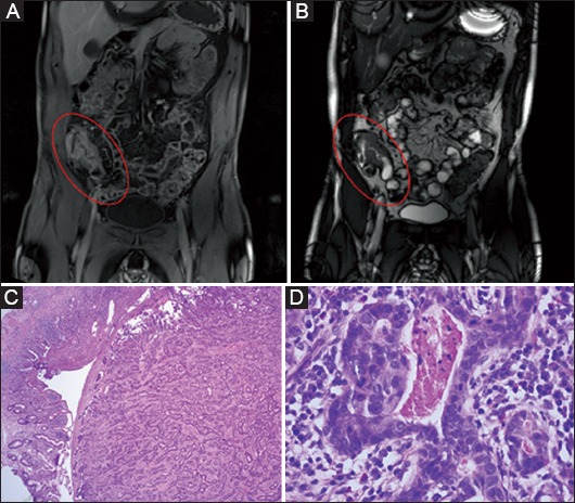
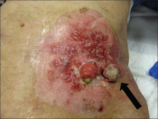
No systematic screening of SBA has been proposed in patients with ileal CD due to the small number of total cases reported. However, in high-risk patients, such as those with longstanding stricturing CD who have not undergone small bowel resection, regular imaging with magnetic resonance enterography and/or ileocolonoscopy with targeted biopsies should be performed and sudden change in symptoms, including onset of systemic symptoms or progression of disease on imaging, should raise suspicion of malignancy. In Fig. 2, a case of ileal adenocarcinoma is presented in a patient with longstanding ileal stenosing CD, who suddenly developed obstructive symptoms and was diagnosed with ileal adenocarcinoma during semi-urgent ileocecal resection. In Fig. 3, an incidental adenocarcinoma of intestinal origin was detected 14 years after end-ileostomy for CD.
Figure 2.

Small bowel adenocarcinoma. A 45-year-old male, with a 26-year duration ileal Crohn’s disease presented with subobstructive symptoms. At magnetic resonance imaging (MRI), severe wall thickening and ulcerative stenosis was observed. The patient underwent a laparoscopic right hemicolectomy and the pathology study of the resected segment revealed an ileal adenocarcinoma. (A) T1-weighted MRI sequence in coronal plane after administration of gadolinium. Severe wall thickening (1 cm) and presence of inflamed lymph nodes, without inflammatory signs in the surrounding tissue can be observed. (B) T2-weighted MRI sequence in coronal plane. Additionally, a reduction of the lumen with ulcerative stenosis can be observed. (C) 25x magnification pathology study. Residual ileal mucosa can be observed in the left lower corner. The rest of the picture shows the transition to the ileal tumor, composed by very irregular and atypical glands in a fibrous stroma. (D) 400x magnification pathology study. Tumor glands can be observed in the resected ileum. They have a very irregular shape and are composed by atypical cells with big hypercromatic overlapping nucleus and necrotic material in the gland
Figure 3.

Adenocarcinoma of intestinal origin adjacent to end-ileostomy. An 80-year-old male diagnosed with Crohn’s disease for more than 50 years, and for which an end-ileostomy for 14 years presented an exophytic lesion at the left side of the ileostomy (arrow). Biopsy of the lesion showed adenocarcinoma of intestinal origin
Cholangiocarcinoma (CC)
CC is a rare malignant tumor arising from the cholangiocytes lining the biliary tree, and has a poor prognosis. Several studies have shown a higher incidence of cholangiocarcinoma in patients with IBD [37-40]. PSC, an extraintestinal manifestation of IBD, is often suggested as a predisposing factor to this malignancy in IBD patients [41,42].
Immunosuppressive treatment-related cancer
Lymphoproliferative disorders (LD)
Organ transplant recipients who receive thiopurines as part of the immunosuppressive therapy are at increased risk of developing LD, with a frequent pathogenic association with Epstein-Barr virus (EBV) that comprises a spectrum of B-cell hyperproliferative states from benign conditions such as infectious mononucleosis-like illnesses and polyclonal lymphoid hyperplasia to B-cell, and occasionally T-cell, lymphomas [43].
The landmark prospective CESAME study assessed the risk of LD in IBD patients [44]. It was observed that the risk of LD was five times higher in patients exposed to thiopurines than in those never exposed to these drugs. Age >65 years, male sex, and longer duration of IBD were also associated with increased risk of incident LD. The reason for the excess risk of LD in IBD patients receiving thiopurines is not well known and it has been postulated that inflammatory process itself, thiopurine exposure, or combination of both could contribute. Patients who discontinued thiopurines in this study had a similar risk to those patients who never received thiopurines. An American nationwide retrospective cohort study with UC patients also showed a four-fold increased risk of lymphoma in patients treated with thiopurines [45].
Few patients in the CESAME study were given biologics and in most of these patients, this was given in combination with thiopurines, so no solid conclusions can be drawn. However, analysis of a nationwide study, a systematic review with meta-analysis and a pooled analysis of 10 clinical trials did not show an increased risk of cancer associated with the use of biologics [46-49].
A special type of lymphoma, the early post-mononucleosis lymphoma, can appear in EBV-negative young males exposed to thiopurines, after developing a severe primary EBV infection [50-53]. In CESAME cohort, two cases of early fatal post-mononucleosis LD in young men receiving thiopurines were recorded [44].
In IBD patients under thiopurines, the suspicion of LD should arise in cases of unexplained headache, fatigue or fever, with adenopathies not attributable to intestinal inflammation, increased size of spleen or liver, unexplained biological inflammatory syndrome, with or without increase in serum lactate dehydrogenase (LDH) and β2-microglobulin levels and hemophagocytic lymphohystiocytosis. The routine workout should be performed, including EBV viral load, and the hematologist’s opinion should be requested [54].
Following these cases of hemophagocytic syndrome, it has been proposed to test EBV serology in IBD patients before starting thiopurines, and to consider avoiding thiopurines in EBV-negative young male patients [54].
Hepatosplenic T-cell lymphoma (HSTCL)
HSTCL is a rare form of lymphoma that affects young men and is lethal in most cases. It is not related to EBV and its pathogenesis is unknown. Up to 20% of patients have a history of chronic immunosuppression, solid organ transplantation, LD or IBD. In patients with IBD, the risk appears to be particularly increased when receiving long-term thiopurine therapy [55]. In the CESAME study population, only one case of HSTCL occurred, after the end of the study period [56].
According to ECCO recommendations, the long-term combination of thiopurines and biologics should be avoided in young people because of the risk of HSTCL [2,54].
Non-melanoma skin cancer (NMSC)
NMSC mainly comprises basal cell carcinoma and squamous cell carcinoma, which are by far the most prevalent malignancies in industrialized countries. UV-radiation exposure has been identified as a risk factor for NMSC, especially squamous cell carcinoma but, on the other hand, exposure to UV-radiation results in cutaneous vitamin D synthesis. Vitamin D deficiency has been associated with various types of cancer (colon, prostate and breast cancer). Thus, dietary vitamin D supplementation has been proposed as chemoprophylaxis for these type of cancers, although the association of vitamin D intake with NMSC has been inconsistent [57-59]. An excess risk of this type of cancers has also been observed in transplant recipients.
In IBD patients, several studies have shown that ongoing and past exposure to thiopurines significantly increases the risk of NMSC in patients with IBD [60-65]. Additional factors can increase the risk of skin cancer: fair skin, atypical moles, advanced age, a history of painful or severe sunburns, outdoor occupation, and a family history of skin cancer [54]. As a general recommendation, IBD patients under immunosuppressive treatments should be protected against UV radiation and receive lifelong dermatologic screening [54].
Melanoma skin cancer (MSC)
Retrospective cohort and nested case-control studies showed an increased risk of MSC among IBD patients under biologics. A meta-analysis including 172,837 patients with IBD reported a total of 179 MSC cases which reflected a 37% higher risk than expected in the general population. There was no increased risk attributable to immunosuppressive medication use [63-66]. This finding supports, together with the increased risk of NMSC, the recommendation for routine dermatologic screening in patients with IBD. Moreover, primary prevention with UV-radiation protective measures should be advised to all patients with IBD, regardless of thiopurine and/or anti-TNF-α use.
Uterine cervical high-grade dysplasia or cancer
Uterine cervical cancer is the third most common cancer in women, with an estimated 529,828 new cases and 275,128 deaths reported worldwide in 2008. HPV is a carcinogenic virus that is sexually transmitted and is the causal risk factor for uterine cervical cancer. It is detected in 99% of cervical tumors, in particular the oncogenic subtypes such as HPV 16 and 18 [67]. A recent meta-analysis has suggested an increased risk of high-grade uterine cervical dysplasia and cervical cancer among patients with IBD who are on immunosuppressive medications compared with the general population (odds ratio 1.34 [95%CI 1.23-1.46]) [68]. ECCO guidelines recommend regular gynecologic screening for uterine cervical cancer for women with IBD, especially if treated with IMM [69]. Young women under immunosuppressive treatment should obtain a Pap test twice in the first year after diagnosis and, if the results are normal, annually thereafter.
Current or past infection with HPV is not a contraindication for IMM therapy, although in patients with extensive cutaneous warts and/or condylomata, discontinuation of IMM should be considered. Routine prophylactic HPV vaccination is recommended for females and males according to national guidelines [54]. The available HPV vaccines are not live vaccines and, as such, can be safely administered to immunosuppressed patients and provide protection against the most two prevalent oncogenic HPV genotypes, namely HPV 16 and 18, which account for about 70% of cervical cancers: the tetravalent HPV4 is directed against HPV types 6, 11, 16, and 18 (Gardasil, Merck and Co, Whitehouse Station, NY), and the bivalent HPV2 is directed against HPV 16 and 18 (Cervarix, GlaxoSmithKline, London, United Kingdom) [70]. Smoking cessation and counseling about family planning and sexual practices should be provided in women with IBD and cervix abnormalities.
Oral cancer
Different risk factors have been associated with oral cancer: tobacco smoking, alcohol consumption, age over 40, male sex, dietary factors and nutritional deficiencies, infections (HPV, human immunodeficiency virus, syphilis, chronic candidiasis, cytomegalovirus, and EBV), chronic irritation in oral cavity, genetic susceptibility and immunosuppressive status. Precancerous lesions in the oral cavity include leukoplakia, erythroplakia, and lichen planus. Histology of oral cancer varies widely but the great majority are squamous cell carcinomas.
Although IBD patients should be considered as a high-risk group because of the increasing HPV prevalence and the increasing use of immunosuppressive drugs, the lack of studies prevents us from knowing the incidence and prevalence of oral cancerous and pre-cancerous lesions in IBD. Data collected in a systematic review show that precancerous lesions have been rarely reported in IBD patients; two cases of oral cancer have been reported in IBD patients under azathioprine and no cases of oral cancer associated with biological therapy have been published [71]. Efforts should be focused on prevention and educating on modifiable risk behaviors in patients with oral cancer. Oral screening should be performed for all IBD patients, especially before starting IMM or biological treatment.
Unrelated cancer occurring in a patient with IBD
Patients with a diagnosis of IBD also have the same risks of developing cancer as the general population. Worldwide, cancer is the second leading cause of death with prostate cancer being the most commonly occurring cancer in males and breast cancer in females, followed by lung cancer and CRC in both sexes [72,73]. In general, one in three people will develop cancer during their lifetime.
Secondary cancer risk in IBD patients
In the CESAME prospective cohort, the overall incidence of cancer was significantly higher in IBD patients with a previous cancer when compared with patients without a history of cancer [56]. Patients with IBD and a history of cancer were more prone to develop a new cancer than to develop a recurrence.
Among the 17,047 followed patients there were 405 patients (2.4%) with a personal history of cancer at study entry and 16 patients with a previous history of two subsequent cancers involving distinct organs. CRCs (21.4% of patients) and breast cancers (20.2%) were the more frequently diagnosed, followed by uterine cervical cancer (7.6%), prostate cancer (5.9%), NMSC (5%), kidney (4%) and non-Hodgkin lymphoma (4%). Among the 405 patients with previous cancer, 93 (23%) were exposed to any immunosuppressant at the time of entry into the observational period, and 312 (77%) were not exposed. During the study period, 316 patients developed an incident cancer: 293 patients did not have a history of previous cancer (293/16,642 patients, 1.8%) and 23 had previous cancer at study entry (23/405, 5.7%). Most of the patients with a previous cancer (16/23, 70%) developed new cancers affecting a different organ, or affecting the same organ but with a different histological type.
In a recently published retrospective study, 333 IBD patients who developed cancer and subsequently received treatment with biologics, IMM, combo or did not receive any immunosuppressive treatment were followed up to detect incident cancers (new or recurrent) over the study period [74]. Ninety patients (27%) developed an incident cancer (44 [13.2%] a new cancer, 48 [14.4%] a recurrent cancer, and 8 [2.4%] a new and recurrent cancer), but exposure to IMM or biologics was not associated with an increased risk of incident cancer.
The use of biologics in patients with cancer
As gastroenterologists and oncologists are regularly confronted with an IBD patient in whom cancer occurs, an important concern is whether or not to start or continue treatment with biologics. As a general rule, we feel all types of immunosuppressive treatments for IBD should best be stopped while patients undergo chemotherapy, and frequently therapy is not restarted after completion of the chemotherapy whilst patients are in remission from cancer, given the theoretical cancer risk associated with continuous immunosuppression. In addition, patients with a history of cancer within five years are generally excluded from randomized controlled trials of biological therapy in IBD. But to date, among the few available clinical data on the management of IBD after a diagnosis of cancer, biologics have not shown an increased risk of incident cancer.
In the CESAME study, the rates of incident cancer were 27/1000 patient-years in patients exposed and 19.2/1000 patient-years in patients not exposed to immunosuppressant at the time of study entry, which shows no significant difference in the incidence of new or recurrent cancers [56]. Of note, all the patients with previous cancer who were exposed to any immunosuppressant at study entry and who developed new or recurrent cancer, were exposed to thiopurines alone and none of them to other IMM or biologics.
In the TREAT registry, a prospective, observational, multicentre, long-term registry of 6,273 North American patients with CD in which safety of drug regimens including infliximab was evaluated, cancer incidences were not significantly increased relative to general population [75]. In a recent retrospective study, the exposure to biologics or IMM in patients with IBD and a history of cancer was not associated with an increased risk of incident cancer, compared with patients who did not receive immunosuppression [74]. Furthermore, the safety and tolerability profile of infliximab at 5 and 10 mg/kg in patients with a range of advanced solid cancers was demonstrated in a clinical study [76]. Although these patients had been previously treated with chemotherapy, no infection was reported in any of the 41 patients included.
Chemotherapy in patients with IBD
The immunosuppressive effect of chemotherapy could theoretically induce remission in patients with IBD. However, there are few data regarding the effect of chemotherapy on the course of IBD. One study investigated the durability of chemotherapy-induced remission and likelihood of IBD activation in a retrospective cohort of 84 patients with IBD and extraintestinal cancer [53]. They concluded that a majority of patients with active IBD might experience IBD remission related to cytotoxic chemotherapy.
One concern of administering chemotherapy in IBD patients might be the increase in gastrointestinal side effects such a diarrhea [77]. One very small retrospective study in 8 patients described grade three diarrhea in 38% of patients [78]. Therefore, caution is needed when chemotherapy regimens known for their enteric side effects are used such as for example 5-fluorouracil and irinotecan [79]. Sometimes it is also difficult to distinguish chemotherapy-induced diarrhea from IBD-associated diarrhea (in case of flare).
Abdominopelvic radiation therapy in IBD patients
Abdominopelvic radiation therapy can be applied for the local treatment of cancers of the rectum, anus, cervix, uterus, prostate, urinary bladder, and testes. The most common radiation therapy approach is to deliver the radiation from a source outside the patient, an external beam radiation therapy (EBRT). EBRT machines produce ionizing radiation, either by radioactive decay of a nuclide such as cobalt-60, or electronically by the acceleration of electrons or other charged particles like protons. However, the potential severe toxicity experienced by patients with IBD undergoing EBRT and the inherent predisposition of these patients to bowel complications have historically limited the use of this modality [80].
A single-center study of IBD and non-IBD patients with CRC has recently compared: the perioperative complications in IBD patients who received and did not receive abdominopelvic radiation therapy with IBD patients with CRC who did not receive it; the acute radiation toxicity of abdominopelvic radiation therapy between IBD patients and non-IBD patients with CRC [81]. There was significantly more postoperative bleeding, wound dehiscence, and perineal infection in the group of IBD patients who received EBRT compared with IBD patients without EBRT. Patients with IBD were significantly more likely to experience severe lower gastrointestinal toxicity after EBRT for CRC and to experience severe lower gastrointestinal toxicity and postoperative wound dehiscence than non-IBD patients.
Immunotherapy
Tumors develop multiple strategies to evade and suppress the immune system. Immunotherapy refers to therapeutic approach that uses the immune system to eliminate tumors. Immunotherapeutic approaches search for increasing the anti-tumor immune response through different strategies. Recent developments of immunotherapy agents have shown promising results for the treatment of cancers as melanoma and non-small lung cancer, and they are being currently investigated in CRC patients [82].
Cancer vaccination therapy is based on the ability of the immune system to recognize altered self-antigens that are present on most of cancer cells (tumor-associated antigens) and aims to elicit an anti-tumor immune response that will eliminate a tumor and provide ongoing surveillance to protect against its regrowth. The major categories of vaccination are: whole tumor vaccine, peptide vaccines, viral vector vaccines, and dendritic cells vaccines.
Adoptive cell transfer therapy aims to restore the cytolytic anti-tumor activity to a patient’s own T cells, taking advantage of the high specificity and targeting ability of T cells. In this therapy, autologous T cells are extracted from patients’ tumors, activated with cytokines, and expanded into large number in vitro before being transferred back to the patient.
Monoclonal antibodies have been clinically effective as tumor therapy for decades. They induce tumor cell death by several mechanisms, including disruption of vital signaling pathways and engaging innate immune effector mechanisms that recognize the Fc portion of the antibody via Fc receptors and induces antibody-dependent cytotoxicity through various cellular mechanisms.
Checkpoint inhibitors are a new class of antibodies that target the suppressive mechanisms in the tumor environment. This programmed death 1 pathway (PD-1) is a negative feedback system that represses Th1 cytotoxic immune responses. Blockade of this pathway with antibodies to PD-1 or its ligands has led to remarkable clinical responses in patients with many different types of cancer, including melanomas, non-small-cell lung cancer, renal-cell carcinoma, bladder cancer, and Hodgkin’s lymphoma [82-84]. A PD-L1 expression is present in 30-50% of digestive cancers. Multiple molecules are currently under evaluation in digestive cancers, although in CRC checkpoint inhibition is only effective in microsatellite instability high tumors, which are only a very small subset of colon cancer patients [83].
Discussion
According to a recent British study, the lifetime risk of cancer for men born since 1950 is higher than 50%; for women it is slightly less [73]. Patients with IBD have an additional risk of cancer due to local chronic inflammation and the use of thiopurines, while biologics do not seem to increase the overall cancer risk. As a consequence of the rising prevalence of IBD in Western countries, more and more patients with IBD will develop cancer or patients with a history of cancer will present an IBD flare that will require treatment. Their physicians will have to make a decision on stopping, continuing or restarting IBD medication.
When a cancer is diagnosed in an IBD patient under IMM or biologics, the risk of IBD exacerbation if medication is stopped, versus the risk of cancer progression if medication is continued, needs to be balanced. If a localized cancer can be eradicated by surgical resection, the immunosuppressive treatment could be maintained but, if chemotherapy must be started for the treatment of a solid tumor, IMM and biologics should temporarily be stopped, as chemotherapy has an immunosuppressive effect and can induce or maintain IBD remission.
While undergoing chemotherapy, the presence of diarrhea in an IBD patient might suggest an IBD relapse, but this suspicion should always be confirmed by endoscopy, as diarrhea can be a side effect of the cancer treatment itself, or represent infectious enteritis. Moreover, it seems prudent to treat chemotherapy-associated diarrhea with 5-ASA and avoid immunosuppressive drugs, as they might increase the hematoxicity of chemotherapy.
After the cancer has been treated, there is no established interval until immunosuppression for IBD can be restarted. In clinical practice, IMM or biologics are preferably held off for at least one year because of the fear of increasing cancer risk. However, there is no scientific evidence to support this recommendation. Although patients with IBD and a previous history of cancer are at increased risk of developing a secondary cancer, more often than not, new cancers affect a different organ, or affect the same organ but with a different histological type than the recurrent cancer. Overall, no association with the use of IMM or biologics has been observed [44,56,74]. Long-term safety of the new biologics agents with a different mode of action such as anti-leukocyte migration agents (vedolizumab) or ustekinumab will still need to be looked at in long-term epidemiological studies. Therefore, individual decisions should still be made in a multidisciplinary manner together with the oncologist, gynecologist, dermatologist, or other specialist, depending on the location, stage and histological type of the previous cancer, and the time since the end of chemotherapy. The treatment with the best risk-benefit ratio should be chosen. Methotrexate should be considered as a valid option in patients in whom thiopurines are not recommended.
Several screening measures have been recommended to prevent IBD-related and immunosuppressive treatment-related cancers. For the detection of dysplasia and prevention of CRC in patients with UC or colonic CD, the use of chemoprophylaxis with 5-ASA and the performance of surveillance colonoscopy intervals have been recommended by international guidelines [27-30]. For the prevention of LD, EBV serology test before starting thiopurines and avoiding thiopurines in EBV-negative young male patients should be performed [54]. In the case of HSTCL, its risk could be diminished by avoiding long-term combination of thiopurines and biologics in young people [2,54]. For skin cancers, protection against UV radiation and lifelong dermatologic screening in patients undergoing immunosuppression is recommended [54]. Regular gynecologic screening for uterine cervical cancer in women with IBD, especially if treated with IMM is recommended [69].
Acknowledgments
Dr Dirk Vanbeckevoort provided the images of the Magnetic Resonance Imaging of the ileal carcinoma. Dr De Hertogh selected the pathology images of the case.
Biography
University Hospitals Leuven, Belgium
Footnotes
Conflict of Interest: Séverine Vermeire received grant support from MSD, Abbvie and Takeda and has received speaker fees/consultancy for Abbvie, MSD, Takeda, Pfizer, Galapagos, Mundipharma, Cellgene, Hospira, Genentech/Roche, Shire, Ferring; Hans Prenen is a senior clinical investigator of the Belgian Foundation against Cancer
References
- 1.Dignass A, Lindsay JO, Sturm A, et al. Second European evidence-based consensus on the diagnosis and management of ulcerative colitis Part 2:Current management. J Crohns Colitis. 2012;6:991–1030. doi: 10.1016/j.crohns.2012.09.002. [DOI] [PubMed] [Google Scholar]
- 2.Dignass A, Van Assche G, Lindsay JO, et al. The second European evidence-based Consensus on the diagnosis and management of Crohn’s disease:Current management. J Crohns Colitis. 2010;4:28–62. doi: 10.1016/j.crohns.2009.12.002. [DOI] [PubMed] [Google Scholar]
- 3.Hanauer SB, Feagan BG, Lichtenstein GR, et al. Maintenance infliximab for Crohn’s disease:The ACCENT I randomised trial. Lancet. 2002;359:1541–1549. doi: 10.1016/S0140-6736(02)08512-4. [DOI] [PubMed] [Google Scholar]
- 4.Rutgeerts P, Sandborn WJ, Feagan BG, et al. Infliximab for induction and maintenance therapy for ulcerative colitis. N Engl J Med. 2005;353:2462–2476. doi: 10.1056/NEJMoa050516. [DOI] [PubMed] [Google Scholar]
- 5.Hanauer SB, Sandborn WJ, Rutgeerts P, et al. Human anti-tumor necrosis factor monoclonal antibody (adalimumab) in Crohn’s disease:The CLASSIC-I trial. Gastroenterology. 2006;130:323–332. doi: 10.1053/j.gastro.2005.11.030. [DOI] [PubMed] [Google Scholar]
- 6.Sandborn WJ, Hanauer SB, Rutgeerts P, et al. Adalimumab for maintenance treatment of Crohn’s disease:results of the CLASSIC II trial. Gut. 2007;56:1232–1239. doi: 10.1136/gut.2006.106781. [DOI] [PMC free article] [PubMed] [Google Scholar]
- 7.Sandborn WJ, Van Assche G, Reinisch W, et al. Adalimumab induces and maintains clinical remission in patients with moderate-to-severe ulcerative colitis. Gastroenterology. 2012;142:257–265. doi: 10.1053/j.gastro.2011.10.032. [DOI] [PubMed] [Google Scholar]
- 8.Sandborn WJ, Feagan BG, Marano C, et al. Subcutaneous golimumab induces clinical response and remission in patients with moderate-to-severe ulcerative colitis. Gastroenterology. 2014;146:85–95. doi: 10.1053/j.gastro.2013.05.048. [DOI] [PubMed] [Google Scholar]
- 9.Sandborn WJ, Feagan BG, Marano C, et al. Subcutaneous golimumab maintains clinical response in patients with moderate-to-severe ulcerative colitis. Gastroenterology. 2014;146:96–109. doi: 10.1053/j.gastro.2013.06.010. [DOI] [PubMed] [Google Scholar]
- 10.Sandborn WJ, Colombel JF, Enns R, et al. Natalizumab induction and maintenance therapy for Crohn’s disease. N Engl J Med. 2005;353:1912–1925. doi: 10.1056/NEJMoa043335. [DOI] [PubMed] [Google Scholar]
- 11.Sandborn WJ, Feagan BG, Rutgeerts P, et al. Vedolizumab as induction and maintenance therapy for Crohn’s disease. N Engl J Med. 2013;369:711–721. doi: 10.1056/NEJMoa1215739. [DOI] [PubMed] [Google Scholar]
- 12.Feagan BG, Rutgeerts P, Sands BE, et al. Vedolizumab as induction and maintenance therapy for ulcerative colitis. N Engl J Med. 2013;369:699–710. doi: 10.1056/NEJMoa1215734. [DOI] [PubMed] [Google Scholar]
- 13.Sandborn WJ, Gasink C, Gao LL, et al. Ustekinumab induction and maintenance therapy in refractory Crohn’s disease. N Engl J Med. 2012;367:1519–28. doi: 10.1056/NEJMoa1203572. [DOI] [PubMed] [Google Scholar]
- 14.Zitvogel L, Tesniere A, Kroemer G. Cancer despite immunosurveillance:immunoselection and immunosubversion. Nat Rev Immunol. 2006;6:715–727. doi: 10.1038/nri1936. [DOI] [PubMed] [Google Scholar]
- 15.Münz C, Moormann A. Immune escape by Epstein-Barr virus associated malignancies. Semin Cancer Biol. 2008;18:381–387. doi: 10.1016/j.semcancer.2008.10.002. [DOI] [PMC free article] [PubMed] [Google Scholar]
- 16.O’Donovan P, Perrett CM, Zhang X, et al. Azathioprine and UVA light generate mutagenic oxidative DNA damage. Science. 2005;309:1871–1874. doi: 10.1126/science.1114233. [DOI] [PMC free article] [PubMed] [Google Scholar]
- 17.Beaugerie L. Inflammatory bowel disease therapies and cancer risk:where are we and where are we going? Gut. 2012;61:476–483. doi: 10.1136/gutjnl-2011-301133. [DOI] [PubMed] [Google Scholar]
- 18.Balkwill F. Tumour necrosis factor and cancer. Nat Rev Cancer. 2009;9:361–371. doi: 10.1038/nrc2628. [DOI] [PubMed] [Google Scholar]
- 19.Jess T, Gamborg M, Matzen P, Munkholm P, Sørensen TI. Increased risk of intestinal cancer in Crohn’s disease:a meta-analysis of population-based cohort studies. Am J Gastroenterol. 2005;100:2724–2729. doi: 10.1111/j.1572-0241.2005.00287.x. [DOI] [PubMed] [Google Scholar]
- 20.Jess T, Rungoe C, Peyrin-Biroulet L. Risk of colorectal cancer in patients with ulcerative colitis:a meta-analysis of population-based cohort studies. Clin Gastroenterol Hepatol. 2012;10:639–645. doi: 10.1016/j.cgh.2012.01.010. [DOI] [PubMed] [Google Scholar]
- 21.Jess T, Loftus EV, Velayos FS, et al. Risk of intestinal cancer in inflammatory bowel disease:A population-based study from Olmsted County, Minnesota. Gastroenterology. 2006;130:1039–1046. doi: 10.1053/j.gastro.2005.12.037. [DOI] [PubMed] [Google Scholar]
- 22.Jess T, Simonsen J, Jørgensen KT, Pedersen BV, Nielsen NM, Frisch M. Decreasing risk of colorectal cancer in patients with inflammatory bowel disease over 30 years. Gastroenterology. 2012;143:375–381. doi: 10.1053/j.gastro.2012.04.016. [DOI] [PubMed] [Google Scholar]
- 23.Winther KV, Jess T, Langholz E, Munkholm P, Binder V. Long-term risk of cancer in ulcerative colitis:a population-based cohort study from Copenhagen County. Clin Gastroenterol Hepatol. 2004;2:1088–1095. doi: 10.1016/s1542-3565(04)00543-9. [DOI] [PubMed] [Google Scholar]
- 24.Baars JE, Looman CW, Steyerberg EW, et al. The risk of inflammatory bowel disease-related colorectal carcinoma is limited:results from a nationwide nested case-control study. Am J Gastroenterol. 2011;106:319–328. doi: 10.1038/ajg.2010.428. [DOI] [PubMed] [Google Scholar]
- 25.Ekbom A, Helmick C, Zack M, Adami HO. Ulcerative colitis and colorectal cancer. N Engl J Med. 1990;323:1228–1233. doi: 10.1056/NEJM199011013231802. [DOI] [PubMed] [Google Scholar]
- 26.Söderlund S, Brandt L, Lapidus A, et al. Decreasing time-trends of colorectal cancer in a large cohort of patients with inflammatory bowel disease. Gastroenterology. 2009;136:1561–1567. doi: 10.1053/j.gastro.2009.01.064. [DOI] [PubMed] [Google Scholar]
- 27.Van Assche G, Dignass A, Bokemeyer B, et al. Second European evidence-based consensus on the diagnosis and management of ulcerative colitis part 3:special situations. J Crohns Colitis. 2013;7:1–33. doi: 10.1016/j.crohns.2012.09.005. [DOI] [PubMed] [Google Scholar]
- 28.Annese V, Daperno M, Rutter MD, et al. European evidence based consensus for endoscopy in inflammatory bowel disease. J Crohns Colitis. 2013;7:982–1018. doi: 10.1016/j.crohns.2013.09.016. [DOI] [PubMed] [Google Scholar]
- 29.Farraye FA, Odze RD, Eaden J, et al. AGA medical position statement on the diagnosis and management of colorectal neoplasia in inflammatory bowel disease. Gastroenterology. 2010;138:738–745. doi: 10.1053/j.gastro.2009.12.037. [DOI] [PubMed] [Google Scholar]
- 30.Cairns SR, Scholefield JH, Steele RJ, et al. Guidelines for colorectal cancer screening and surveillance in moderate and high risk groups (update from 2002) Gut. 2010;59:666–689. doi: 10.1136/gut.2009.179804. [DOI] [PubMed] [Google Scholar]
- 31.Derikx LA, Nissen LH, Smits LJ, Shen B, Hoentjen F. Risk of neoplasia after colectomy in patients with inflammatory bowel disease:a systematic review and meta-analysis. Clin Gastroenterol Hepatol. 2015 doi: 10.1016/j.cgh.2015.08.042. Epub ahead of print. [DOI] [PubMed] [Google Scholar]
- 32.Connell WR, Sheffield JP, Kamm MA, Ritchie JK, Hawley PR, Lennard-Jones JE. Lower gastrointestinal malignancy in Crohn’s disease. Gut. 1994;35:347–352. doi: 10.1136/gut.35.3.347. [DOI] [PMC free article] [PubMed] [Google Scholar]
- 33.Slesser AA, Bhangu A, Bower M, Goldin R, Tekkis PP. A systematic review of anal squamous cell carcinoma in inflammatory bowel disease. Surg Oncol. 2013;22:230–237. doi: 10.1016/j.suronc.2013.08.002. [DOI] [PubMed] [Google Scholar]
- 34.Glynne-Jones R, Nilsson PJ, Aschele C, et al. Anal cancer:ESMO-ESSO-ESTRO clinical practice guidelines for diagnosis, treatment and follow-up. Eur J Surg Oncol. 2014;40:1165–1176. doi: 10.1016/j.ejso.2014.07.030. [DOI] [PubMed] [Google Scholar]
- 35.Palascak-Juif V, Bouvier AM, Cosnes J, et al. Small bowel adenocarcinoma in patients with Crohn’s disease compared with small bowel adenocarcinoma de novo. Inflamm Bowel Dis. 2005;11:828–832. doi: 10.1097/01.mib.0000179211.03650.b6. [DOI] [PubMed] [Google Scholar]
- 36.Elriz K, Carrat F, Carbonnel F, Marthey L, Bouvier AM, Beaugerie L. Incidence, presentation, and prognosis of small bowel adenocarcinoma in patients with small bowel Crohn’s disease:a prospective observational study. Inflamm Bowel Dis. 2013;19:1823–1826. doi: 10.1097/MIB.0b013e31828c84f2. [DOI] [PubMed] [Google Scholar]
- 37.Huai JP, Ding J, Ye XH, Chen YP. Inflammatory bowel disease and risk of cholangiocarcinoma:evidence from a meta-analysis of population-based studies. Asian Pacific J Cancer Prev. 2014;15:3477–3482. doi: 10.7314/apjcp.2014.15.8.3477. [DOI] [PubMed] [Google Scholar]
- 38.Welzel TM, Graubard BI, El-Serag HB, et al. Risk factors for intrahepatic and extrahepatic cholangiocarcinoma in the United States:a population-based case-control study. Clin Gastroenterol Hepatol. 2007;5:1221–1228. doi: 10.1016/j.cgh.2007.05.020. [DOI] [PMC free article] [PubMed] [Google Scholar]
- 39.Erichsen R, Jepsen P, Vilstrup H, Ekbom A, Sørensen HT. Incidence and prognosis of cholangiocarcinoma in Danish patients with and without inflammatory bowel disease:A national cohort study 1978-2003. Eur J Epidemiol. 2009;24:513–520. doi: 10.1007/s10654-009-9365-4. [DOI] [PubMed] [Google Scholar]
- 40.Shaib YH, El-Serag HB, Davila JA, Morgan R, McGlynn KA. Risk factors of intrahepatic cholangiocarcinoma in the United States:a case-control study. Gastroenterology. 2005;128:620–626. doi: 10.1053/j.gastro.2004.12.048. [DOI] [PubMed] [Google Scholar]
- 41.Boberg KM, Bergquist A, Mitchell S, et al. Cholangiocarcinoma in primary sclerosing cholangitis:risk factors and clinical presentation. Scand J Gastroenterol. 2002;37:1205–1211. doi: 10.1080/003655202760373434. [DOI] [PubMed] [Google Scholar]
- 42.Manninen P, Karvonen AL, Laukkarinen J, Aitola P, Huhtala H, Collin P. Colorectal cancer and cholangiocarcinoma in patients with primary sclerosing cholangitis and inflammatory bowel disease. Scand J Gastroenterol. 2015;50:423–428. doi: 10.3109/00365521.2014.946085. [DOI] [PubMed] [Google Scholar]
- 43.Loren AW, Porter DL, Stadtmauer EA, Tsai DE. Post-transplant lymphoproliferative disorder:a review. Bone Marrow Transplant. 2003;31:145–155. doi: 10.1038/sj.bmt.1703806. [DOI] [PubMed] [Google Scholar]
- 44.Beaugerie L, Brousse N, Bouvier AM, et al. Lymphoproliferative disorders in patients receiving thiopurines for inflammatory bowel disease:a prospective observational cohort study. Lancet. 2009;374:1617–1625. doi: 10.1016/S0140-6736(09)61302-7. [DOI] [PubMed] [Google Scholar]
- 45.Khan N, Abbas AM, Lichtenstein GR, Loftus EV, Bazzano LA. Risk of lymphoma in patients with ulcerative colitis treated with thiopurines:A nationwide retrospective cohort study. Gastroenterology. 2013;145:1007–1015. doi: 10.1053/j.gastro.2013.07.035. [DOI] [PubMed] [Google Scholar]
- 46.Lichtenstein GR, Rutgeerts P, Sandborn WJ, et al. A pooled analysis of infections, malignancy, and mortality in infliximab- and immunomodulator-treated adult patients with inflammatory bowel disease. Am J Gastroenterol. 2012;107:1051–1063. doi: 10.1038/ajg.2012.89. [DOI] [PMC free article] [PubMed] [Google Scholar]
- 47.Williams CJ, Peyrin-Biroulet L, Ford AC. Systematic review with meta-analysis:malignancies with anti-tumour necrosis factor-αtherapy in inflammatory bowel disease. Aliment Pharmacol Ther. 2014;39:447–458. doi: 10.1111/apt.12624. [DOI] [PubMed] [Google Scholar]
- 48.Nyboe Andersen N, Pasternak B, Basit S, et al. Association between tumor necrosis factor-αantagonists and risk of cancer in patients with inflammatory bowel disease. JAMA. 2014;23:2406–2413. doi: 10.1001/jama.2014.5613. [DOI] [PubMed] [Google Scholar]
- 49.Peyrin-Biroulet L, Deltenre P, de Suray N, Branche J, Sandborn WJ, Colombel JF. Efficacy and safety of tumor necrosis factor antagonists in Crohn’s disease:meta-analysis of placebo-controlled trials. Clin Gastroenterol Hepatol. 2008;6:644–653. doi: 10.1016/j.cgh.2008.03.014. [DOI] [PubMed] [Google Scholar]
- 50.Dayharsh GA, Loftus EV, Jr, Sandborn WJ, et al. Epstein-Barr virus-positive lymphoma in patients with inflammatory bowel disease treated with azathioprine or 6-mercaptopurine. Gastroenterology. 2002;122:72–77. doi: 10.1053/gast.2002.30328. [DOI] [PubMed] [Google Scholar]
- 51.Beaugerie L. Lymphoma:The bête noire of the long-term use of thiopurines in adult and elderly patients with inflammatory bowel disease. Gastroenterology. 2013;145:927–930. doi: 10.1053/j.gastro.2013.09.035. [DOI] [PubMed] [Google Scholar]
- 52.Swoger JM, Regueiro M. Stopping, continuing, or restarting immunomodulators and biologics when an infection or malignancy develops. Inflamm Bowel Dis. 2014;20:926–935. doi: 10.1097/MIB.0000000000000002. [DOI] [PubMed] [Google Scholar]
- 53.Axelrad JE, Fowler SA, Friedman S, Ananthakrishnan AN, Yajnik V. Effects of cancer treatment on inflammatory bowel disease remission and reactivation. Clin Gastroenterol Hepatol. 2012;10:1021–1027. doi: 10.1016/j.cgh.2012.06.016. [DOI] [PubMed] [Google Scholar]
- 54.Magro F, Peyrin-Biroulet L, Sokol H, et al. Extra-intestinal malignancies in inflammatory bowel disease:Results of the 3rd ECCO Pathogenesis Scientific Workshop (III) J Crohns Colitis. 2014;8:31–44. doi: 10.1016/j.crohns.2013.04.006. [DOI] [PubMed] [Google Scholar]
- 55.Kotlyar DS, Osterman MT, Diamond RH, et al. A systematic review of factors that contribute to hepatosplenic T-cell lymphoma in patients with inflammatory bowel disease. Clin Gastroenterol Hepatol. 2011;9:36–41. doi: 10.1016/j.cgh.2010.09.016. [DOI] [PubMed] [Google Scholar]
- 56.Beaugerie L, Carrat F, Colombel JF, et al. Risk of new or recurrent cancer under immunosuppressive therapy in patients with IBD and previous cancer. Gut. 2014;63:1416–1423. doi: 10.1136/gutjnl-2013-305763. [DOI] [PubMed] [Google Scholar]
- 57.Eide MJ, Johnson DA, Jacobsen GR, et al. Vitamin D and nonmelanoma skin cancer in a health maintenance organization cohort. Arch Dermatol. 2011;147:1379–1384. doi: 10.1001/archdermatol.2011.231. [DOI] [PubMed] [Google Scholar]
- 58.Gandini S, Raimondi S, Gnagnarella P, Doré JF, Maisonneuve P, Testori A. Vitamin D and skin cancer:a meta-analysis. Eur J Cancer. 2009;45:634–641. doi: 10.1016/j.ejca.2008.10.003. [DOI] [PubMed] [Google Scholar]
- 59.Tang JY, Fu T, LeBlanc E, et al. Calcium plus vitamin D supplementation and the risk of nonmelanoma and melanoma skin cancer:Post hoc analyses of the women’s health initiative randomized controlled trial. J Clin Oncol. 2011;29:3078–3084. doi: 10.1200/JCO.2011.34.5967. [DOI] [PMC free article] [PubMed] [Google Scholar]
- 60.Peyrin-Biroulet L, Khosrotehrani K, Carrat F, et al. Increased risk for nonmelanoma skin cancers in patients who receive thiopurines for inflammatory bowel disease. Gastroenterology. 2011;141:1621–1628. doi: 10.1053/j.gastro.2011.06.050. [DOI] [PubMed] [Google Scholar]
- 61.Ariyaratnam J, Subramanian V. Association between thiopurine use and nonmelanoma skin cancers in patients with inflammatory bowel disease:a meta-analysis. Am J Gastroenterol. 2014;109:163–169. doi: 10.1038/ajg.2013.451. [DOI] [PubMed] [Google Scholar]
- 62.Singh H, Nugent Z, Demers AA, Bernstein CN. Increased risk of nonmelanoma skin cancers among individuals with inflammatory bowel disease. Gastroenterology. 2011;141:1612–1620. doi: 10.1053/j.gastro.2011.07.039. [DOI] [PubMed] [Google Scholar]
- 63.Riddell J, Hillman L, Chiragakis L, Clarke A. Collagenous colitis:oral low-dose methotrexate for patients with difficult symptoms:Long-term outcomes. J Gastroenterol Hepatol. 2007;22:1589–1593. doi: 10.1111/j.1440-1746.2007.05128.x. [DOI] [PubMed] [Google Scholar]
- 64.Long MD, Martin CF, Pipkin CA, Herfarth HH, Sandler RS, Kappelman MD. Risk of melanoma and nonmelanoma skin cancer among patients with inflammatory bowel disease. Gastroenterology. 2012;143:390–399. doi: 10.1053/j.gastro.2012.05.004. [DOI] [PMC free article] [PubMed] [Google Scholar]
- 65.McKenna MR, Stobaugh DJ, Deepak P. Melanoma and non-melanoma skin cancer in inflammatory bowel disease patients following tumor necrosis factor- inhibitor monotherapy and in combination with thiopurines:analysis of the food and drug administration adverse event reporting system. J Gastrointestin Liver Dis. 2014;23:267–271. doi: 10.15403/jgld.2014.1121.233.mrmk. [DOI] [PubMed] [Google Scholar]
- 66.Singh S, Nagpal SJ, Murad MH, et al. Inflammatory bowel disease is associated with an increased risk of melanoma:a systematic review and meta-analysis. Clin Gastroenterol Hepatol. 2014;12:210–218. doi: 10.1016/j.cgh.2013.04.033. [DOI] [PubMed] [Google Scholar]
- 67.Colombo N, Carinelli S, Colombo A, Marini C, Rollo D, Sessa C. Cervical cancer:ESMO clinical practice guidelines for diagnosis, treatment and follow-up. Ann Oncol. 2012;23(Suppl 7):vii27–vii32. doi: 10.1093/annonc/mds268. [DOI] [PubMed] [Google Scholar]
- 68.Allegretti JR, Barnes EL, Cameron A. Are patients with inflammatory bowel disease on chronic immunosuppressive therapy at increased risk of cervical high-grade dysplasia/cancer? A meta-analysis. Inflamm Bowel Dis. 2015;21:1089–1097. doi: 10.1097/MIB.0000000000000338. [DOI] [PMC free article] [PubMed] [Google Scholar]
- 69.Rahier JF, Magro F, Abreu C, et al. Second European evidence-based consensus on the prevention, diagnosis and management of opportunistic infections in inflammatory bowel disease. J Crohns Colitis. 2014;6:443–468. doi: 10.1016/j.crohns.2013.12.013. [DOI] [PubMed] [Google Scholar]
- 70.Tjalma WA. There are two prophylactic human papillomavirus vaccines against cancer, and they are different. J Clin Oncol. 2015;33:964–965. doi: 10.1200/JCO.2014.59.0331. [DOI] [PubMed] [Google Scholar]
- 71.Katsanos KH, Roda G, Brygo A, Delaporte E, Colombel JF. Oral cancer and oral precancerous lesions in inflammatory bowel diseases:a systematic review. J Crohns Colitis. 2015;9:1043–1052. doi: 10.1093/ecco-jcc/jjv122. [DOI] [PubMed] [Google Scholar]
- 72.Annese V, Beaugerie L, Egan L, et al. European Evidence-based Consensus:inflammatory bowel disease and malignancies. J Crohns Colitis. 2015;9:945–965. doi: 10.1093/ecco-jcc/jjv141. [DOI] [PubMed] [Google Scholar]
- 73.Ahmad AS, Ormiston-Smith N, Sasieni PD. Trends in the lifetime risk of developing cancer in Great Britain:comparison of risk for those born from 1930 to 1960. Br J Cancer. 2015;112:943–947. doi: 10.1038/bjc.2014.606. [DOI] [PMC free article] [PubMed] [Google Scholar]
- 74.Axelrad J, Bernheim O, Colombel JF, et al. Risk of new or recurrent cancer in patients with inflammatory bowel disease and previous cancer exposed to immunosuppressive and anti-TNF agents. Clin Gastroenterol Hepatol. 2016;14:58–64. doi: 10.1016/j.cgh.2015.07.037. [DOI] [PubMed] [Google Scholar]
- 75.Lichtenstein GR, Feagan BG, Cohen RD, et al. Drug therapies and the risk of malignancy in Crohn’s disease:results from the TREATTM registry. Am J Gastroenterol. 2014;109:212–223. doi: 10.1038/ajg.2013.441. [DOI] [PubMed] [Google Scholar]
- 76.Brown ER, Charles KA, Hoare SA, et al. A clinical study assessing the tolerability and biological effects of infliximab, a TNF-alpha inhibitor, in patients with advanced cancer. Ann Oncol. 2008;19:1340–1346. doi: 10.1093/annonc/mdn054. [DOI] [PubMed] [Google Scholar]
- 77.Stein A, Voigt W, Jordan K. Chemotherapy-induced diarrhea:pathophysiology, frequency and guideline-based management. Ther Adv Med Oncol. 2010;2:51–63. doi: 10.1177/1758834009355164. [DOI] [PMC free article] [PubMed] [Google Scholar]
- 78.Naito A, Mizushima T, Takeyama H, et al. Feasibility of chemotherapy in patients with inflammatory bowel disease-related gastrointestinal cancer. Hepatogastroenterology. 2014;61:942–946. [PubMed] [Google Scholar]
- 79.Alimonti A, Satta F, Pavese I, Burattini E, Zoffoli V, Vecchione A. Prevention of irinotecan plus 5-fluorouracil/leucovorin-induced diarrhoea by oral administration of neomycin plus bacitracin in first-line treatment of advanced colorectal cancer. Ann Oncol. 2003;14:805–806. doi: 10.1093/annonc/mdg193. [DOI] [PubMed] [Google Scholar]
- 80.Willett CG, Ooi CJ, Zietman AL, et al. Acute and late toxicity of patients with inflammatory bowel disease undergoing irradiation for abdominal and pelvic neoplasms. Int J Radiat Oncol Biol Phys. 2000;46:995–998. doi: 10.1016/s0360-3016(99)00374-0. [DOI] [PubMed] [Google Scholar]
- 81.Chang BW, Kumar AM, Koyfman SA, Kalady M, Lavery I, Abdel-Wahab M. Radiation therapy in patients with inflammatory bowel disease and colorectal cancer:Risks and benefits. Int J Color Dis. 2015;30:403–408. doi: 10.1007/s00384-014-2103-8. [DOI] [PubMed] [Google Scholar]
- 82.Singh PP, Sharma PK, Krishnan G, Lockhart AC. Immune checkpoints and immunotherapy for colorectal cancer. Gastroenterol Rep (Oxf) 2015;3:289–297. doi: 10.1093/gastro/gov053. [DOI] [PMC free article] [PubMed] [Google Scholar]
- 83.Le DT, Uram JN, Wang H, et al. PD-1 Blockade in tumors with mismatch-repair deficiency. N Engl J Med. 2015;372:2509–2520. doi: 10.1056/NEJMoa1500596. [DOI] [PMC free article] [PubMed] [Google Scholar]
- 84.De Guillebon E, Roussille P, Frouin E, Tougeron D. Anti program death-1/anti program death-ligand 1 in digestive cancers. World J Gastrointest Oncol. 2015;7:95–101. doi: 10.4251/wjgo.v7.i8.95. [DOI] [PMC free article] [PubMed] [Google Scholar]


