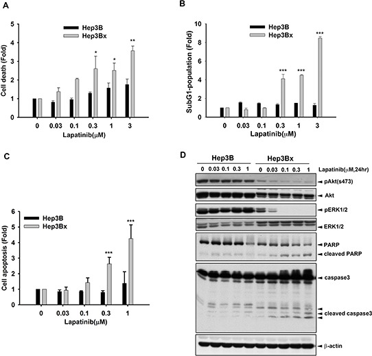
Figure 2. HBx enhanced the sensitization of Hep3B cells to lapatinib-induced apoptosis.

A–C. Hep3B and Hep3Bx were treated with indicated concentration of lapatinib for 5 days. The cells were trypsinized for cell number counting (A), PI staining for determining sub-G1 population (B), and PI and Annexin V double staining for determining apoptosis (C). D. Total lysate prepared from lapatinib-treated Hep3B and Hep3Bx cells were subjected to Western blot analysis with anti-pAkt(s473), anti-Akt, anti-pERK, anti-ERK, anti-PARP, anti-caspase3, and anti-β-actin antibodies. Statistical analysis was performed by Student's t test. *, p < 0.05; **, p < 0.01; ***, p < 0.001 as compared to control group.
