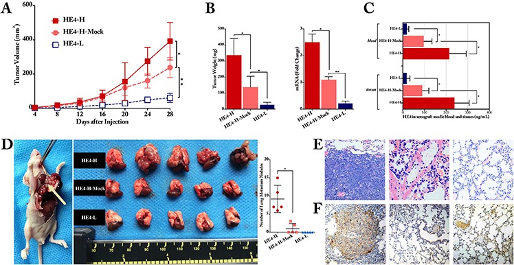
Figure 3. HE4 contributed to ovarian cancer cell progression and metastasis in vivo.

The xenograft tumor formation assay showed that, compared with the mock group, high-expressed HE4 enhanced tumor growth (P < 0.05), whereas low-expressed HE4 inhibited the tumor size formation significantly (P < 0.01, panel A.) At the end of the experiment, compared with the mock group, the mean tumor weight was remarkably heavier in the HE4-H group and lighter in HE4-L group (P < 0.05, panel B). The relative mRNA expression of HE4 in tumors was significantly higher in HE4-H group and lower in HE4-L group than the mock group (P < 0.01 and P < 0.05, respectively, panel B.). ELISA measurement revealed that HE4 protein level was obviously higher in the HE4-H group and lower in the HE4-L group than the mock group both in tumor tissues and blood of the nude mice (all P < 0.05, panel C). For metastasis assay, lung surface metastatic nodules (arrow in panel D.) after inoculation with cancer cells were observed and quantified, the metastatic nodules in HE4-H group were remarkable more than the mock group, and there was no nodule in HE4-L group (panel D) Representative H&E staining of lung tissues showed that metastatic tumor cells in lung tissues section (panel E, from left to right: HE4-H, HE4-H-Mock and HE4-L group). Immunohistochemical staining of HE4 for lung tissues further confirmed the H&E findings (panel F, from left to right: HE4-H, HE4-H-Mock and HE4-L group) the images in panel E and F are partly from Zhuang, H.,et al., Mol Cancer, 2014. 13:p243
