

Introduction
Keratosis follicularis spinulosa decalvans (KFSD) is an inherited rare disorder characterized by diffuse keratosis pilaris and scarring alopecia. Palmoplantar keratoderma, ocular abnormalities, and atopy can also be present.1, 2 Most cases occur in males and have a X-linked pattern of inheritance, although autosomal dominant and sporadic cases have been reported.2 The pathogenesis of this intriguing disease is not well understood. Substance P (SP), an important marker of neurogenic inflammation, is widely distributed in the central and peripheral nervous systems. In the skin, SP-positive nerve fibers can be normally present in the dermis and epidermis.3 SP is considered a potent neuropeptide vasodilator and has been associated with many inflammatory processes, such as mast cell degranulation and neutrophil/lymphocyte chemotaxis. SP can also lower itch thresholds and stimulate fibrosis and healing processes through fibroblast activation.4, 5 Increased levels of SP have been reported in many inflammatory skin conditions, including alopecia areata, atopic dermatitis, and psoriasis.3, 5, 6 We describe a Brazilian girl with a generalized severely pruritic scalp and typical features of KFSD whose lesional skin scalp showed increased levels of the neuropeptide SP. To our knowledge, this is the first time this neuropeptide is reported in the setting of KFSD.
Case report
A 10-year-old white Brazilian girl with a 2-year history of a severely pruritic scalp and progressive hair loss was referred to the Hair Disease Clinic at the University of Sao Paulo. At the time of presentation, she was taking 5 mg of folic acid since age 5 for a congenital meningomielocele related to methylenetetrahydrofolate reductase gene deficiency. On clinical examination, a scarring alopecia with perifollicular and diffuse scale on the vertex area was noted (Fig 1). Keratosis pilaris was evident on the face, limbs, and chest (Fig 2). Teeth, nails, palms, and soles were normal. Ophthalmologic examination and routine laboratory examination found no abnormalities. No family members were affected. Two 4-mm punch scalp biopsy sections were collected for histopathologic analysis, one from the most affected area (vertex) and one from a less affected area (occipital). Histologic examination of vertical sections from the vertex area found perifollicular lymphocytic chronic inflammatory infiltrate, hyperkeratosis in the follicular ostium and fibrosis. Mild perifollicular inflammation could also be noted in the occipital biopsy. Direct immunofluorescence results were negative for both sites. Because of the severe pruritus in this patient, we decided to examine SP expression using an enzyme-linked immunosorbent assay technique on specimens obtained adjacent to the previously biopsied sites. All scalp hairs were plucked from tissue samples before homogenization and centrifugion. The enzyme-linked immunosorbent assay analysis was performed using enzyme immunoassay human SP kits from Cayman (Ann Arbor, MI) and protocols validated by Societè de Pharmacologie ET d'Immunologie–BIO. The most affected and symptomatic scalp area showed 37.2 pg/mL of SP compared with 31.3 pg/mL of SP in the less affected area. Blood samples were also studied and compared with scalp skin and blood samples of 11 control subjects. All serum and control samples showed undetectable results.
Fig 1.
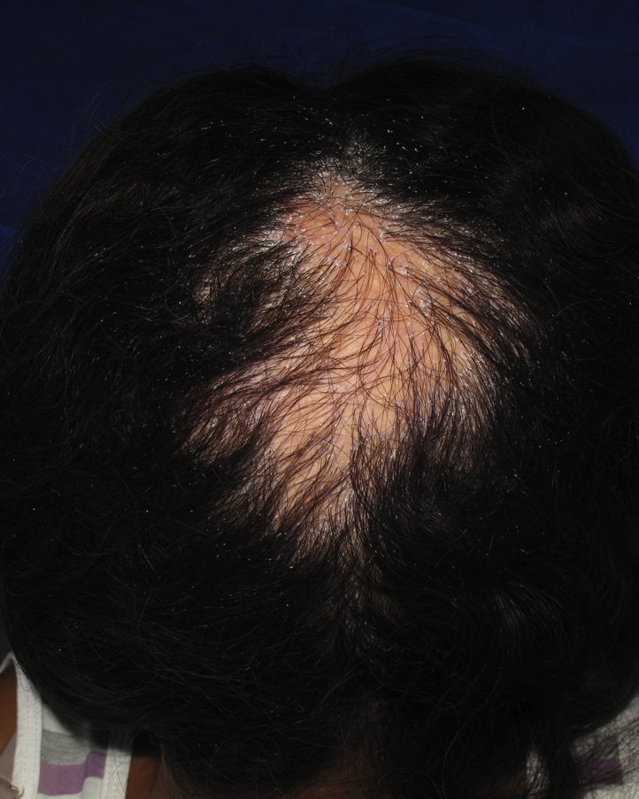
KFSD. Scarring alopecia plaque on the vertex area of the scalp with moderate perifollicular and diffuse scale.
Fig 2.
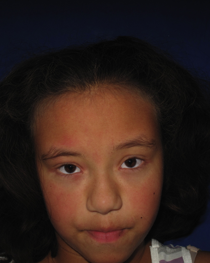
KFSD. Note characteristic keratosis pilaris on the face.
Discussion
The role of neuropeptides in cutaneous innervation is well estabilished by in vitro, in vivo, and clinical studies, and there is substantial evidence that sensory neuropeptides contribute to the development and maintenance of many inflammatory skin conditions and symptoms such as pruritus.4 SP may be related to the overt pruritus experienced by this patient. Because treatment of KFSD is often unrewarding, the finding of increased SP levels in this patient suggests more studies of neurogenic inflammation are needed to assess the pathogenesis of this rare variant of cicatricial alopecia. New medications that target neurogenic inflammation, such as a NK-1R antagonist, may in the future benefit KFSD patients.
Acknowledgments
The authors thank the Universities of São Paulo (Brazil) and Minnesota (USA) for laboratory assistance.
Footnotes
Funding sources: None.
Conflicts of interest: None declared.
References
- 1.Rand R., Baden H.P. Keratosis follicularis spinulosa decalvans: report of two cases and literature review. Arch Dermatol. 1983;119:22–26. doi: 10.1001/archderm.119.1.22. [DOI] [PubMed] [Google Scholar]
- 2.Castori M., Covaciu C., Paradisi M. Clinical and genetic heterogeneity in keratosis follicularis spinulosa decalvans. Eur J Med Genet. 2009;52:53–58. doi: 10.1016/j.ejmg.2008.09.005. [DOI] [PubMed] [Google Scholar]
- 3.Hordinsky M.K., Kennedy W., Wendelschafer-Crabb G. Structure and function of cutaneous nerves in alopecia areata. J Invest Dermatol. 1995;104(5 Suppl):28S–29S. doi: 10.1038/jid.1995.48. [DOI] [PubMed] [Google Scholar]
- 4.Peters E., Ericson M.E., Hosoi J. Neuropeptide control mechanisms in cutaneous biology: physiological and clinical significance. J Invest Dermatol. 2006;126:1937–1947. doi: 10.1038/sj.jid.5700429. [DOI] [PubMed] [Google Scholar]
- 5.Teresiak-Mikolajczak E., Czarnecka-Operacz M., Jenerowicz D. Neurogenic markers of the inflammatory process in atopic dermatitis: relation to the severity and pruritus. Postep Derm Alergol. 2013;5:286–292. doi: 10.5114/pdia.2013.38357. [DOI] [PMC free article] [PubMed] [Google Scholar]
- 6.Saraceno R., Kleyn C.E., Terenghi G. The role of neuropeptides in psoriasis. Br J Dermatol. 2006;154:876–882. doi: 10.1111/j.1365-2133.2006.07518.x. [DOI] [PubMed] [Google Scholar]