

Abstract
We describe a percutaneous technique for repair of type II SLAP lesions. Through the Neviaser portal, a spinal needle is used to pass a FiberStick suture (Arthrex, Naples, FL) through the labrum to create 2 mattress sutures that are secured with PushLock anchors (Arthrex). This technique is simple, reproducible, and knotless and requires no cannulas. At the end of the procedure, minimal suture material remains in the joint.
Superior labral tears extending anterior to posterior were first described by Andrews et al.1 in 1985. SLAP lesions can occur as a result of either compression injuries or traction injuries2 and may be acutely traumatic or occur as a result of continuous or repetitive overhead athletic activity, particularly throwing sports.1
There is evidence that the number of SLAP repairs being undertaken each year is rising.3 The arthroscopic technique for repairing the superior labrum does affect the outcome. Yang et al.2 showed a better range of motion after a horizontal mattress suture repair compared with a conventional vertical knot. We describe a simple, percutaneous knotless technique that minimizes trauma to the rotator cuff.
Surgical Technique
Positioning
The procedure is performed with the patient in the lateral decubitus position. The arm is suspended at an approximately 30° angle of abduction and 10° of forward flexion. Three to five kilograms of skin traction is applied to distract the arm.
Portals
Three portals are used for this procedure: a standard posterior portal, an anterior portal in the rotator interval, and a lateral portal adjacent to the anterolateral edge of the acromion. No cannulas are used.
SLAP Repair
Step 1: Diagnosis
An anterior portal is created in the rotator interval under direct vision, and the diagnosis of a SLAP tear is confirmed by probing the superior labrum.
Step 2: Glenoid Neck Preparation
Viewing from the posterior portal, the surgeon prepares the neck of the glenoid with a tissue elevator, rasps, and a shaver, exposing the subchondral bone. If a shaver is used, we recommend careful use of suction to avoid labrum damage.
Step 3: Suture Passage
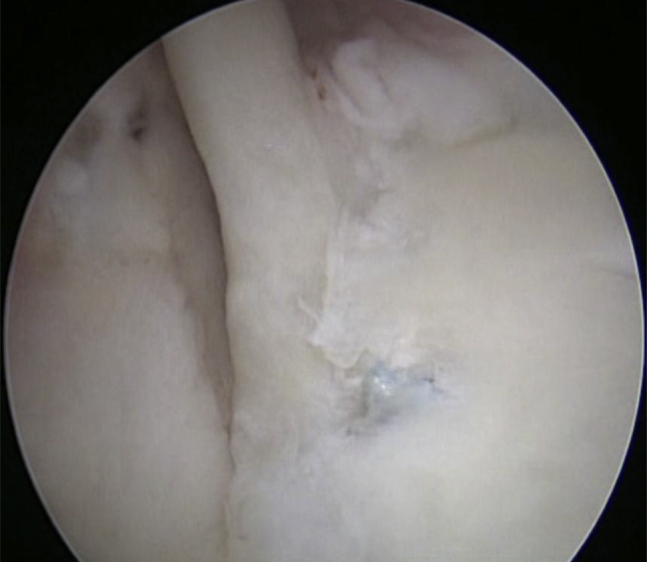
A 19-gauge spinal needle is passed into the joint through the Neviaser portal (Table 1). The trajectory of the needle is such that it enters the joint almost parallel to the face of the glenoid behind the insertion of the long head of the biceps. The needle is then passed through the superior labrum at the 1-o'clock position and exists just below the free edge of the labrum (Fig 1A). A No. 2 FiberStick suture (Arthrex, Naples, FL) is passed through the needle and retrieved through the anterior portal using a suture retriever. The spinal needle is then carefully withdrawn until the tip is clear of the labrum but remains within the joint in the superior recess. The needle is repositioned approximately 0.5 cm posteriorly and passed through the labrum a second time (Fig 1B). The tip of the needle is seen again just below the free edge of the labrum, this time with a loop of suture.
Table 1.
Tips and Pearls
| 1. A needle is used to determine the perfect position of the Neviaser portal. The needle should enter the joint nearly parallel to the glenoid and just behind the long head of the biceps insertion. |
| 2. The surgeon should not use any suction with the shaver when preparing the superior part of glenoid. It should only be used when away from the tissues to clear debris. |
| 3. When the first suture has been retrieved, the needle should be carefully withdrawn until the tip is clear of the labrum but remains within the joint in the superior recess. |
| 4. When the needle has been passed for the second time, it should be advanced as far as possible through the labrum into the joint. If the needle is then withdrawn slightly, a loop of suture is created, which is easier to retrieve through the anterior portal. |
| 5. A spinal needle is used to confirm the optimum position for the anterolateral portal for anchor insertion. |
| 6. The surgeon should drill both anchor pilot holes before removing the drill from the joint; this minimizes trauma to the rotator cuff. |
| 7. Both suture limbs should be retrieved at the same time to avoid soft-tissue bridges before anchor insertion. |
Fig 1.

(A) Viewing from the posterior portal, the needle is passed through the superior labrum at the 1-o'clock position. A suture is then passed through the needle and retrieved through the anterior portal. (B) Viewing from the posterior portal, the needle is carefully withdrawn to remain in the joint and is repositioned 0.5 cm posteriorly and passed through the labrum a second time. The loop of suture is retrieved through the anterior portal.
Step 4: Suture Retrieval
The loop of suture is retrieved through the anterior portal. The needle is withdrawn, and the rest of the posterior suture limb is pulled out of the anterior portal. This leaves a horizontal mattress suture between the 12- and 1-o'clock positions.
The surgeon repeats the process, passing a second mattress suture between the 12- and 11-o'clock positions. It is advisable to use a suture of a different color.
Step 5: Socket Preparation
A spinal needle is passed from a point adjacent to the anterolateral edge of the acromion toward the glenoid face to determine the best trajectory for anchor placement. This needle passes through the muscular portion of the supraspinatus and then the capsule. A stab incision is made, and a drill guide with a pointed sharp trocar is passed into the joint along the same path as the needle. The trocar is withdrawn, and the drill guide is positioned at the edge of the glenoid (Fig 2). Two holes are drilled, one at the 11:30 clock-face position and one at the 12:30 clock-face position.
Fig 2.

Viewing from the posterior portal, the drill guide is inserted and 2 holes are drilled at the 11:30 clock-face position and 12:30 clock-face position. The sutures are then retrieved and the anchors placed.
Step 6: Anchor Insertion
The drill guide is withdrawn, and a suture retriever is passed along the same track to retrieve both limbs of the posterior suture. It is essential that both limbs are withdrawn at the same time to avoid producing a soft-tissue bridge between them. The ends of the suture are passed through the eyelet of a 2.9-mm BioComposite PushLock anchor (Arthrex), and if some tension is applied to the suture limbs, the anchor will slide easily down the previous track. Once the anchor is in place, the sutures can be tensioned and the anchor driven home. The suture tails are cut flush. The process is repeated to secure the anterior suture pair. At the completion of the procedure, 2 mattress sutures lie behind the superior labrum with almost no exposed suture material (Fig 3, Video 1).
Fig 3.
Completed repair, viewing from posterior portal.
Discussion
Our technique combines established arthroscopic elements in a unique way to produce a number of advantages. The technique minimizes trauma to the supraspinatus when passing the suture through the Neviaser portal4 because of the use of a spinal needle rather than a bulkier suture passer. The anchors are passed through a 3.5-mm hole in the muscular portion of the supraspinatus, again causing minimal trauma.
Knotless fixation5, 6 and the use of mattress rather than simple sutures minimize the total amount of suture material in the joint because there is no bulky knot and minimize the amount of exposed suture material that could potentially abrade the humeral head. Rhee and Ha7 reported a case of glenoid and humeral head erosion induced by a knot after a SLAP repair procedure. New techniques are regularly being published, but some still adhere to the use of multiple cannula knots in the repair.8
The described technique is not technically demanding but requires careful portal placement. The guide for the anchors needs to be passed through the muscular portion of the supraspinatus, and the anchors must be directed into the body of the glenoid to avoid damage to the suprascapular nerve.
Footnotes
The authors report the following potential conflict of interest or source of funding: D.T. receives support from Arthrex. E.P. receives support from Arthrex.
Supplementary Data
The technique is shown in the right shoulder of a patient positioned in the lateral decubitus position with 5 kg of skin traction, viewing from the posterior portal. Glenoid preparation and suture management are performed through an anterior rotator interval portal. Needle introduction is performed through the Neviaser portal, and anchor insertion is performed through a portal lateral to the acromion.
References
- 1.Andrews J.R., Carson W.G., McLeod W.D. Glenoid labrum tears related to the long head of the biceps. Am J Sports Med. 1985;13:338–341. doi: 10.1177/036354658501300508. [DOI] [PubMed] [Google Scholar]
- 2.Yang H.J., Yoon K., Jin H., Song H.S. Clinical outcome of arthroscopic SLAP repair: Conventional vertical knot versus knotless mattress sutures. Knee Surg Sports Traumatol Arthrosc. 2016;24:464–469. doi: 10.1007/s00167-014-3449-8. [DOI] [PubMed] [Google Scholar]
- 3.Zhang A.L., Kreulen C., Ngo S.S., Hame S.L., Wang J.C., Gamradt S.C. Demographic trends in arthroscopic SLAP repair in the United States. Am J Sports Med. 2012;40:1144–1147. doi: 10.1177/0363546512436944. [DOI] [PubMed] [Google Scholar]
- 4.Neviaser T.J. Arthroscopy of the shoulder. Orthop Clin North Am. 1987;18:361–372. [PubMed] [Google Scholar]
- 5.Thal R. Arthroscopic Bankart repair using knotless suture anchors. Arthroscopy. 2007;23:566–567. doi: 10.1016/j.arthro.2007.02.012. [DOI] [PubMed] [Google Scholar]
- 6.Dines J.S., Elattrache N.S. Horizontal mattress with a knotless anchor to better recreate the normal superior labrum anatomy. Arthroscopy. 2008;12:1422–1425. doi: 10.1016/j.arthro.2008.06.012. [DOI] [PubMed] [Google Scholar]
- 7.Rhee Y.G., Ha J.H. Knot-induced glenoid erosion after arthroscopic fixation for unstable superior labrum anterior-posterior lesion: Case report. J Shoulder Elbow Surg. 2006;15:391–393. doi: 10.1016/j.jse.2005.03.010. [DOI] [PubMed] [Google Scholar]
- 8.Castagna A., De Giorgi S., Garofalo R., Tafuri S., Conti M., Moretti B. A new anatomic technique for type II SLAP lesions repair. Knee Surg Sports Traumatol Arthrosc. 2016;24:456–463. doi: 10.1007/s00167-014-3440-4. [DOI] [PubMed] [Google Scholar]
Associated Data
This section collects any data citations, data availability statements, or supplementary materials included in this article.
Supplementary Materials
The technique is shown in the right shoulder of a patient positioned in the lateral decubitus position with 5 kg of skin traction, viewing from the posterior portal. Glenoid preparation and suture management are performed through an anterior rotator interval portal. Needle introduction is performed through the Neviaser portal, and anchor insertion is performed through a portal lateral to the acromion.