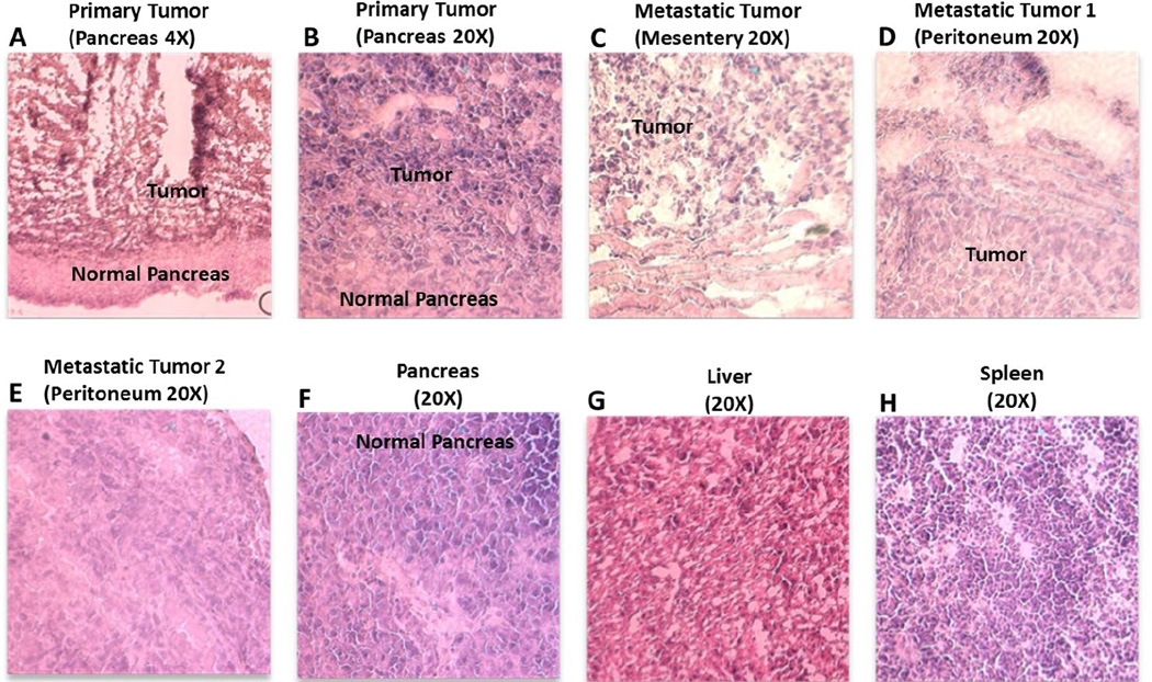
Figure 7.
Hematoxylin and eosin (H&E) stained imaging of tumor and tissue section of orthotopic pancreatic adenocarcinoma tumor bearing mice. The tumor from primary site of injection (A, B) and those resected from mesentery (C) and peritoneum (D, E) showed cancer cells surrounded with normal cells unlike a normal pancreas (F) while sections from liver (G) and spleen (H) did not show any metastatic sites or neoplastic cells.

