Abstract
Cerebrospinal fluid (CSF) leak occurs due to an abnormal communication between the subarachnoid space and sinonasal tract. We reported a retrospective case series of five patients of spontaneous CSF rhinorrhea. These patients were undergone successful repair with a single transnasal endoscopic procedure. This is seen in anterior part of the cribriform plate of middle aged obese females. HRCT paranasal sinus (1 mm cuts) was an effective modality of investigation in our study with ancillary investigations been CT cisternography. Endoscopic repair of CSF rhinorrhea carries a high success rate with very low morbidity rate.
Keywords: Spontaneous CSF rhinorrhoea, Endoscopic repair, Case series
Introduction
Cerebrospinal fluid (CSF) leak occurs as a result of an abnormal communication between the subarachnoid space and a pneumatized area in the skull base that includes the sinonasal tract. This communication or fistula must involve a breech of the arachnoid and dura matter, the bone of skull base, and the underlying mucosa [1]. CSF rhinorrhea can be classified according to the etiology into traumatic and non-traumatic [2]. Traumatic fistulae are subdivided into accidental, which compose the majority (up to 80 %), and iatrogenic or post-surgical (16 %). The non-traumatic or primary CSF fistula accounts for <4 % of all CSF fistulae. The non traumatic CSF fistula can be further classified into high pressure or normal pressure. Because of the serious potential complications of CSF rhinorrhea, (e.g. meningitis, brain abscess, pneumocephalus) prompt management and repair of all CSF rhinorrhea cases should be attempted. Different approaches have been described for the repair of sinonasal CSF fistula, including the intracranial approach which has a high failure rate (up to 70 %) [3]. Multiple extra cranial approaches have been used for repair of CSF leak [4]. Mattox and Kennedy [5] were the first to explain the details of endoscopic repair, reporting an initial success rate of 88 % with a final success rate of 100 %. Careful review of the literature reveals a minor emphasis on non-traumatic CSF leak. Most of the published studies discuss CSF leaks of traumatic origin as it is considered the most common. Therefore, the present study emphasizes on a case series of spontaneous CSF rhinorrhoea.
Materials and Methods
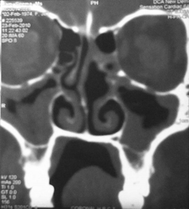
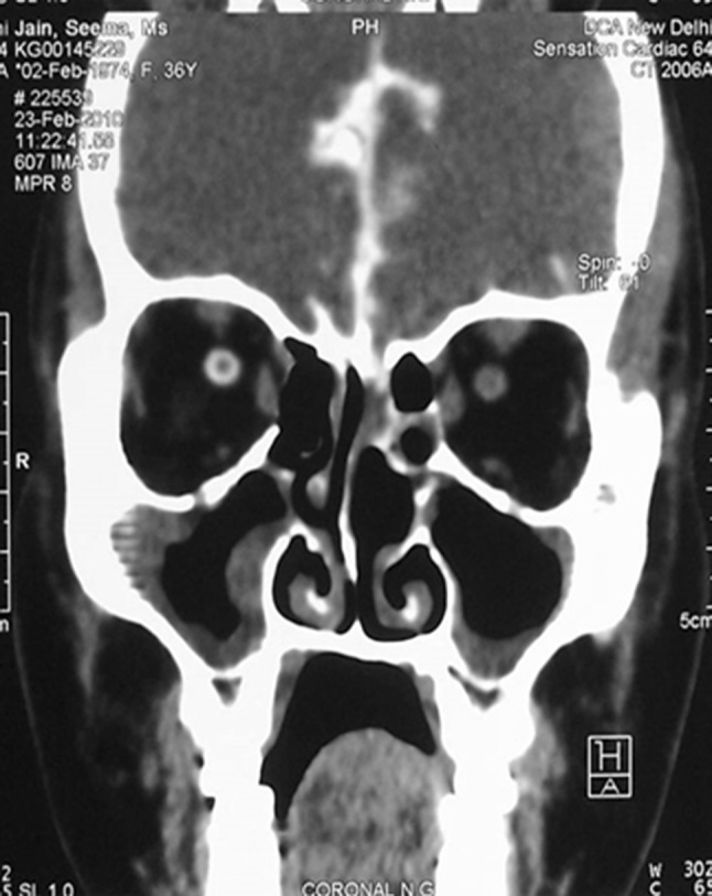
We reviewed the patients who presented in our department with CSF rhinorrhea. Patients with spontaneous CSF leak were included in our retrospective study, while those with history of trauma, previous nasal surgery, congenital malformations of the skull base were excluded from the study. Five patients (Table 1) were included in the study. The most common presenting symptom was continuous watery nasal discharge. All the patients underwent a diagnostic nasal endoscopic examination. The presence of CSF was confirmed by CSF analysis. CT scan coronal cuts (1 mm) (Fig. 1) of the paranasal sinuses were obtained for all the patients. Additional investigations included CT cisternography (Fig. 2), MRI paranasal sinus. The most common site for CSF leak was the anterior part of cribriform plate according to the study. The repair of the CSF leak was done by the extracranial transnasal endoscopic approach under GA. Pre operatively lumbar drain was kept, CSF pressure was raised in all the patients. Patient under general anaesthesia, adequate exposure to the site of leak was obtained. Once the site of leak was identified, the mucosa from the edges of the bony defect was denuded. Fascia lata graft harvested from the thigh and trimmed to appropriate size. Fascia lata is placed by the underlay technique laterally and overlay technique medially followed by another layer of Fascia lata over the initial graft, followed by gel foam and merocel nasal pack. Merocel pack was removed on the third post operative day. Lumbar drain was kept for 2 days post operatively.
Table 1.
Description of the clinical details of the patients in the study
| Our data | |||||
|---|---|---|---|---|---|
| Patient | 1 | 2 | 3 | 4 | 5 |
| Age/sex | 40/male | 39/female | 36/female | 28/male | 54/female |
| Weight (Kg) | 70 | 74 | 97 | 80 | 79 |
| Chief complain | Watery nasal discharge (nd) | Watery nasal discharge | Watery nasal discharge + headache | Watery nasal discharge | Watery nasal discharge + sneezing |
| Duration of chief complain | 1 months | 4 months | 20 days | 3 months | 2 months |
| Side | Right | Right | Left | Left | Right |
| Site of leak | Right cribriform | Right cribriform | Left cribriform | Left cribriform | Right cribriform |
| Olfaction | Normal | Normal | Normal | Normal | Normal |
| Csf pressure | Raised | Raised | Raised | Raised | Raised |
| Surgery | Sep + Fess + Tner | Sep + Fess + Tner | Sep + Fess + Tner | Sep + Fess + Tner | Sep + Fess + Tner |
| Site of leak on table | Right cribriform | Right cribriform | Left cribriform | Left cribriform | Right cribriform |
| Size of the leak | 2 mm | 2.5 mm | 3 mm | 2 mm | 3 mm |
| Graft | Fascia lata | Fascia lata | Fascia lata | Fascia lata | Fascia lata |
| Leak free post op period | 9 months | 7 months | 9 months | 2 years | 8 months |
| Investigation | CSF analysis + CT + MRI | CSF analysis + CT + MRI | CSF analysis + CT + CT cisternography | CSF analysis + CT + CT cisternography | CSF analysis + CT cisternography |
Sep septoplasty, Fess functional endoscopic sinus surgery, Tner transnasal endoscopic repair of CSF leak
Fig. 1.

High resolution computerised tomography (HRCT) nose paranasal sinuses coronal images showing the defect in the cribriform plate on left side in Case-3
Fig. 2.

CT cisternography coronal images showing CSF leak into the left nasal cavity from the left cribriform region in Case-3
Results
Out of the five, three were females and two were males, their ages ranged between 28 and 54 years and weight between 70 and 97 kg, the follow up period ranged between 7 months and 2 years. The most common site of leak was the anterior cribriform plate with size of the leak in five patients ranged between 2 and 3 mm. The clear fluids collected from the patients were sent for CSF analysis and was confirmed to be CSF in all the five patients. CSF pressure was elevated in all our patients. HRCT scan paranasal sinus studies were positive in all of the five patients. CT cisternography was done and positive in three patients, MRI (T2) were done in two patients and was used to localize the defect where the leak was not active during the study. Endonasal endoscopic approach under general anaesthesia was used in all the patients to repair the CSF leak with auto graft fascia lata. All the patients had successful cessation of the rhinorrhea after a single procedure with post operative leak free periods ranging from 7 months to 2 years. No major complications occurred secondary to surgical management.
Discussion
Non-traumatic CSF rhinorrhea can be spontaneous, caused by a bony defect. The causes of spontaneous CSF rhinorrhea are variable and not very well understood. Obesity is a very important risk factor. Overweight increases intra abdominal and intra thoracic pressure. This may effect blood circulation in cranial venous collectors and lead to development of permanent benign intracranial hypertension [6]. The causes of elevated intracranial pressure (ICP) can be multifactorial; nevertheless, once elevated ICP develops, the pressure exerted on areas of the anterior skull base (e.g., cribriform, lateral recess of the sphenoid sinus) result in remodeling and thinning of the bone. Ultimately, the bone is weakened until a defect is formed. If a defect is large, brain parenchyma may be herniated as well Since the communication between a sterile intracranial compartment and a nonsterile sinonasal cavity can lead to life threatening complications(e.g., meningitis, pneumocephalus, or brain abscess, prompt diagnosis and management is demanded. Chemical analysis of the collected samples of the rhinorrhea is important to confirm the nature of the CSF fluid. Radiological investigations play a pivotal role in identifying the underlying etiology of CSF rhinorrhea and in detecting the site, side, and size of the leaking fistula. With thin 1 mm cuts of coronal section, a CT scan was helpful in detecting bony dehiscence [7]. High resolution CT scan has an overall sensitivity of 70 % in detecting bony dehiscence. However, the rate of detection is much lower in cases of spontaneous CSF rhinorrhea with <2 mm skull base defects, due to partial volume averaging, resulting in false positive and false negative results. MRI is a valuable non-invasive test for detecting and localizing CSF leak. Modification of MRI technique by using both T2 MRI images with fluid attenuated inversion recovery (FLAIR) imaging was very helpful in localizing CSF rhinorrhea. CT cisternography is another valuable radiographic study that is considered the procedure of choice in detecting CSF leak [8]. It requires administration of a low osmolar non-ionic iodine contrast agent into the subarachnoid space with a subsequent search for agent egress through the fistula site [7]. The sensitivity can reach up to 90 % in active leaks. However, this technique is invasive and the sensitivity can be as low as 40 % in cases of intermittent or inactive leaks [9]. Surgical management of CSF rhinorrhea can be achieved by an intracranial or extracranial approach. Management of CSF rhinorrhea by an intracranial approach carries morbidity and failure rates between 20 and 40 % [10]. On the other hand, an endoscopic approach is less morbid and has a success rate of 90–100 % [5]. In the present study endoscopic approach for closure of the leak was done with combined overlay and underlay technique. This technique is useful for defects localized to the cribriform area since this combined technique doesn’t cause alteration of olfaction post operatively. Adjunctive techniques that can be used to increase the success rate of CSF fistula repair include perioperative antibiotics, diuretics, lumbar drain, and complete bed rest with elevation of the head. We did not obtain results with respect to the use or withholding the use of these techniques due to ethical considerations and the results have to be viewed in this perspective. The contribution of adjunctive techniques to the overall success needs further evaluation.
Conclusion
Spontaneous CSF rhinorrhea is a relatively rare condition occurring due to different etiologies mostly seen in middle aged obese female patients. Prompt diagnosis and management of the condition is necessary because of the potential risky complications. Proper history taking, preoperative examination, precise preoperative localization is essential for successful repair. Among the multiple techniques available for localisation include HRCT paranasal sinus, CT Cisternography, MRI paranasal sinus. In the absence of a large skull base lesion or tumor, endoscopic repair of a CSF fistula with auto graft fascia lata carries a high success rate with a very high margin of safety and very low morbidity rate. All our cases were cribriform plate defects 2–3 mm in size where the dura cannot be elevated medially. Fascia lata was placed as an overlay graft medially, and in an underlay fashion posteriorly, anteriorly, and laterally. Cartilage is not suitable at this site due to its inelasticity. Our maximum follow up is for 2 years with an average follow up of 11 months. No recurrences were seen.
Compliance with Ethical Standards
Conflict of interest
The authors declare that there is no conflict of interests.
Ethical Approval
This article does not contain any studies with animals performed by any of the authors. All procedures performed in this study involving human participant was in accordance with the ethical standards of the institutional and/or national research committee and with the 1964 Helsinki declaration and its later amendments or comparable ethical standards.
Informed Consent
Informed consent was obtained from the participants included in the study.
Contributor Information
Aniruddha Sarkar, Email: drsarkar17@gmail.com.
Nishi Sharma, Email: drsharmanish@yahoo.co.in.
References
- 1.Zapalac JS, Marple BF, Schwade ND. Skull base cerebrospinalfluid fistulas: comprehensive diagnostic algorithm. Otolaryngol Head Neck Surg. 2002;126:669–676. doi: 10.1067/mhn.2002.125755. [DOI] [PubMed] [Google Scholar]
- 2.Ommaya AK, DiChiro G, Baldwin M, et al. Nontraumatic cerebrospinal fluid rhinorrhea. J Neurol Neurosurg Psychiatry. 1968;31:214–255. doi: 10.1136/jnnp.31.3.214. [DOI] [PMC free article] [PubMed] [Google Scholar]
- 3.Park JI, Strezlow VV, Freidman WH. Current management of cerebrospinal fluid rhinorrhea. Laryngoscope. 1983;93:1294–1300. doi: 10.1002/lary.1983.93.10.1294. [DOI] [PubMed] [Google Scholar]
- 4.Dohlman G. Spontaneous cerebrospinal rhinorrhea. Acta Otolaryngol Suppl (Stockh) 1948;67:20–23. doi: 10.3109/00016484809129635. [DOI] [PubMed] [Google Scholar]
- 5.Mattox DE, Kennedy DW. Endoscopic management of cerebrospinal fluid leaks and cephaloceles. Laryngoscope. 1990;100:857–862. doi: 10.1288/00005537-199008000-00012. [DOI] [PubMed] [Google Scholar]
- 6.Badia L, Loughran S, Lund V. Primary spontaneous cerebrospinal fluid rhinorrhea and obesity. Am J Rhinol. 2001;15:117–119. doi: 10.2500/105065801781543736. [DOI] [PubMed] [Google Scholar]
- 7.Wax MK, Ramadan HH, Ortiz O, et al. Contemporary management of cerebrospinal fluid rhinorrhea. Otolaryngol Head Neck Surg. 1997;116:442–449. doi: 10.1016/S0194-5998(97)70292-4. [DOI] [PubMed] [Google Scholar]
- 8.Johnson DBS, Toland BJ, O’Dwyer AJ. Magnetic resonance imaging in the evaluation of cerebrospinal fluid fistulae. Clin Radiol. 1996;51:837–841. doi: 10.1016/S0009-9260(96)80079-1. [DOI] [PubMed] [Google Scholar]
- 9.EL Jamel MS, Pidgeon CN, Toland J, et al. MR cisternography and the localization of CSF fistulae. Br J Neurosurg. 1994;8:433–437. doi: 10.3109/02688699408995111. [DOI] [PubMed] [Google Scholar]
- 10.Nachtigal D, Frenkiel S, Yoskovitch A, Mohr G. Endoscopic repair of cerebrospinal fluid rhinorrhea: is it the treatment of choice? J Otolaryngol Head Neck Surg. 1999;28:129–133. [PubMed] [Google Scholar]


